Core structure of gp41 from the HIV envelope glycoprotein
a glycoprotein and core structure technology, applied in the field of core structure of gp41 from the hiv envelope glycoprotein, can solve the problem that it has not been possible to obtain a detailed structure for gp41, and achieve the effect of preventing or inhibiting gp41 activity
- Summary
- Abstract
- Description
- Claims
- Application Information
AI Technical Summary
Benefits of technology
Problems solved by technology
Method used
Image

Examples
example 1
Production of Crystals of N36 / C34
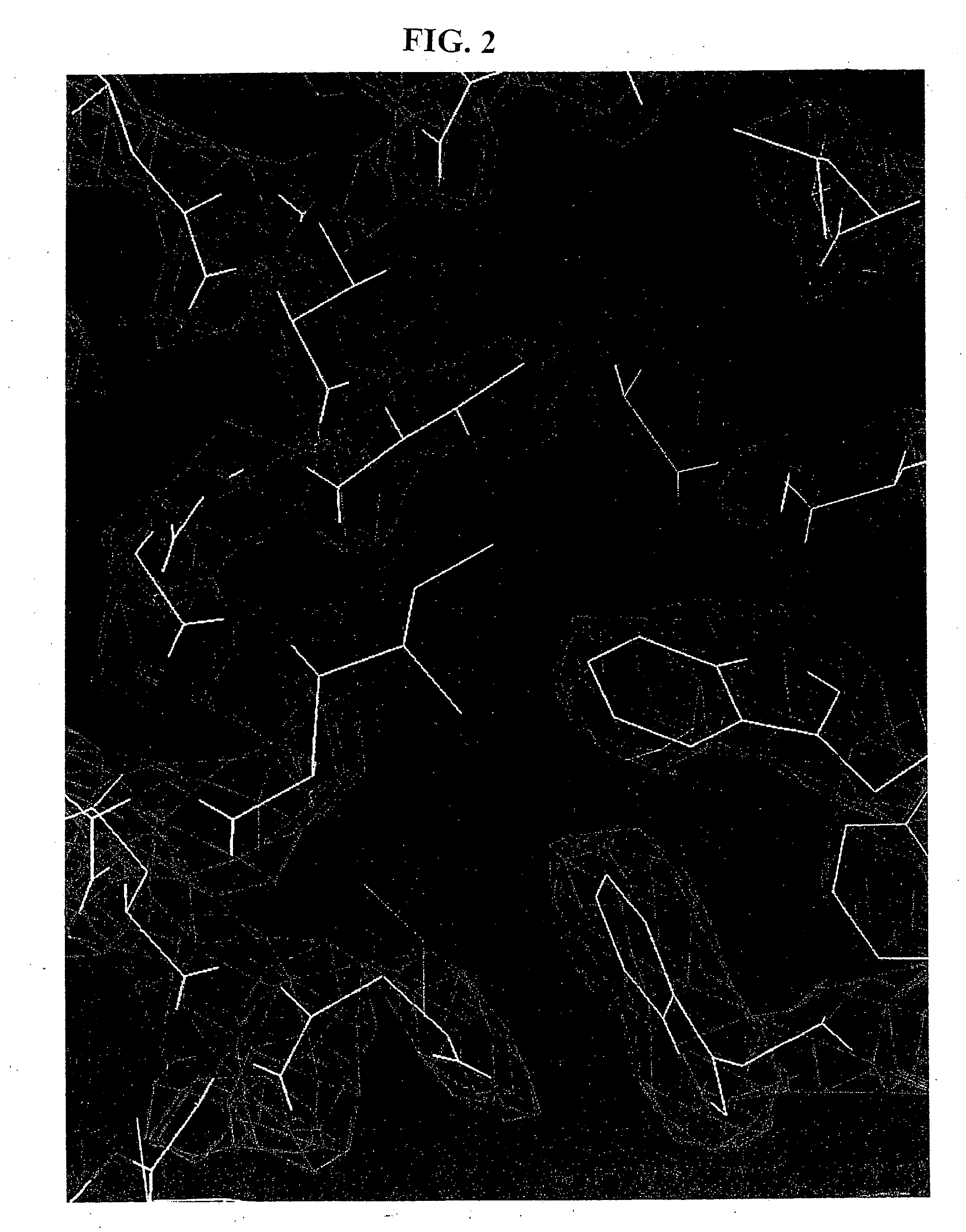
[0070] Crystals of N36 / C34 were grown by sitting-drop vapor diffusion (see Methods). An initial model of the complex was built into an electron density map generated by multi-wavelength anomalous dispersion (MAD) analysis (Hendrickson, W. A., Science 254:51-58 (1991)) of an osmium-derivatized crystal. Details of data collection and MAD phasing statistics are listed in the Table. A representative portion of the solvent-flattened electron density map used for building the initial model is shown in FIG. 2. The structure was refined against data to 2.0 Å from a native crystal to yield an Rfree of 0.266 and an Rcryst of 0.238 (Table).
TABLECrystallographic arid refinement statisticsData collectionCrystalλ (Å)% completeRsym1 (%)Resol. (Å)Native1.541896.55.52.0OsO4 λ11.139896.44.32.7OsO4 λ21.139695.44.32.7OsO4 λ31.134496.84.52.7OsO4 λ41.140693.44.52.7Phasing statistics (12-2.7 Å)Riso2Rdiff3 (%)Rcullis4Rcullis4Derivative(%)(weight)AcentricCentricOsO44.46.7...
example 2
Assessment of the Structure of the N36 / C34 Complex
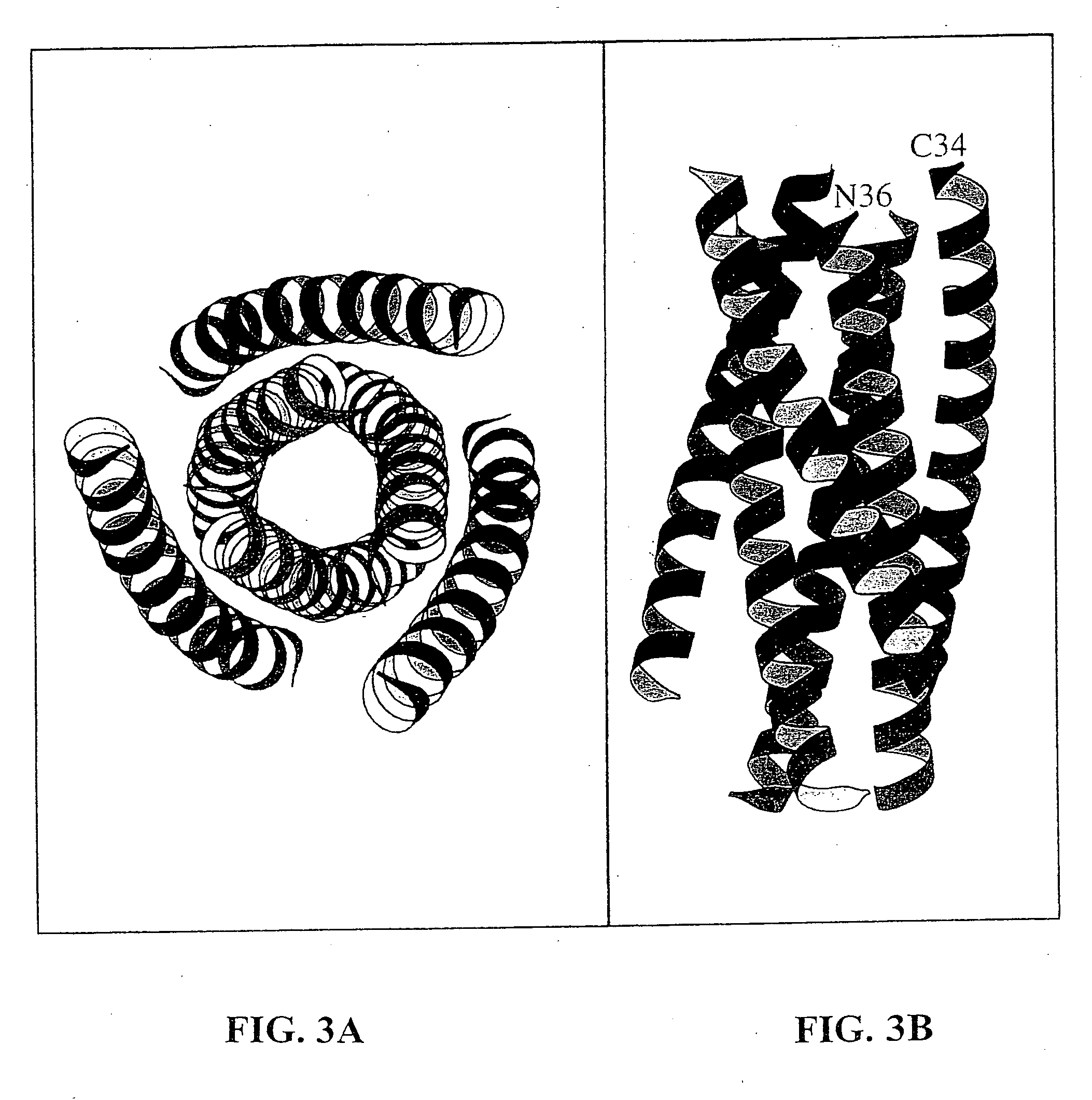
[0071] The N36 / C34 complex is a six-stranded helical bundle (FIG. 3). The center of this bundle consists of a parallel, trimeric coiled coil of three N36 helices wrapped in a gradual left-handed superhelix. Three C34 helices wrap antiparallel to the N36 helices in a left-handed direction around the outside of the central coiled-coil trimer. The complex is a cylinder measuring ˜35 Å in diameter and ˜55 Å in height.
[0072] As in other naturally-occurring coiled coils (Cohen, C. et al., Proteins 7: 1-15 (1990)), the interior residues at the a and d positions of the N36 heptad repeat are predominantly hydrophobic, although occasional buried polar interactions are also present in the central three-stranded coiled coil (FIG. 4). A sequence comparison of HIV-1 (HXB2 strain) and SIV (Mac239 strain) gp41 shows that the residues at these two heptad-repeat positions are highly conserved (FIG. 4). The characteristic “knobs-into-holes” packing o...
example 3
Determination of Interactions Between the—and C-Peptide Helices
[0074] Three C34 helices pack obliquely against the outside of the N36 coiled-coil trimer in an antiparallel orientation. These C34 helices interact with N36 mainly through hydrophobic residues in three grooves on the surface of the central coiled-coil trimer. Sequence comparisons between HIV and SIV gp41 shows that the residues lining these grooves are highly conserved. In contrast, the N36 residues flanking the C34 helices are divergent between HIV and SIV.
[0075] This pattern of sequence conservation is also apparent on a helical wheel representation of three N36 helices and one C34 helix (FIG. 4). In this diagram, the residue positions in C34 are depicted as ellipses to indicate the oblique tilt of the C34 helix relative to the N36 superhelix and to emphasize that C34 is not part of a coiled coil. Residues at the e and g positions of the N36 helices lie on the outside of the central coiled coil and point into the tr...
PUM
| Property | Measurement | Unit |
|---|---|---|
| melting temperature | aaaaa | aaaaa |
| Core Structure | aaaaa | aaaaa |
| structure | aaaaa | aaaaa |
Abstract
Description
Claims
Application Information