

Pancreatic small cells and uses thereof
a technology of pancreatic cancer and small cells, applied in the field of pancreatic cancer and progenitor cells, can solve the problem of limited evidence of this
- Summary
- Abstract
- Description
- Claims
- Application Information
AI Technical Summary
Benefits of technology
Problems solved by technology
Method used
Image

Examples
example 1
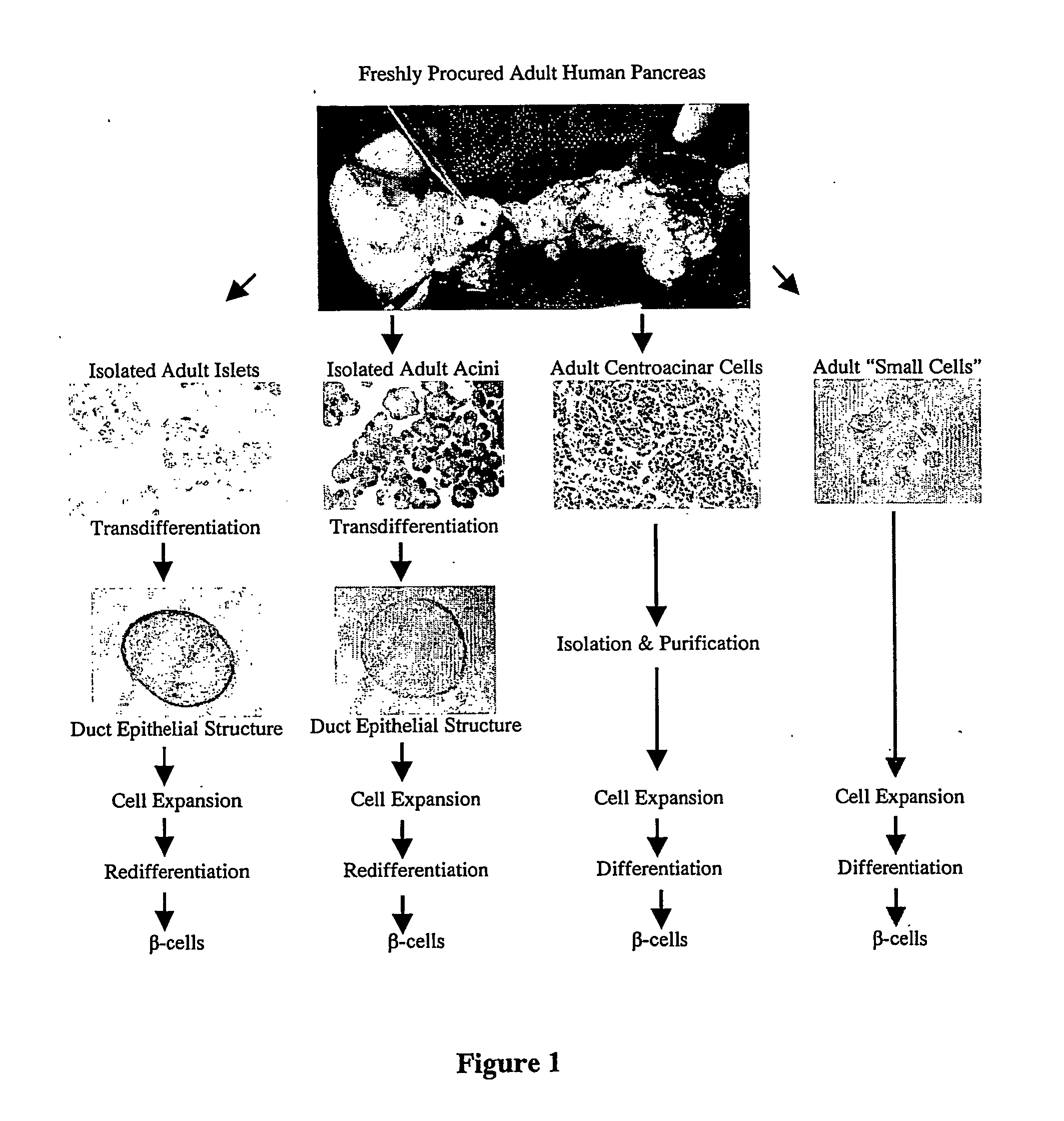
In Vivo Model for Islet Cell Neogenesis
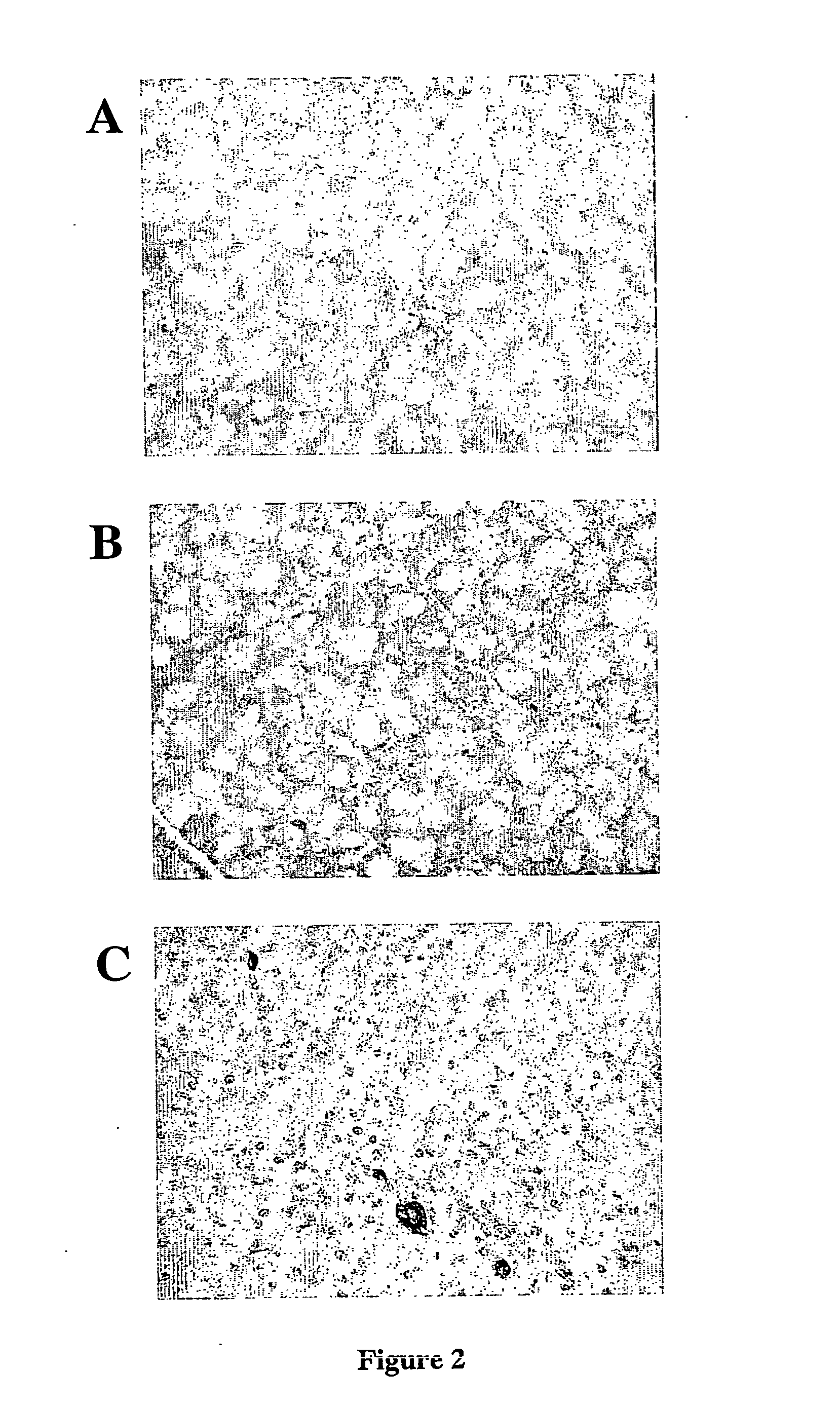
[0085] An in vivo model for islet cell neogenesis and islet formation has been developed [Rosenberg et al., J. Surg. Res, 35:63-72 (1983)], in which new islets appeared to be derived from cells associated with the ductal epithelium (FIG. 2A).
[0086] In the model, islet cell neogenesis is mediated by the novel acinar cell protein INGAP [Rafaeloff et al., JCI, 99:2100-2109 (1997)]. While many of the new islets clearly bud from adjacent ductal epithelium, a careful re-analysis of the tissues from over one thousand hamster pancreata, and more recently a study of canine pancreata exposed to INGAP peptide, have demonstrated that new β-cells also appear (and small islets form) amidst the exocrine tissue (FIG. 2B, C), independent of duct epithelium.
[0087] The appearance of these new β-cells indicates that previously unrecognised progenitor cells are present in the pancreas, which give rise to the new β-cells. While immature cells have been shown to b...
example 2
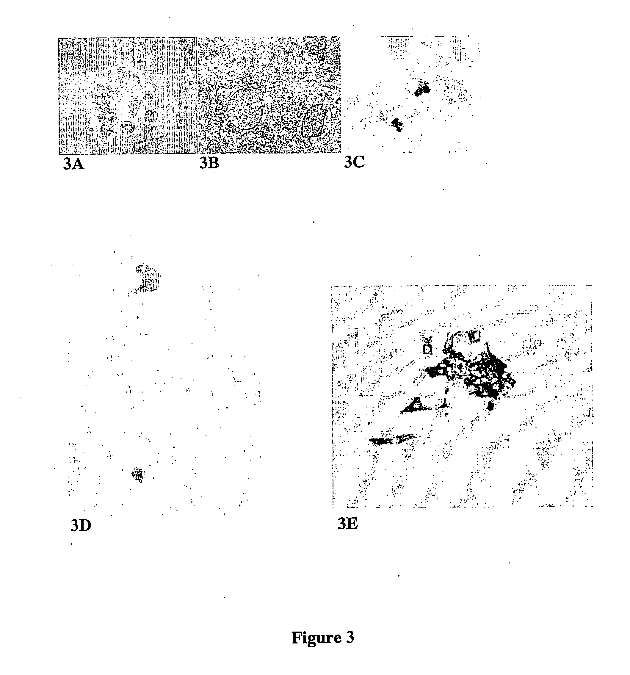
Preliminary Isolation, Purification and Characterisation of Pancreatic Small Cells
[0088] Small cells were first identified in cellular digests of cadaveric human pancreata that had been processed for islet isolation for transplantation. Human islet isolation was performed by the semi-automated method originally proposed by Ricordi [Ricordi, et. al., Diabetes, 37:413-420 (1988)]. Procured pancreases are distended by intra-ductal infusion of Liberase® HI (Roche Molecular Biochemicals, Indianopolis, Ind.) or Serva Collagenase (Cresent Chemical, Brooklyn, N.Y.) [Linetsky et al., Diabetes, 46:1120-1123 (1997)], and then dissociated using the automated method [Ricordi, et. al, Diabetes, 37:413-420 (1988)]. The separation occurs during a process of continuous digestion lasting approximately 12-30 minutes, after which the digestion circuit was cooled and the tissue collected into approximately 8 litres of cold Hanks solution and washed. Liberated islets were separated from non-islet tissue...
example 3
Isolation and Propagation of Canine and Human Pancreatic Small Cells
[0100] Pancreata from mongrel dogs (2-4 years old with body weight 20-25 kg) were removed under general anaesthesia in accordance with Canadian Council for Animal Care (CCAC) guidelines. Human pancreata were obtained from heart-beating cadaveric donors following in situ flush with University of Wisconsin solution at the time of multi-organ harvest for transplantation. Prior consent for organ donation was obtained by the local procurement organisation Quebec-Transplant. Cold ischemia time varied between 4 to 8 hours.
[0101] Islet isolation from both canine and human pancreas was carried out using enzymatic digestion with Liberase® CI and Liberase® HI (Roche Diagnostics, Laval, Que., Canada) for canine and human pancreas respectively, mixed with 0.1 mg / ml DNase I (Roche Diagnostics, Laval, Que., Canada). Digestion was followed by semi-automated dissociation and EuroFicoll purification as previously described [Paraske...
PUM
| Property | Measurement | Unit |
|---|---|---|
| diameter | aaaaa | aaaaa |
| temperature | aaaaa | aaaaa |
| temperature | aaaaa | aaaaa |
Abstract
Description
Claims
Application Information