Method and apparatus for time gating of medical images

a technology of medical images and time gating, applied in the field of medical imaging technology, can solve the problems of limited functionality and capability of existing tools, and the need for medical imaging technology

Active Publication Date: 2005-04-14
ST JUDE MEDICAL ATRIAL FIBRILLATION DIV
View PDF94 Cites 228 Cited by
  • Summary
  • Abstract
  • Description
  • Claims
  • Application Information

AI Technical Summary

Benefits of technology

[0008] In an embodiment of the present invention, a medical imaging system includes a signal generator configured to obtain a trigger signal corresponding to a timing of interest, imaging equipment configured to obtain a plurality of images of a feature of interest, such as an intra-cardial chamber, and a processor programmed to correlate the plurality of images with the trigger signal. The signal generator may include at least one sensor, such as an intra-cardiac electrocardiograph (ECG) sensor, adapted and configured to sense a physiological condition of a patient, the trigger signal corresponding to a physiological condition sensed by the at least one sensor (e.g., a particular ECG signal or a periodic atrial fibrillation feature) designated automatically or by user selection. The imaging eq

Problems solved by technology

Further development in the area of medical imaging technology is needed,

Method used


Image

  • Method and apparatus for time gating of medical images
  • Method and apparatus for time gating of medical images
  • Method and apparatus for time gating of medical images

Examples


Embodiment Construction

[0015] Reference will now be made in detail to exemplary embodiments of the present invention. Wherever possible, the same reference numbers will be used throughout the drawings to refer to the same or like parts.

[0016] A. Exemplary Medical Imaging Systems

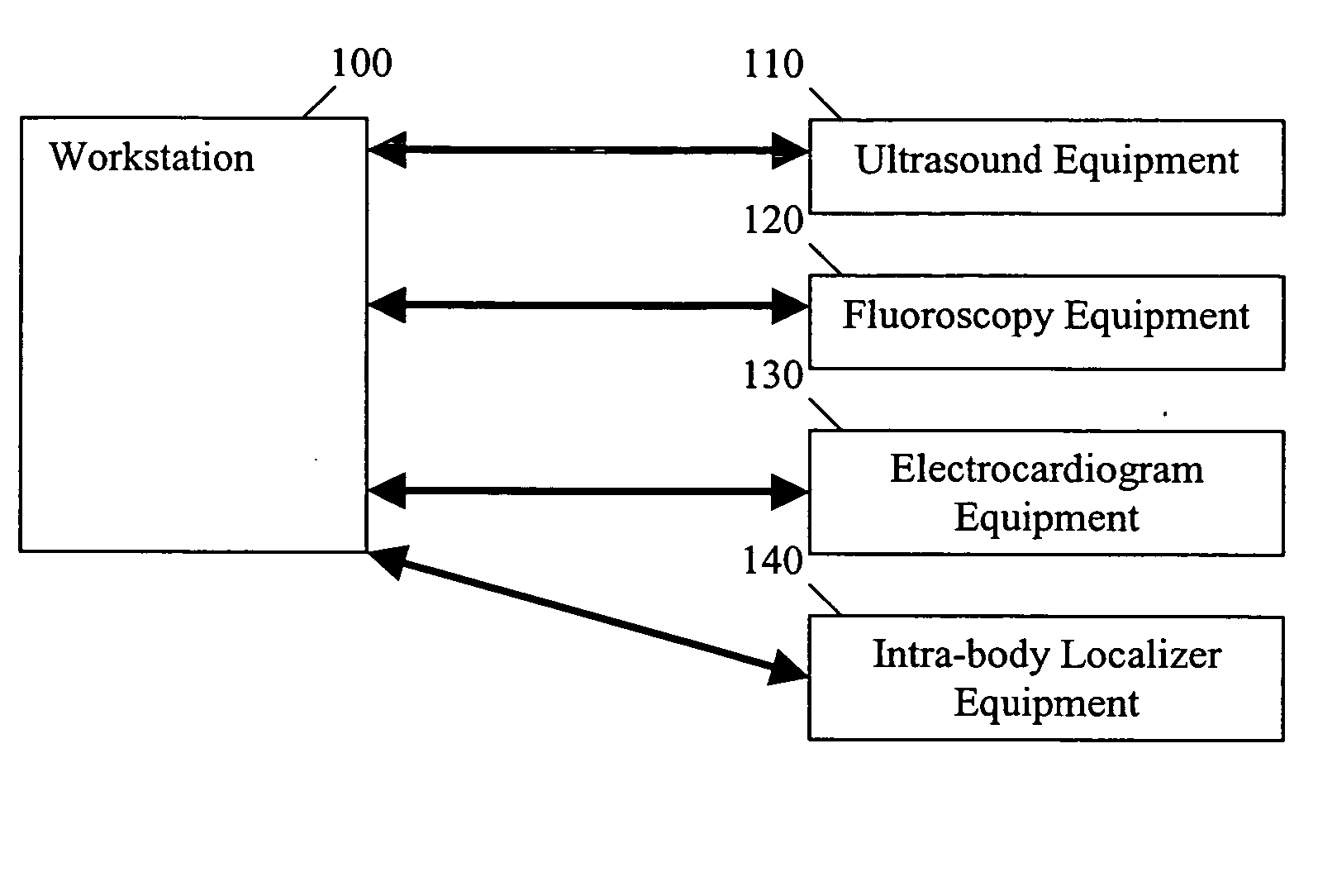
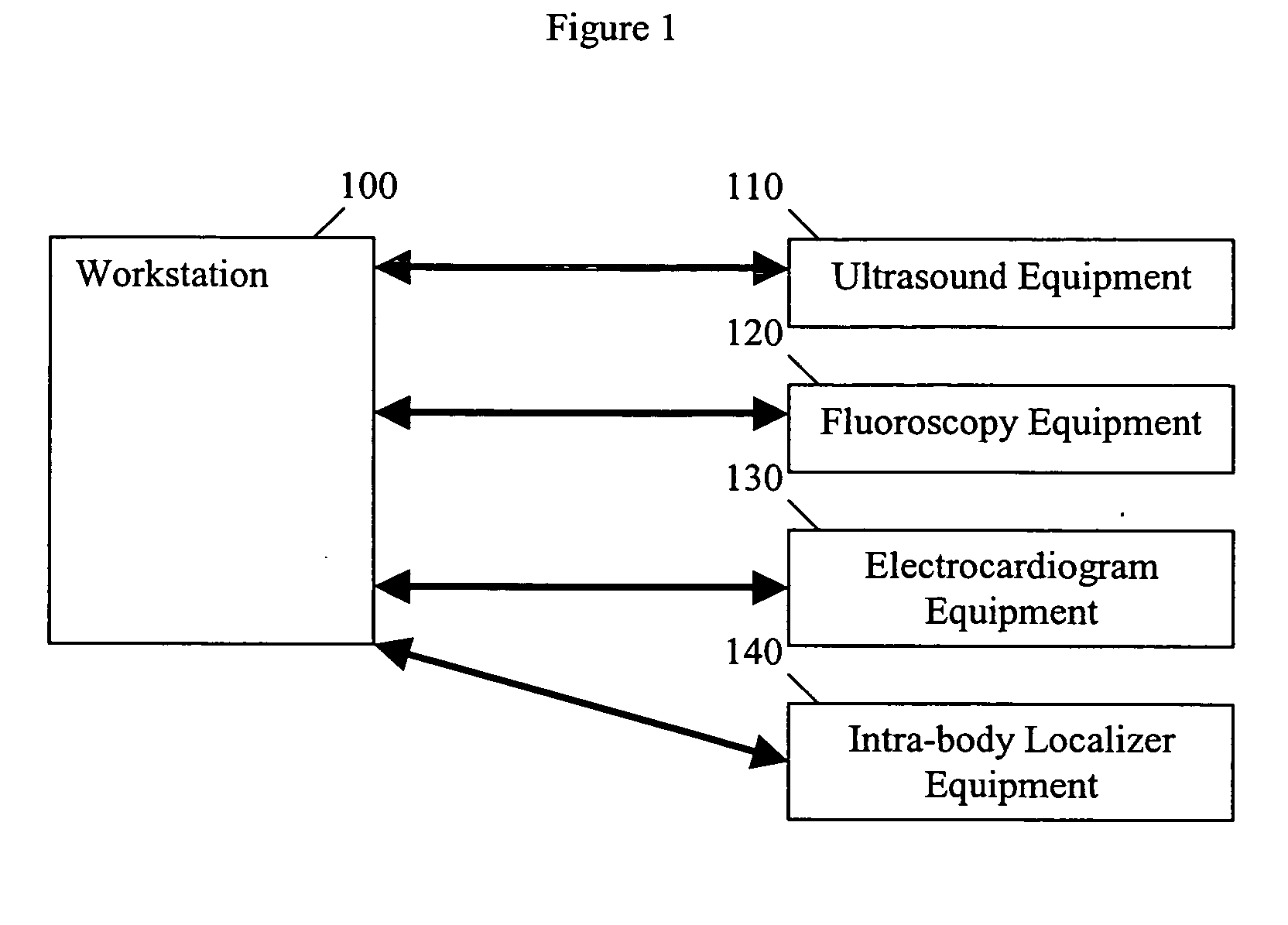
[0017] An exemplary medical imaging system useable with various embodiments of the present invention is shown in the block diagram of FIG. 1. The medical imaging system comprises workstation 100, ultrasound equipment 110 (a first imaging equipment), fluoroscopy equipment 120 (a second imaging equipment), electrocardiogram (ECG) equipment 130 (a physiological measuring equipment), and intra-body localizer equipment 140. Additional components may also be provided and / or some of the depicted components may be combined or eliminated in some embodiments, as would be readily apparent to one of ordinary skill in the art after reading this disclosure.

[0018] Preferably, first imaging equipment 110 includes a percutaneous ultrasound imagi...


PUM


Abstract

A medical imaging system is provided which includes a signal generator configured to obtain a trigger signal corresponding to a timing of interest, imaging equipment configured to obtain a plurality of images of a feature of interest, and a processor programmed to correlate the plurality of images with the trigger signal. Also provided is a method of correlating a plurality of medical images by obtaining a trigger signal of a timing of interest, obtaining a plurality of images of a feature of interest, and correlating the plurality of images with the trigger signal.

Description

CORRESPONDING RELATED APPLICATIONS [0001] The present invention is a continuation in part of U.S. patent application Ser. No. 10 / 620,517 filed on Jul. 16, 2003, entitled Method And System For Using Ultrasound In Cardiac Diagnosis And Therapy claiming priority to Provisional Application Ser. No. 60 / 397,653 filed on Jul. 22, 2002. The entire contents of both these applications are incorporated by reference herein in their entirety.BACKGROUND OF THE INVENTION [0002] 1. Field of the Invention [0003] The present invention is directed at medical imaging technology, and more particularly, to methods and apparatuses for time gating of medical images. [0004] 2. Description of the Related Art [0005] Medical imaging technology is used to improve the diagnosis and treatment of medical conditions. Presently available medical imaging technology includes a wide variety of imaging systems, such as fluoroscopy, angiography, echocardiography, computed tomography, ultrasound, x-ray, nuclear, magnetic ...

Claims


Application Information

Patent Timeline
IPC IPC(8): A61B8/06
CPCA61B8/065A61B8/543A61B8/0883
Inventor BYRD, CHARLES BRYANDALA-KRISHNA, PRAVEEN
Owner ST JUDE MEDICAL ATRIAL FIBRILLATION DIV
Why Eureka
Try Eureka