Robotic surgical device

a robotic and surgical technology, applied in the field of robotic devices, can solve the problems of difficult hand-eye coordination, significant decrease in tactile perception, and the technique itself introduces other things, so as to reduce the time necessary, reduce the pre-surgical prep time, and minimize the trauma to the patient

Inactive Publication Date: 2005-05-05
CEDARS SINAI MEDICAL CENT
View PDF23 Cites 766 Cited by
  • Summary
  • Abstract
  • Description
  • Claims
  • Application Information

AI Technical Summary

Benefits of technology

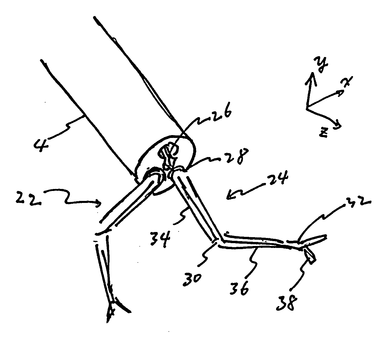
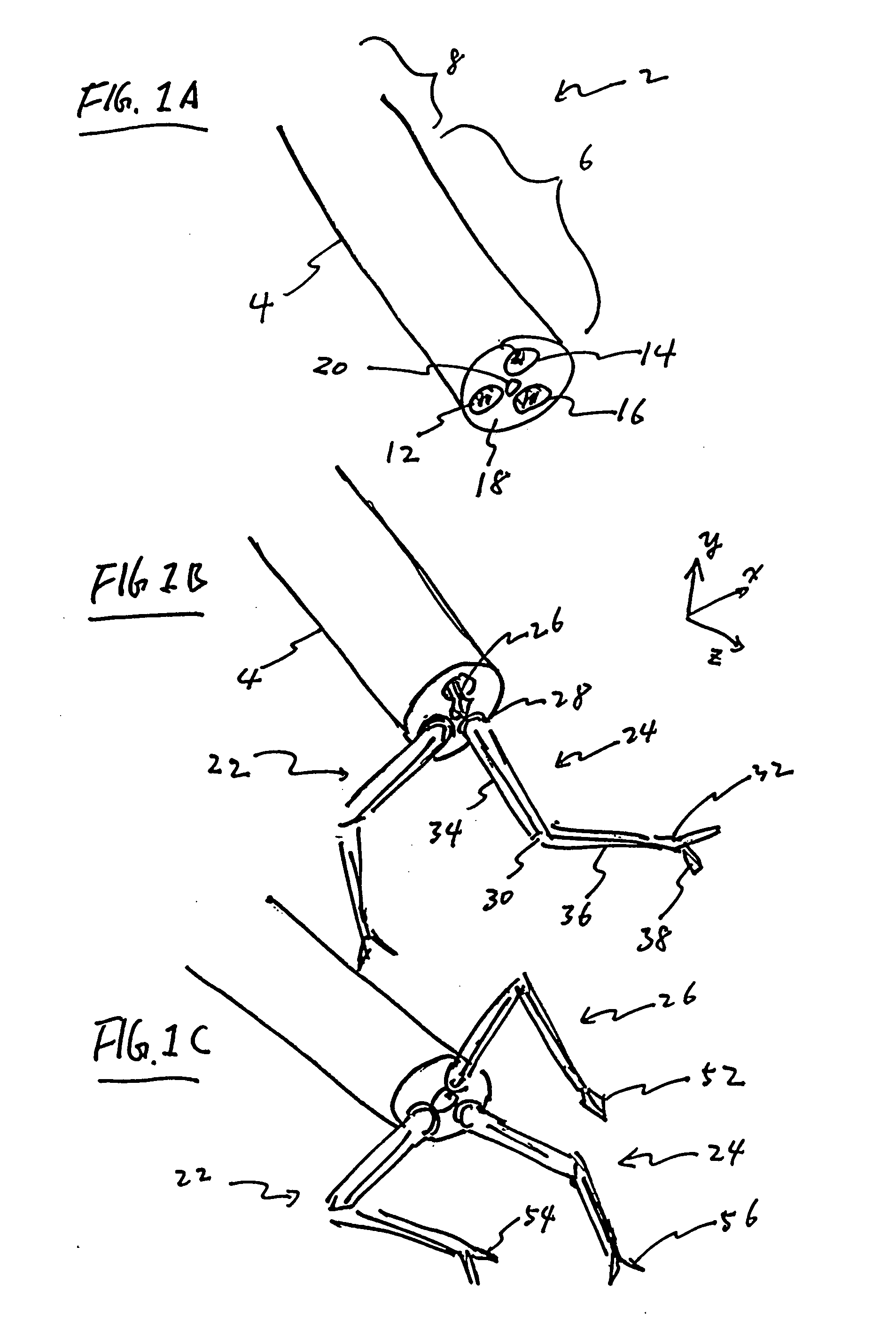
[0016] The ability to introduce multiple robotic arms through a signal incision and having the plurality of arms function in a coordinated manner to accomplish a surgical task inside a patient's body may minimize trauma to patient, decrease pre-surgical prep time, and reduced the time necessary to accomplish the surgery. As the consequence, these benefits may reduce patient recovery time, improve procedure accuracy, and decrease overall cost of the procedure.

Problems solved by technology

However, the technique itself also introduces other disadvantages for the surgeon.
These complications include difficult hand-eye coordination and significant decrease in tactile perception.
In addition, because the elongated instruments are inserted into the body from various directions, they tend to be difficult to handle.
Further more, the confined space within the abdomen makes it even harder to maneuver the tools at the distal end of the instrument through the long and rigid rods.
To prepare the multiple instruments and their corresponding electromechanical supporting devices for surgery increases the complexity of the pre-surgical set-up process and also increases the prep time for the surgery.
These additional processes tend to increase the complexity of the surgical procedure and extend the time needed to complete the procedure.

Method used


Image

  • Robotic surgical device
  • Robotic surgical device
  • Robotic surgical device

Examples


Embodiment Construction

[0044] Before describing the present invention, it is to be understood that unless otherwise indicated this invention need not be limited to a device for performing surgical procedures. Surgical procedures are used herein as examples. It is under stood that some variation of the invention may be applied to various tasks where it would be desirable to deploy multiple robotic arms inside a mammalian body through a single incision. For example, the device may be utilized to accomplish a diagnostic task, such as taking physical or chemical measurements, or extracting a tissue sample from inside the patient's body.

[0045] Laparoscopic surgeries, such as cholecystectomy, are used herein as example applications to illustrate the functionality of the different aspects of the invention disclosed herein. It will be understood that embodiments of the present invention may be applied in a variety of minimally invasive surgical procedures and need not be limited to laparoscopic surgery. For exam...


PUM


Abstract

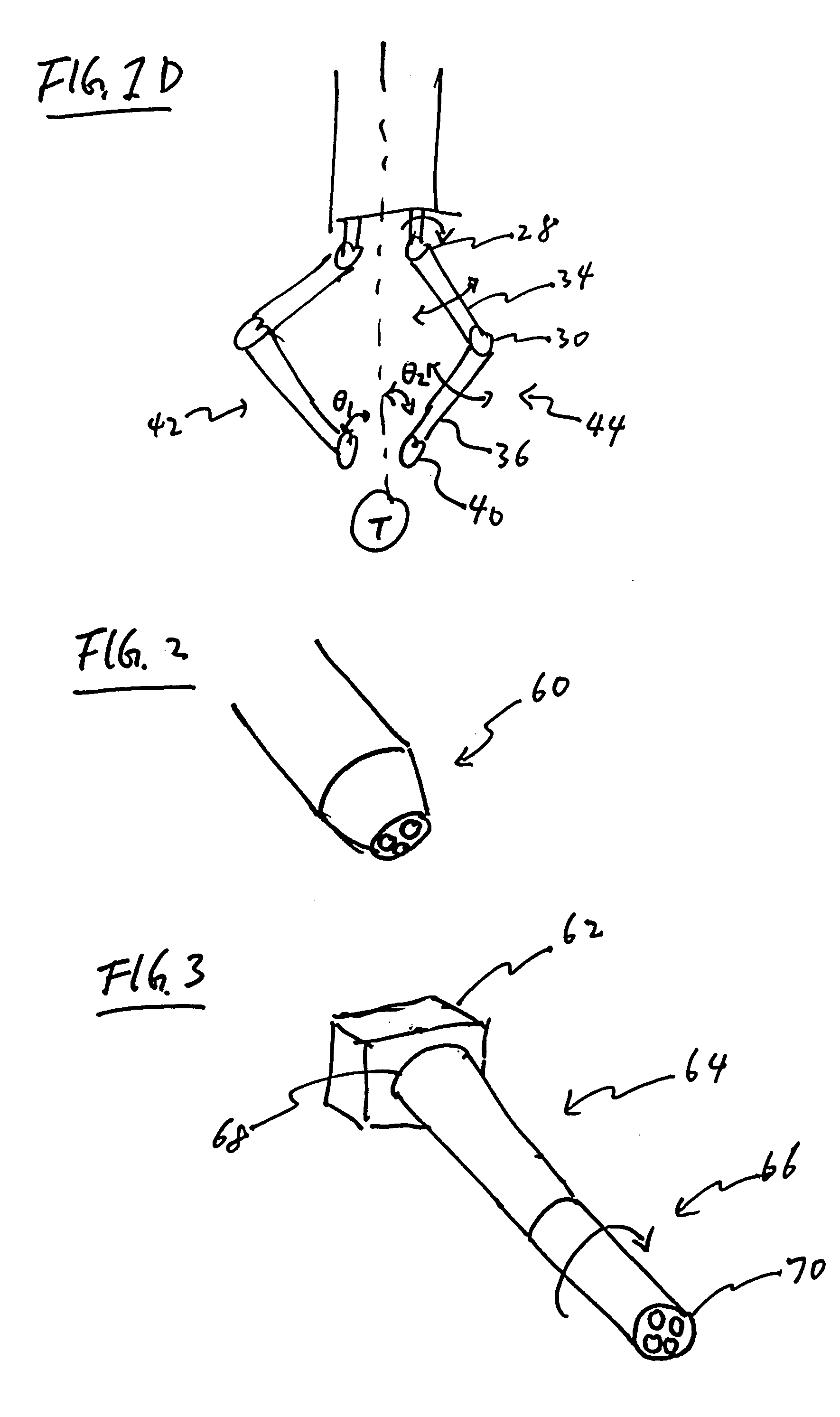
Described herein is a robotic surgical device configured for performing minimally invasive surgical procedures. The robotic surgical device comprises an elongated body for insertion into a patient's body through a small incision. In one variation, the elongated body houses a plurality of robotic arms. Once the distal portion of the elongated body is inserted into the patient body, the operator may then deploy the plurality of robotic arms to perform surgical procedures within the patient's body. An image detector may be positioned at the distal portion of the elongated body or on one of the robotic arms to provide visual feedback to the operator of the device. In another variation, each of the robotic arms comprises two or more joints, allowing the operator to maneuver the robotic arms in a coordinated manner within a region around the distal end of the device.

Description

FIELD OF THE INVENTION [0001] The present invention generally relates to devices and methods for performing minimally invasive surgery. In particular, the invention relates to robotic devices designed for performing minimally invasive surgery. DESCRIPTION OF RELATED ART [0002] Minimally invasive surgery has become more and more common nowadays. The surgical procedure is performed through multiple small incisions on the patient's body to minimize tissue damage and blood loss during surgery. The success of various minimally invasive surgical procedures in decreasing patient pain and improve recovering time has driven the trend to develop devices and procedures that would allow less invasive surgical procedure to be performed. [0003] Most of the minimally invasive surgical procedures are performed with the help of a small endoscopic camera and several long, thin, and rigid instruments. The camera and the instruments are inserted into the patient's body through natural body openings or ...

Claims


Application Information

Patent Timeline
Patent Type & Authority Applications(United States)
IPC IPC(8): A61B1/018A61B1/313A61B19/00
CPCA61B1/018A61B1/313A61B19/22A61B2019/2273A61B19/5212A61B2019/2234A61B2019/2249A61B19/2203A61B34/30A61B34/70A61B34/72A61B90/361A61B2034/305A61B2034/741
Inventor KHALILI, THEODORE M.
Owner CEDARS SINAI MEDICAL CENT
Why Eureka
Try Eureka