
Method and system for patient identification in 3D digital medical images

a technology of 3d digital medical images and patient identification, applied in image enhancement, instruments, applications, etc., can solve the problems of no scheme for automatic patient identification, two tags that are not very reliable, and the patient name does not match, so as to achieve high reliability and robustness

Inactive Publication Date: 2005-05-26
SIEMENS MEDICAL SOLUTIONS USA INC
View PDF4 Cites 14 Cited by
  • Summary
  • Abstract
  • Description
  • Claims
  • Application Information

AI Technical Summary

Benefits of technology

The patent describes a method for automatically determining if two sets of medical data belong to the same patient using only anatomical information, rather than foreign information like patient names and id's. This method can extract anatomical features that are most distinctive from the data, eliminating the possibility of attaching wrong names to data. This can be used alone or in combination with patient information to make the decisions more reliable and robust. The method can be applied to datasets of cross-modality, such as CT-MRI, if the extracted anatomical features are invariant among modalities. The patent also provides a program storage device that can be executed by a computer to perform the method steps for identifying patients from digital medical images.

Problems solved by technology

Those two tags appear to be not very reliable.
It happens often that the patient name does not match because of mistyping.
Currently, there is no scheme for automatic patient identification.
Obviously, this could be error prone.
If the user mistakenly loaded two data sets from two different patients, the conclusion of diagnosis will be completely irrelevant and wrong.

Method used


Image

  • Method and system for patient identification in 3D digital medical images
  • Method and system for patient identification in 3D digital medical images
  • Method and system for patient identification in 3D digital medical images

Examples


Embodiment Construction

[0022] Three-dimensional digital medical images are usually generated by computerized medical equipment such as CT scanners and MRI machines. Selected anatomic information and structures can be extracted as features from this type of volume data. These features, if unique enough, can be used to identify the patient. The anatomic information and structures to be extracted to be features for identification should be reliable and stable. For instance, they should be relatively immune to segmentation errors, body pose, and inhalation levels. This will enable the correct identification under most conditions.

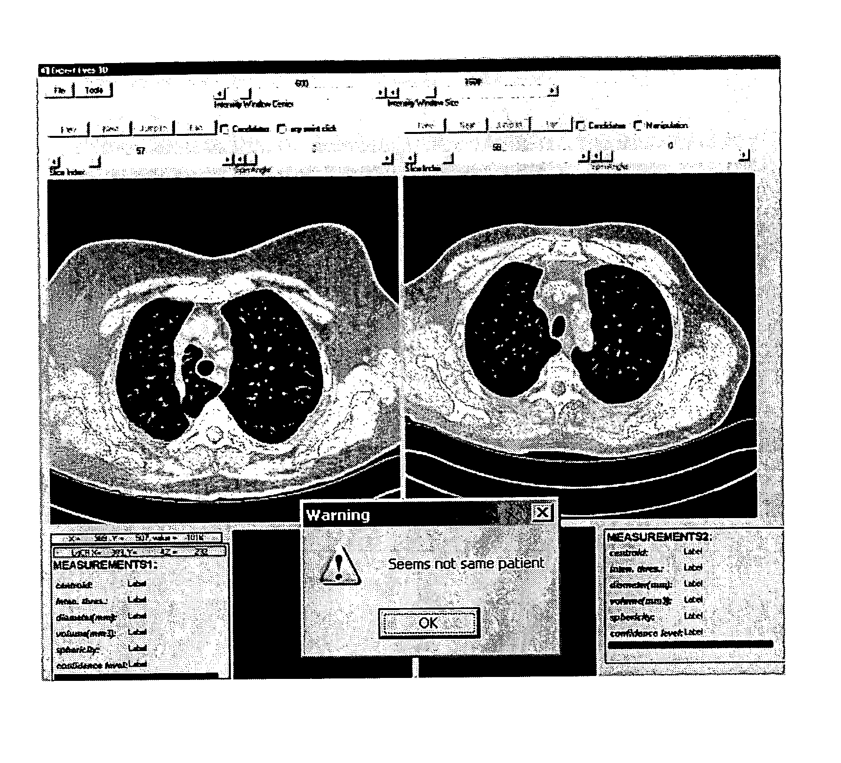
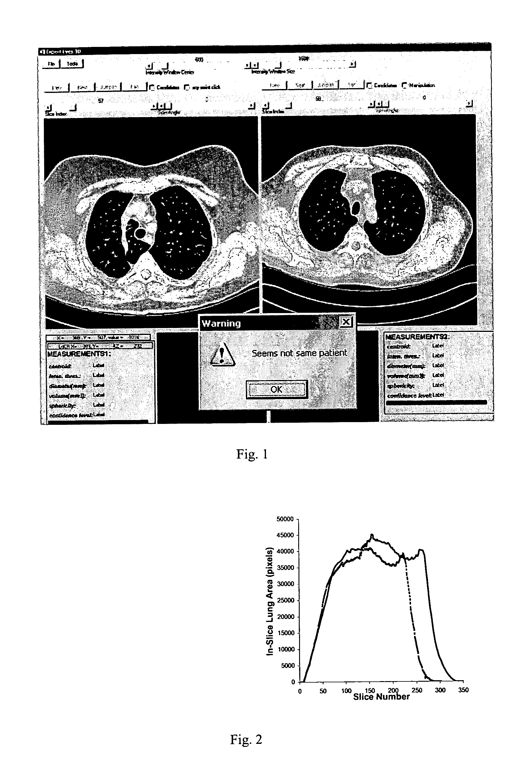
[0023] The methods and systems disclosed herein can be used to identify individuals using biological traits. The software application and algorithm disclosed herein can employ 2-D and 3-D renderings and images of an organ or organ system. As an example, two loaded datasets in a chest CT follow-up study are compared to determine if in fact they belong to the same patient, as shown in ...


PUM


Abstract

A method of identifying a patient from digital medical images includes providing a first digital image volume of an organ of a patient and a second digital image volume of the same organ, segmenting each slice of the first image volume and calculating a cross-sectional area of the organ in each slice to form a first area profile, segmenting each slice of the second image volume and calculating a cross-sectional area of the organ in each slice to form a second area profile, and comparing the first area profile with the second area profile to determine a correlation value for the two profiles. Based on the correlation value between the first area profile and the second area profile, it is determined whether the first digital image volume of the organ and the second digital image volume of the same organ came from the same patient.

Description

CROSS REFERENCE TO RELATED UNITED STATES APPLICATIONS [0001] This application claims priority from “PATIENT IDENTIFICATION IN 3D DIGITAL MEDICAL IMAGES”, U.S. Provisional Application No. 60 / 516,955 of Shen, et al., filed Nov. 3, 2003, the contents of which are incorporated herein by reference.TECHNICAL FIELD [0002] This invention is directed to the automatic extraction of anatomic features from 3D digital medical images for patient identification. DISCUSSION OF THE RELATED ART [0003] Computerized medical equipment, such as computerized tomography (CT) scanners and magnetic resonance imaging (MRI) machines, has been widely used in the screening and diagnosis of many diseases in all human body parts. For example, lung cancer screening and diagnosis is one such application for CT scanners. [0004] In many cases, it is desirable to be able to identify the patient from the data itself, without resorting to patient records and names, which could be unreliable and erroneous. On the other ha...

Claims


Application Information

Patent Timeline
Patent Type & Authority Applications(United States)
IPC IPC(8): G06K9/00G06T7/00
CPCA61B6/5294G06T2207/30061G06T7/0012Y10S378/901
Inventor SHEN, HONGODRY, BENJAMINQING, SHUPING
Owner SIEMENS MEDICAL SOLUTIONS USA INC
Learn More