Cardiopulmonary bypass extracorporeal blood circuit apparatus and method

a blood circuit apparatus and extracorporeal technology, applied in the direction of haemofiltration, suction devices, other medical devices, etc., can solve the problems of air leakage, large space occupied by distribution of components and lines about the operating table, and inability to adjust the volume, etc., to achieve rapid and easy replacement, small volume, and rapid prime

Inactive Publication Date: 2005-06-02
MEDTRONIC INC
View PDF0 Cites 48 Cited by
  • Summary
  • Abstract
  • Description
  • Claims
  • Application Information

AI Technical Summary

Benefits of technology

[0010] While the AVR extracorporeal blood circuit illustrated in FIGS. 3 and 4, and particularly the use of the AAR method and system, represents a significant improvement in extracorporeal circuits, its implementation can be further refined and improved. A need remains for an AAR system and method that optimizes the air sensor and its functions and that detects and responds to error conditions and faults that can arise over the course of prolonged surgical use.
[0011] Moreover, the typical prior art extracorporeal blood circuit, e.g. the above-described extracorporeal blood circuits of FIGS. 1-3, has to be assembled in the operating room from the above-described components, primed, and monitored during the surgical procedure while the patient is on bypass. This set-up of the components can be time-consuming and cumbersome and can result in missteps that have to be corrected. Therefore, a need remains for an extracorporeal blood circuit having standardized components and that can be set up for use using standardized setup procedures minimizing the risk of error.
[0015] Occasionally, it becomes necessary to “change out” one or more of the components of the extracorporeal blood circuit during the procedure. For example, it may be necessary to replace a blood pump or oxygenator. It may be necessary to prime and flush the newly constituted extracorporeal blood circuit after replacement of the malfunctioning component. The arrangement of lines and connectors may make this very difficult to accomplish. A need therefore remains for a compact extracorporeal blood circuit that can be rapidly and easily substituted for a malfunctioning extracorporeal blood circuit and that can be rapidly primed.
[0016] Consequently, a need remains for a extracorporeal blood circuit that is compactly arranged in the operating room, that takes advantage of kinetic assist, and is small in volume to minimize the required prime volume and to minimize the blood contacting surface area and blood-air interfaces. Moreover, a need remains for such an extracorporeal blood circuit that is simple to assemble and prime, provides for automatic monitoring of blood flow and other operating parameters, and facilitates change-out of components during the procedure.
[0019] One embodiment of the invention includes a method of priming an extracorporeal blood circuit. The method can include connecting a venous return line to an arterial line using a pre-bypass loop, preventing flow of prime solution into a venous air return device and a blood filter, and filling a pump and an oxygenator with prime solution in order to drive air bubbles upward and out of the pump and the oxygenator. The method can also include allowing prime solution to fill the venous return line and to pass into the venous return line after the pump and the oxygenator are filled with prime solution, allowing prime solution to rise upward through the venous return line into the blood filter, and coupling a vacuum source to a purge line coupled to the venous air removal device.

Problems solved by technology

This set-up of the components can be time-consuming and cumbersome and can result in missteps that have to be corrected.
The resulting distribution of the components and lines about the operating table can take up considerable space and get in the way during the procedure.
The connections that have to be made can also introduce air leaks introducing air into the extracorporeal blood circuit.
The lengths of the interconnected lines are not optimized to minimize prime volume and attendant hemodilution and to minimize the blood contacting surface area.
A large blood contacting surface area increases the incidences of embolization of blood cells and plasma traversing the extracorporeal blood circuit and complications associated with immune response, e.g., as platelet depletion, complement activation, and leukocyte activation.

Method used


Image

  • Cardiopulmonary bypass extracorporeal blood circuit apparatus and method
  • Cardiopulmonary bypass extracorporeal blood circuit apparatus and method
  • Cardiopulmonary bypass extracorporeal blood circuit apparatus and method

Examples


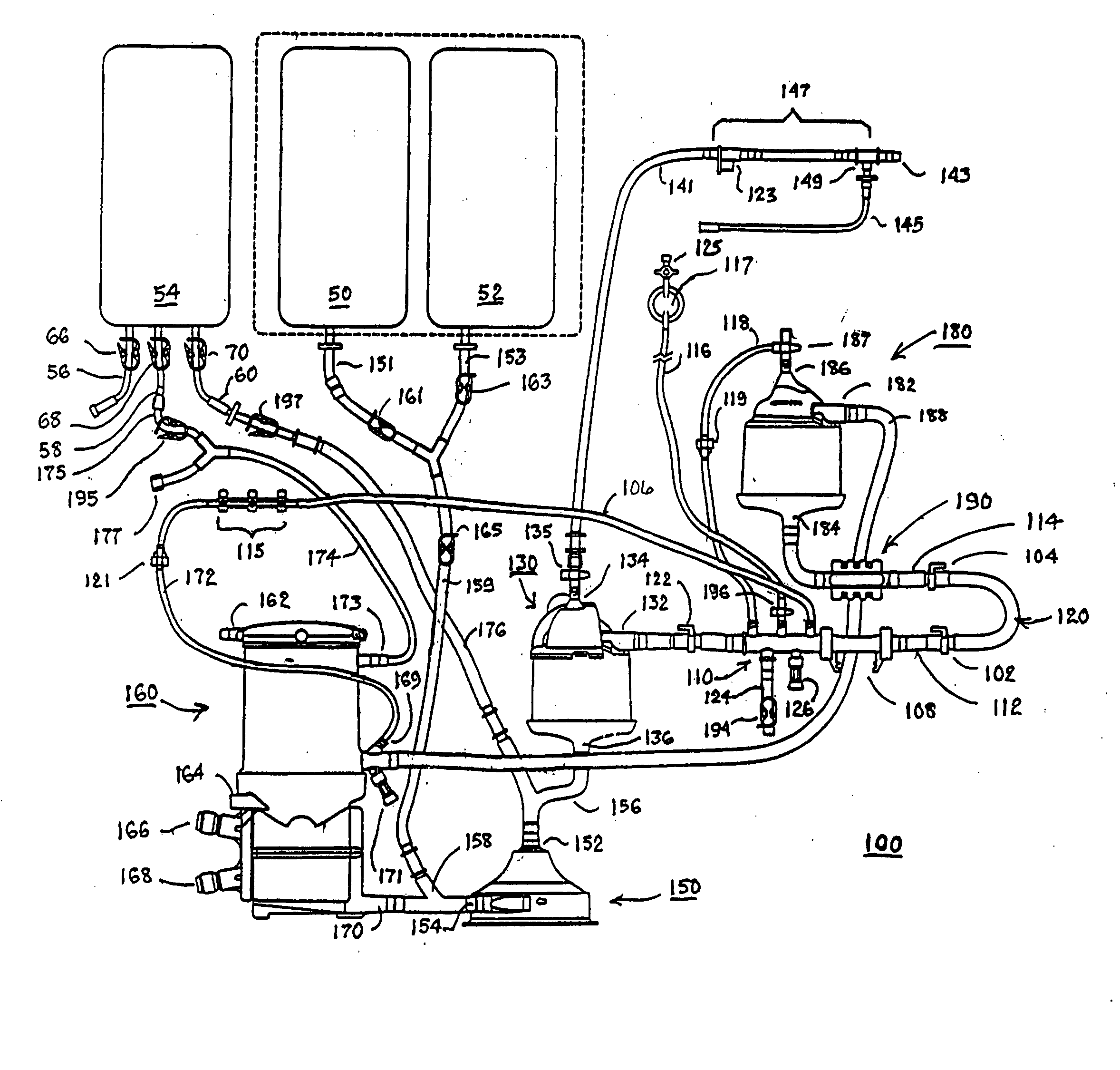
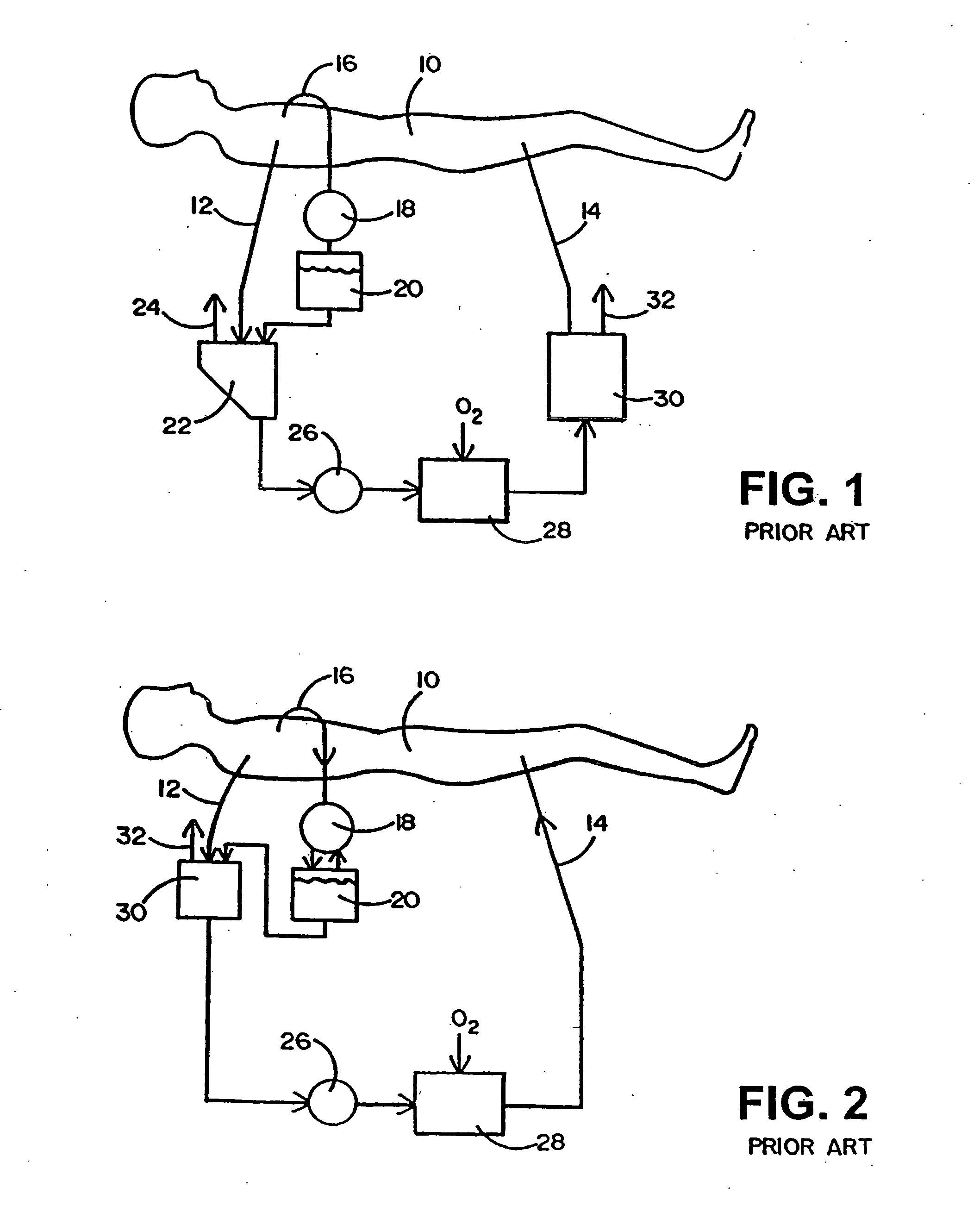
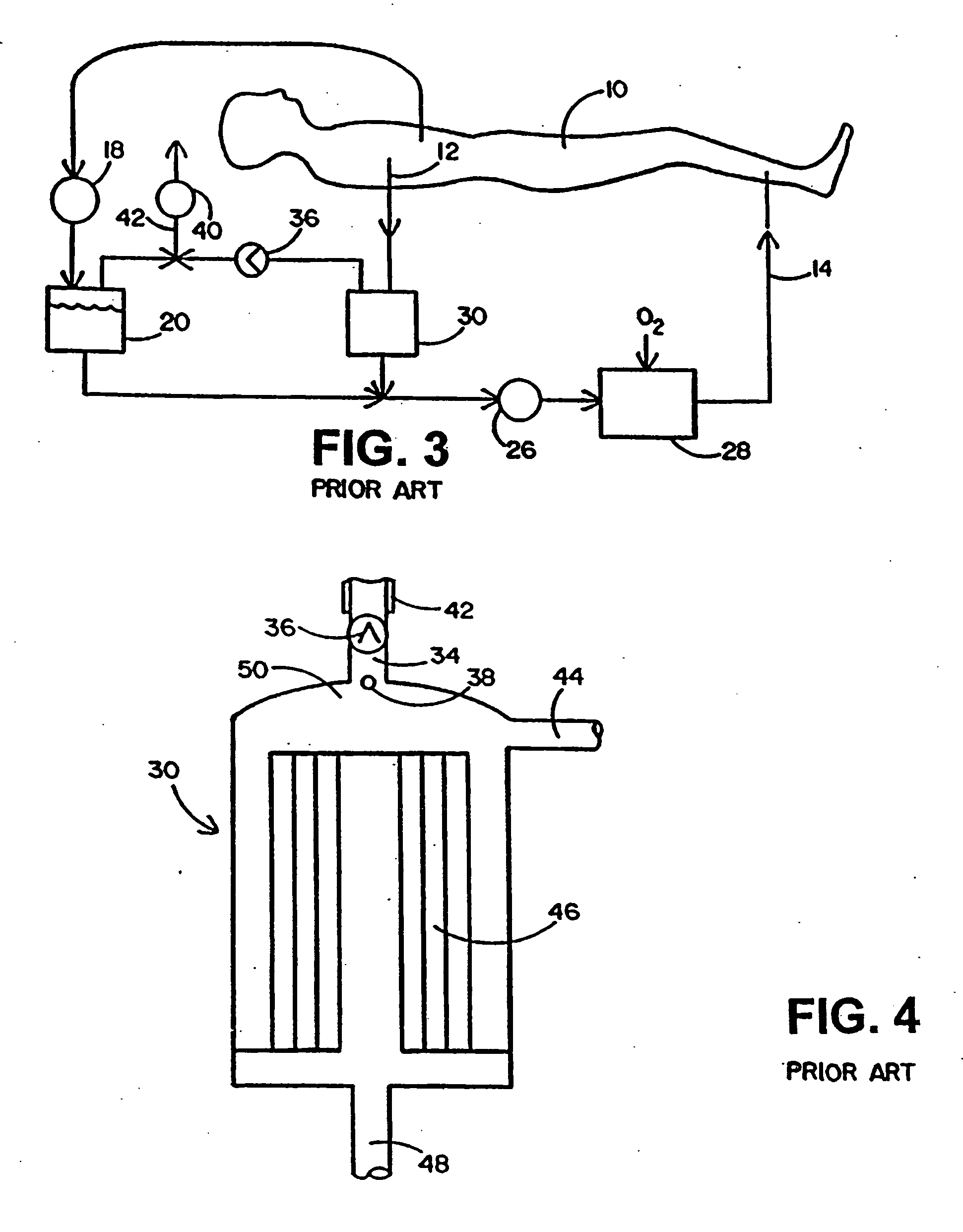
Embodiment Construction

[0037] Before any embodiments of the invention are explained in detail, it is to be understood that the invention is not limited in its application to the details of construction and the arrangement of components set forth in the following description or illustrated in the following drawings. The invention is capable of other embodiments and of being practiced or of being carried out in various ways. Also, it is to be understood that the phraseology and terminology used herein is for the purpose of description and should not be regarded as limited. The use of “including,”“comprising” or “having” and variations thereof herein is meant to encompass the items listed thereafter and equivalents thereof as well as additional items. The terms “mounted,”“connected” and “coupled” are used broadly and encompass both direct and indirect mounting, connecting and coupling. Further, “connected” and “coupled” are not restricted to physical or mechanical connections or couplings, and can include el...


PUM


Abstract

An extracorporeal blood circuit for use with a venous return line and an arterial line coupled to a patient. The extracorporeal blood circuit can include a venous air removal device coupled to the venous return line. The venous air removal device can perform an active air removal function. The extracorporeal blood circuit can include a sensor that determines a blood level in the venous air removal device, a purge line coupled to the venous air removal device, and a controller connected to the sensor. The controller can cause the venous air removal device to perform the active air removal function through the purge line when the blood level is less than a threshold. The extracorporeal blood circuit can further include a pump coupled to the venous air removal device, an oxygenator coupled to the pump, and a blood filter coupled to the oxygenator and the arterial line.

Description

BACKGROUND OF THE INVENTION [0001] Conventional cardiopulmonary bypass uses an extracorporeal blood circuit that is coupled between arterial and venous cannulae and includes a venous drainage line, a venous blood reservoir, a blood pump, an oxygenator, an arterial filter, and blood transporting tubing or lines, ports, and valves interconnecting these components. Prior art, extracorporeal blood circuits as schematically depicted in FIGS. 1-3 and described in commonly assigned U.S. Pat. No. 6,302,860, draw venous blood of a patient 10 during cardiovascular surgery through the venous cannulae (not shown) coupled to venous return line 12, oxygenates the blood, and returns the oxygenated blood to the patient 10 through an arterial line 14 coupled to an arterial cannulae (not shown). Cardiotomy blood and surgical field debris that is aspirated by a suction device 16 is pumped by cardiotomy pump 18 into a cardiotomy reservoir 20. [0002] Air can enter the extracorporeal blood circuit from a...

Claims


Application Information

Patent Timeline
Patent Type & Authority Applications(United States)
IPC IPC(8): A61M1/34A61M1/36A61M37/00
CPCA61M1/3626A61M1/3627A61M1/3643A61M1/3666A61M1/3667A61M2205/502A61M2205/505A61M2205/8206A61M2209/082A61M2205/3379A61M1/3606A61M1/3652A61M1/3644A61M1/3603A61M1/3623
Inventor OLSEN, ROBERT W.CARPENTER, WALTER L.DICKEY, JOHN B.SHOREY, FREDERICK A. JR.YONCE, LAURA A.
Owner MEDTRONIC INC
Why Eureka
Try Eureka