

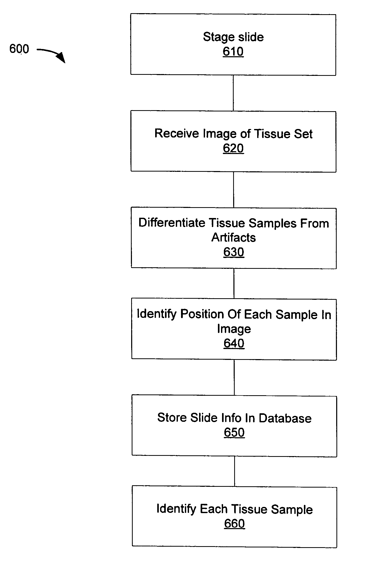
Automated microscope slide tissue sample mapping and image acquisition
a microscope slide and tissue sample technology, applied in the field of automatic microscope slide tissue sample mapping and image acquisition, can solve the problems of inability to handle the present volume inability to use the current process relying on time-consuming visual tissue analysis, and inability to meet the needs of tissue samples requiring identification
- Summary
- Abstract
- Description
- Claims
- Application Information
AI Technical Summary
Problems solved by technology
Method used
Image

Examples
working example
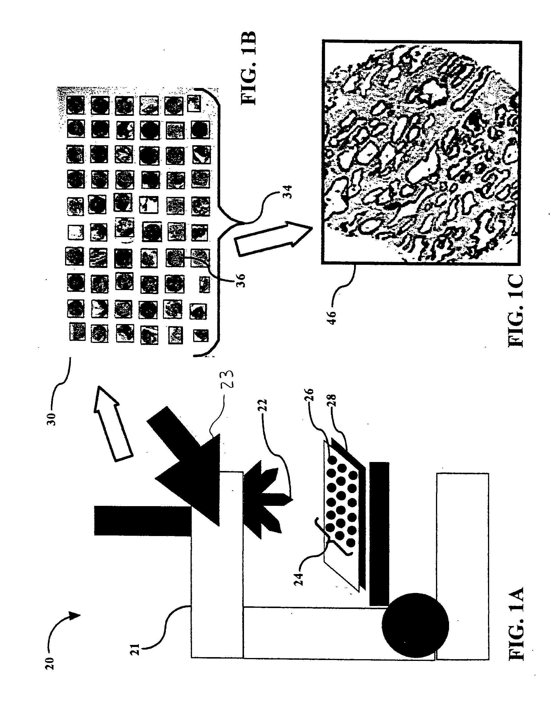
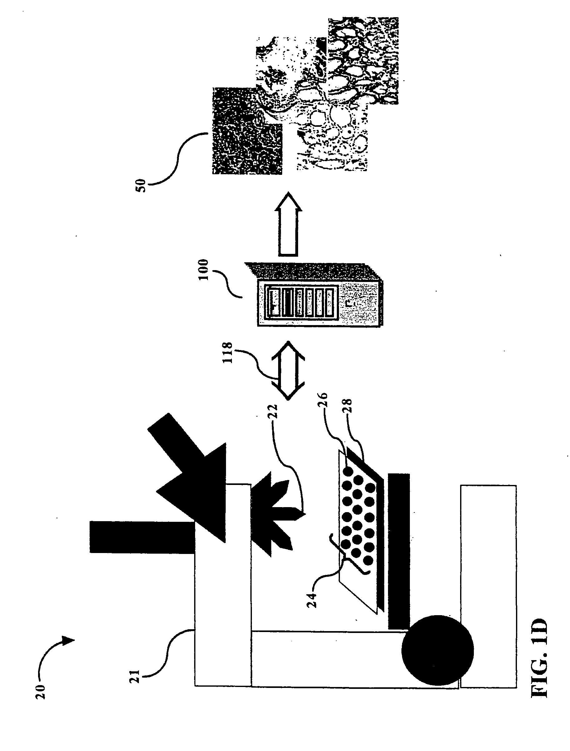
[0047] The following describes the hardware and software components employed in a working example of a system for automated microscope slide tissue sample mapping. This description is merely illustrative and is not to be considered limiting. The hardware components included: [0048] Leica DMLA automated microscope with 2.5×, 5×, 10×, 20× and 40×; [0049] Diagnostic Instruments Spot InSight 4 camera for microscope image capture; [0050] Three color LED light source; [0051] 300 slide auto loading and motorized stage; [0052] Computer hardware including a 2+GHz PC with at least 512 MB of memory and a large (30+MB) hard drive, display screen, and an MS-Windows operating system (2000, NT or 98). A bank of 16 such computers are all loaded with the software. They communicate with each other over a network, using MSMQ (Microsoft Messaging Queue) and messages written in XML format. The software utilizes all of the processing capacity of the PC, so the machines are dedicated to this one purpose. ...
PUM
 Abstract
Description
Claims
Application Information