Transesophageal ultrasound using a narrow probe

Active Publication Date: 2005-06-30
IMACOR INC
View PDF82 Cites 24 Cited by
  • Summary
  • Abstract
  • Description
  • Claims
  • Application Information

AI Technical Summary

Benefits of technology

[0003] Transesophageal ultrasound imaging is implemented using a miniature transversely oriented transducer that is preferably small enough to fit in a 7.5 mm diameter probe, and

Problems solved by technology

This type of transducer, however, is not well suited for transesophageal echocardiography because the transducer elements are oriented longitudinally instead of transversely, which limits the types of

Method used


Image

  • Transesophageal ultrasound using a narrow probe
  • Transesophageal ultrasound using a narrow probe
  • Transesophageal ultrasound using a narrow probe

Examples


Embodiment Construction

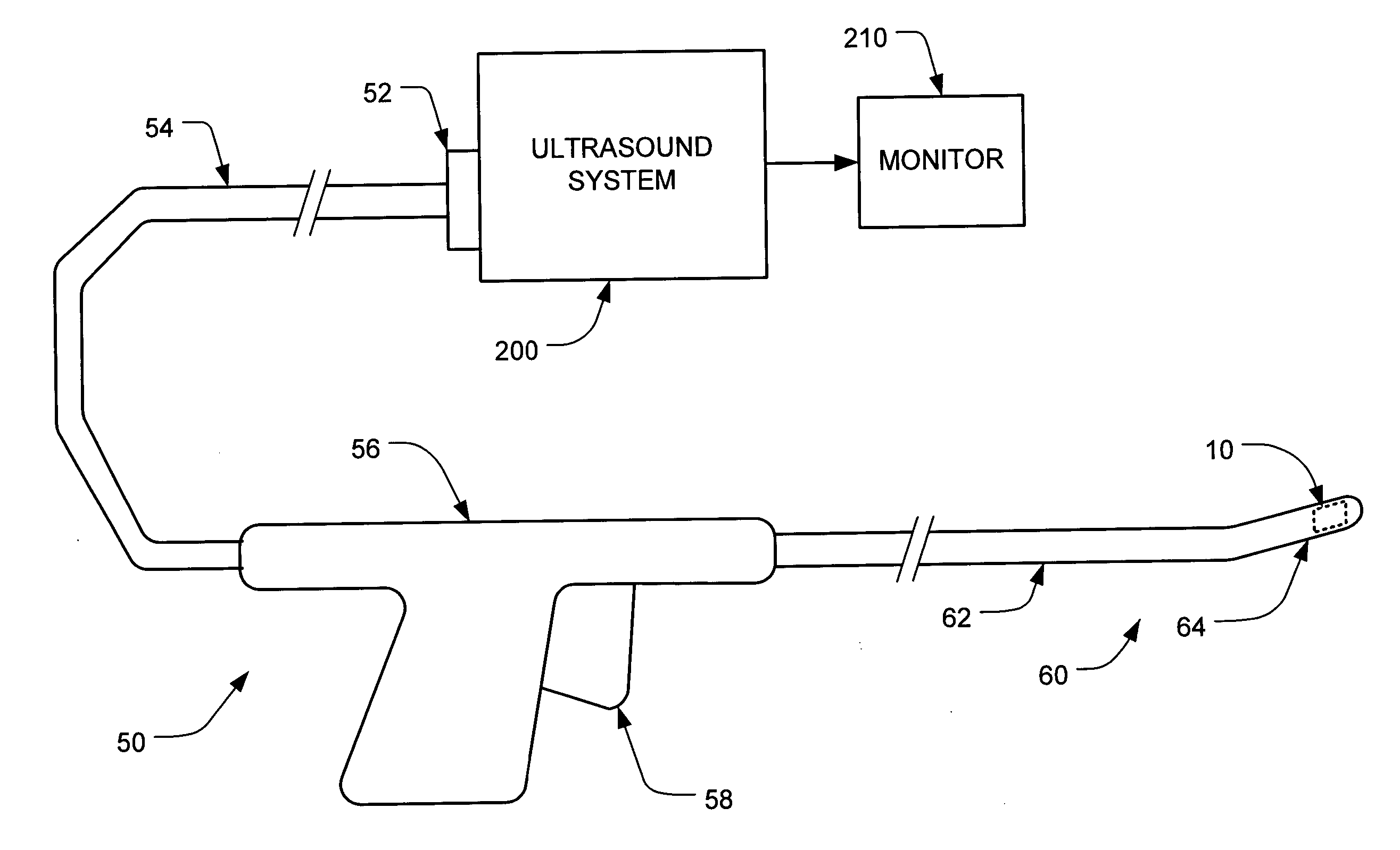
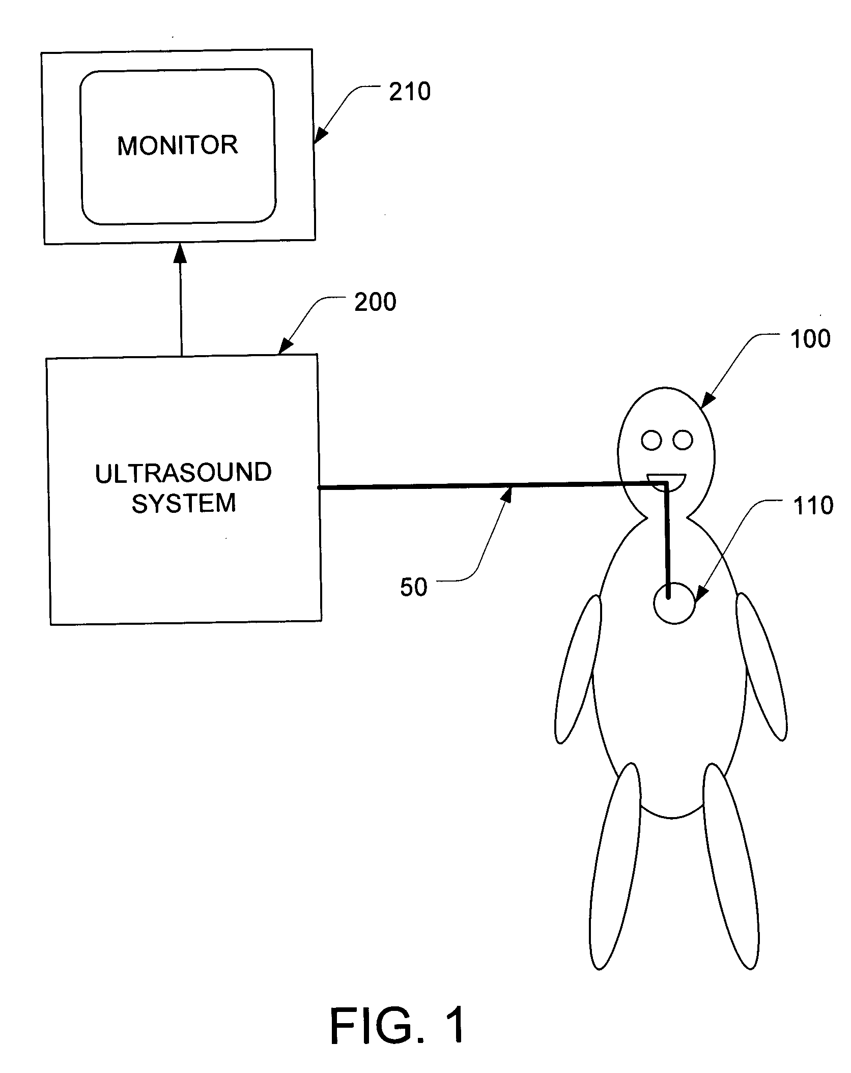
[0021]FIG. 1 is an overall block diagram of a system that may be used for continuous long term monitoring of cardiac function by direct visualization of the heart. An ultrasound system 200 is used to monitor the heart 10 of the patient 100 by sending driving signals into a probe 50 and processing the return signals received from the probe into images, using the image processing algorithms described below. The images generated by those algorithms are then displayed on a monitor 210, in any conventional manner.

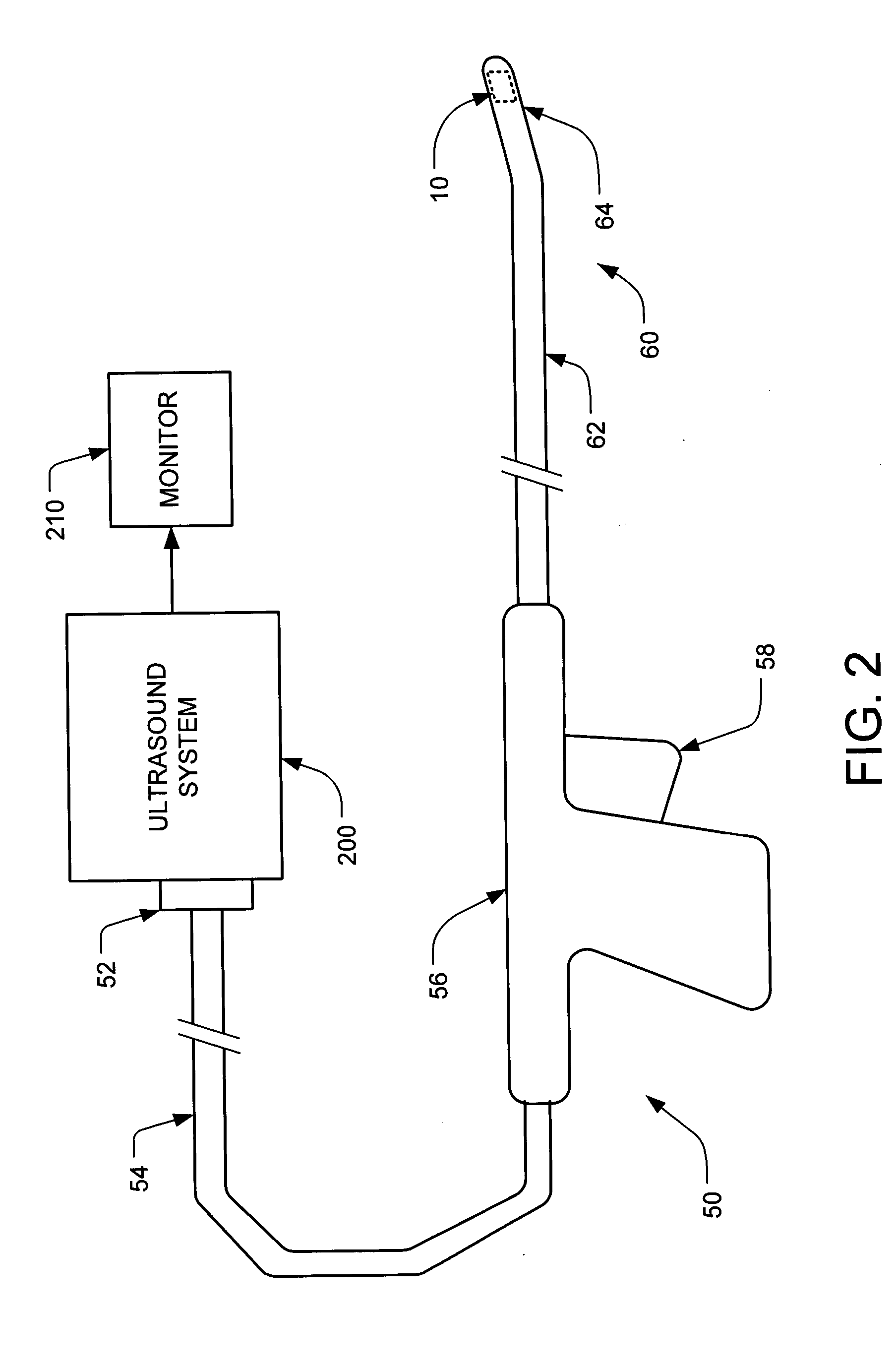
[0022]FIG. 2 shows more details of the probe 50, which is connected to the ultrasound system 200. At the distal end of the probe 50 there is a housing 60, and the ultrasound transducer 10 is located in the distal end 64 of the housing 60. The next portion is the flexible shaft 62, which is positioned between the distal end 64 and the handle 56. This shaft 62 should be flexible enough so that the distal end 64 can be positioned past the relevant anatomical structures to the desi...


PUM


Abstract

Transesophageal echocardiography is implemented using a miniature transversely oriented transducer that is preferably small enough to fit in a 7.5 mm diameter probe, and most preferably small enough to fit in a 5 mm diameter probe. Signal processing techniques improve the depth of penetration to the point where the complete trans-gastric short axis view of the left ventricle can be obtained, despite the fact that the transducer is so small. The reduced diameter of the probe (as compared to prior art probes) reduces risks to patients, reduces or eliminates the need for anesthesia, and permits long term direct-visualization monitoring of patients' cardiac function.

Description

CROSS REFERENCE TO RELATED APPLICATIONS [0001] This application claims priority to U.S. provisional application No. 60 / 525,330, filed Nov. 26, 2003.BACKGROUND [0002] In the medical field, monitoring heart function impacts critical decisions that relate to patient care. One type of prior art heart monitor is the intravascular / intracardiac ultrasound transducer (such as the Accunav™ transducer). This type of transducer, however, is not well suited for transesophageal echocardiography because the transducer elements are oriented longitudinally instead of transversely, which limits the types of images that can be obtained. A second type of prior art heart monitor is the transesophageal echocardiography (TEE) transducer, which is transversely oriented. However, in order to produce repeatedly usable images, the azimuthal aperture of these transducers must be quite large (e.g., 10-15 mm in diameter for adults), which requires a correspondingly large probe. Because of this large probe, conv...

Claims


Application Information

Patent Timeline
IPC IPC(8): A61B8/12G01S7/52G01S7/521G01S15/89
CPCA61B8/12A61B8/4488G01S7/52033G01S7/52036G01S7/52079A61B8/445G01S15/8934G01S15/8977G01S15/8915A61B8/0883G01S15/8925
Inventor ROTH, SCOTT L.HASTINGS, HAROLD M.
Owner IMACOR INC
Why Eureka
Try Eureka