Automated method and system for the detection of lung nodules in low-dose CT image for lung-cancer screening

Inactive Publication Date: 2005-08-04
CHICAGO UNIV OF
View PDF7 Cites 32 Cited by
  • Summary
  • Abstract
  • Description
  • Claims
  • Application Information

AI Technical Summary

Benefits of technology

[0042] According to a still further aspect of the present invention, the calculating step comprises: (1) determining, for each candidate nodule in the first plurality of candidate nodules, at least one respective morphological feature value, including at least one of effective diameter, circularity, and irregularity; and (2) determ

Problems solved by technology

However, it is a difficult and time-consuming task for radiologists to detect

Method used


Image

  • Automated method and system for the detection of lung nodules in low-dose CT image for lung-cancer screening
  • Automated method and system for the detection of lung nodules in low-dose CT image for lung-cancer screening
  • Automated method and system for the detection of lung nodules in low-dose CT image for lung-cancer screening

Examples


Embodiment Construction

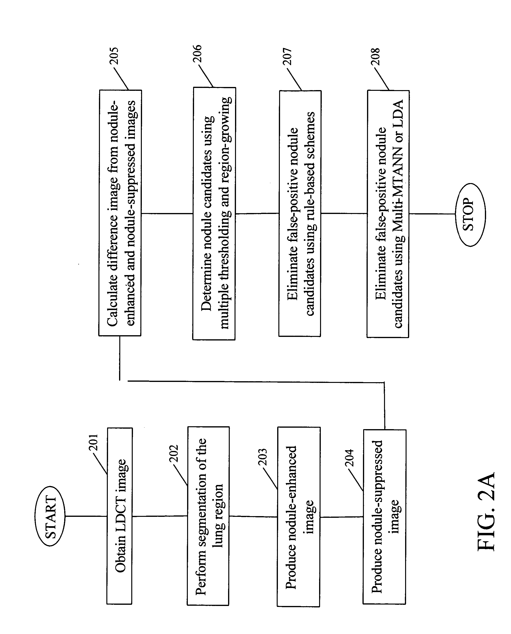
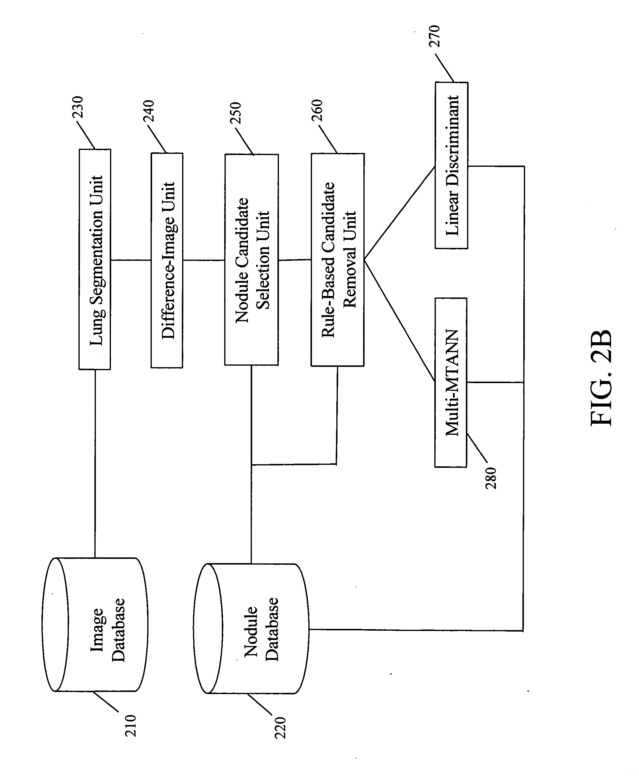
[0069] Referring now to the drawings, wherein like reference numerals designate identical or corresponding parts throughout the several views, FIG. 2A illustrates the method for detecting pulmonary nodules on LDCT images according to the present invention. The present method is based on a difference-image technique [15-17] in which structures similar to nodules are enhanced, and most of the background normal structures, such as small vessels or background noise, are suppressed.

[0070] In step 201, a medical image (e.g., a LDCT slice image) of the lungs is obtained. The medical image may be obtained directly from a CT scanner or from a medical image database.

[0071] In step 202, the left or right lung region of the medical image obtained in step 201 is segmented by use of a linear discriminant method on the histogram of CT values [20], wherein a threshold CT level is automatically determined for dividing a body region into lung regions and other tissue regions. For smoothing the outl...


PUM


Abstract

A method, system, and computer program product for detecting at least one nodule in a medical image of a subject, including identifying, in the medical image, an anatomical region corresponding to at least a portion of an organ of interest; filtering the medical image to obtain a difference image; detecting, in the difference image, a first plurality of nodule candidates within the anatomical region; calculating respective nodule feature values of the first plurality of nodule candidates based on pixel values of at least one of the medical image and the difference image; removing false positive nodule candidates from the first plurality of nodule candidates based on the respective nodule feature values to obtain a second plurality of nodule candidates; and determining the at least one nodule by classifying each of the second plurality of nodule candidates as a nodule or a non-nodule based on at least one of the pixel values and the respective nodule feature values. True-positive nodules are identified using linear discriminant analysis and/or a Multi-MTANN.

Description

BACKGROUND OF THE INVENTION [0001] Field of the Invention [0002] The invention relates generally to the computerized, automated assessment of computed tomography (CT) scans or images, and more particularly, to methods, systems, and computer program products for detecting lung nodules in low-dose helical CT scans of the thorax to assess lung cancer. [0003] The present invention also generally relates to computerized techniques for the automated analysis of digital images, for example, as disclosed in one or more of U.S. Pat. Nos. 4,839,807; 4,841,555; 4,851,984; 4,875,165; 4,907,156; 4,918,534; 5,072,384; 5,133,020; 5,150,292; 5,224,177; 5,289,374; 5,319,549; 5,343,390; 5,359,513; 5,452,367; 5,463,548; 5,491,627; 5,537,485; 5,598,481; 5,622,171; 5,638,458; 5,657,362; 5,666,434; 5,673,332; 5,668,888; 5,732,697; 5,740,268; 5,790,690; 5,832,103; 5,873,824; 5,881,124; 5,931,780; 5,974,165; 5,982,915; 5,984,870; 5,987,345; 6,011,862; 6,058,322; 6,067,373; 6,075,878; 6,078,680; 6,088,473; ...

Claims


Application Information

Patent Timeline
IPC IPC(8): G06T7/00G21K1/12H05G1/60
CPCG06T2207/30061G06T7/0012
Inventor ARIMURA, HIDETAKALI, FENGSHIRAISHI, JUNJIDOI, KUNIO
Owner CHICAGO UNIV OF
Why Eureka
Try Eureka