Minimally invasive orthopaedic apparatus and methods

a minimally invasive, orthopaedic technology, applied in the direction of prosthesis, surgical forceps, osteosynthesis devices, etc., can solve the problems of long-since viewed as prohibitive the size of such plates, the difficulty of achieving the effect and the inability to significantly improve the accuracy of minimally invasive techniques

Active Publication Date: 2005-08-11
BIOMET CV
View PDF68 Cites 16 Cited by
  • Summary
  • Abstract
  • Description
  • Claims
  • Application Information

AI Technical Summary

Problems solved by technology

However, despite growing use in other surgical fields, minimally invasive techniques have not been significantly developed for use in orthopaedic procedures.
The size of such plates has long since been viewed as prohibitive in regard to the use of minimally invasive techniques for the implantation thereof.
In particular, orthopaedic procedures commonly include complicated fractures which require precision in regard to the installation of fixation devices (e.g., screws and the like) and the reduction of such fractures.
As a result of such continued use of “open” procedures, soft tissue surrounding the bone continues to be compromised thereby impairing normal blood circulation to the treated bone, potentially delaying fracture healing, and potentially increasing the risk of infection.
Indeed, although the majority of patients treated with such procedures heal without complication, there are certain occasions in which complications such as infection or non-union occur thereby prolonging healing rates and, in certain cases, increasing the rates of secondary revisions.
However, such assemblies suffer from a number of drawbacks.
In addition to the fundamental limitations relating to the resolution associated with fluoroscopy, many surgeons may also be reluctant to embrace the use of large amounts of fluoroscopy in order to minimize radiation exposure to themselves, the other members of the surgical staff, and the patient.

Method used


Image

  • Minimally invasive orthopaedic apparatus and methods
  • Minimally invasive orthopaedic apparatus and methods
  • Minimally invasive orthopaedic apparatus and methods

Examples


Embodiment Construction

[0063] While the concepts of the present disclosure are susceptible to various modifications and alternative forms, specific embodiments thereof have been shown by way of example in the drawings and will herein be described in detail. It should be understood, however, that there is no intent to limit the concepts of the present disclosure to the particular forms disclosed, but on the contrary, the intention is to cover all modifications, equivalents, and alternatives falling within the spirit and scope of the disclosure.

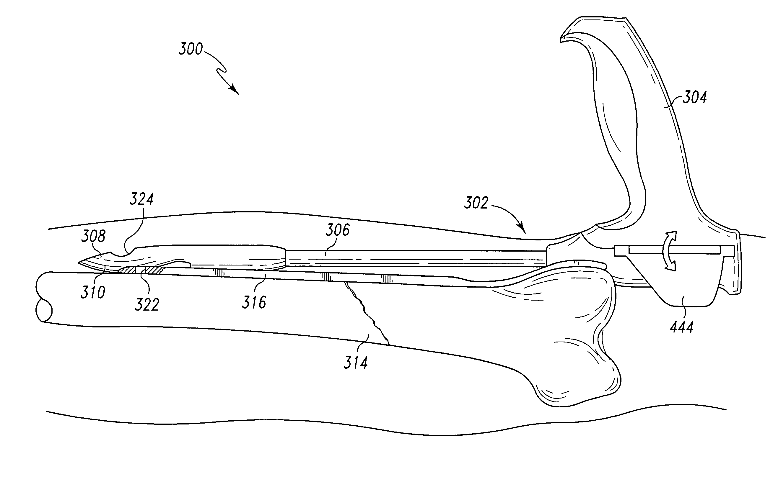
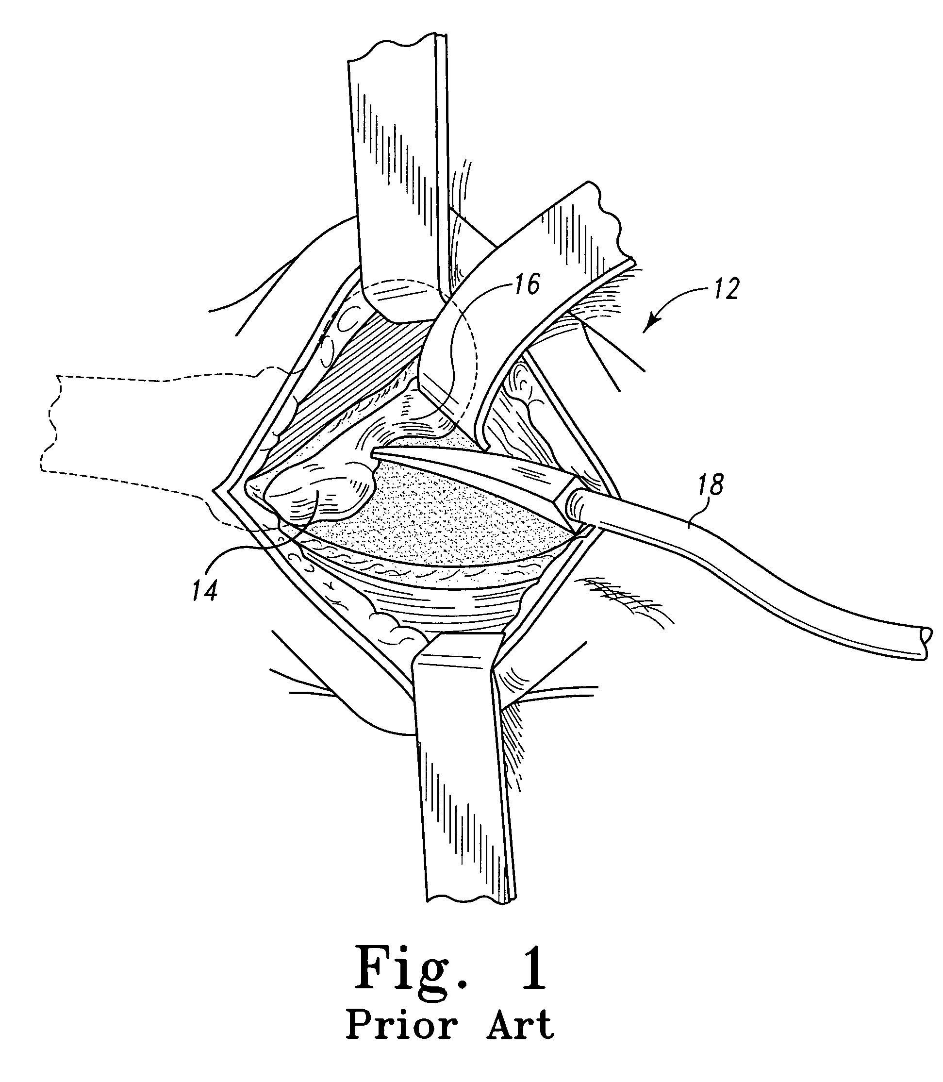
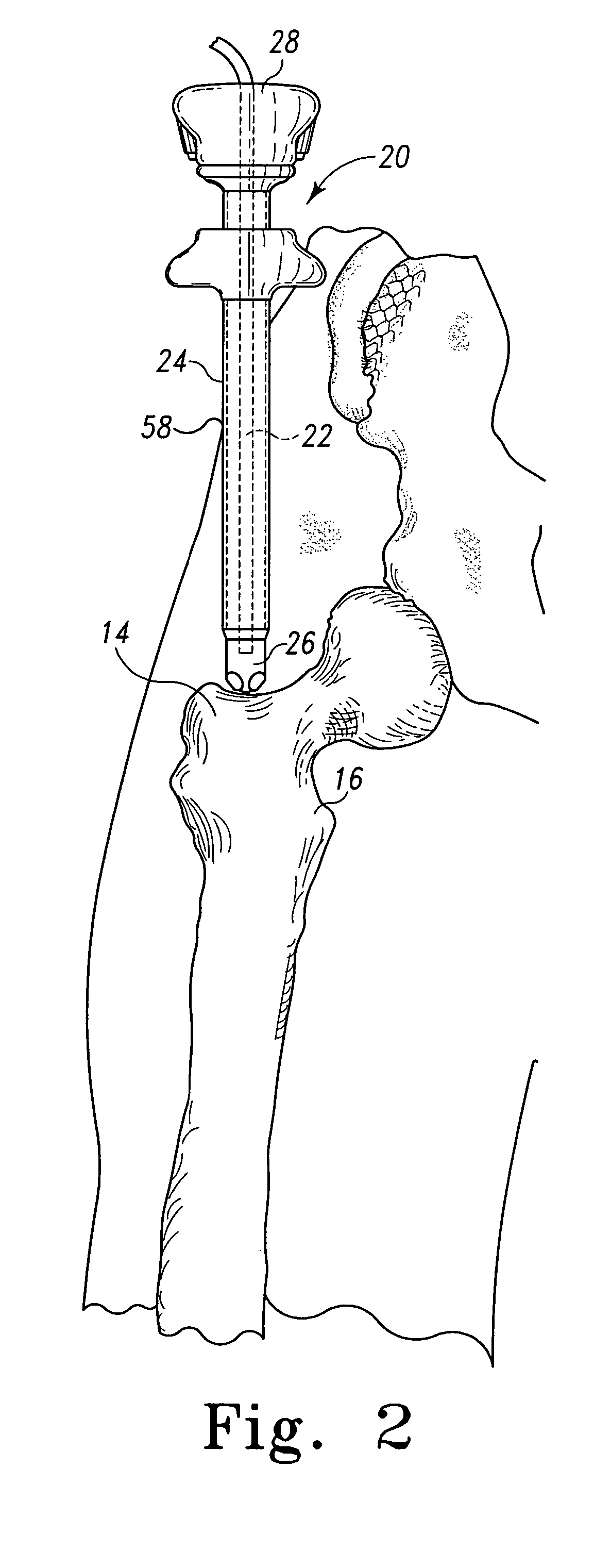
[0064] Referring now to FIGS. 1-53, there is shown a number of apparatus and methods which may be utilized to perform a minimally invasive orthopaedic surgical procedure. Common to many of the concepts disclosed herein is the notion of utilizing endoscopic instruments to provide the surgeon with enhanced viewing capabilities in the form of direct visualization of the surgical site. The concepts of the present disclosure may be utilized in a wide variety of orthopaed...


PUM


Abstract

Apparatus and methods for use in the performance of minimally invasive orthopaedic procedures, including apparatus and methods for use in the performance of such procedures under the visualization of an endoscope, are herein disclosed. Such procedures include a minimally invasive intramedullary nailing procedure, a minimally invasive bone graft harvesting procedure, a minimally invasive pelvic osteotomy procedure, an orthopaedic implant revision procedure, and a minimally invasive percutaneous bone plating procedure.

Description

[0001] This application is a continuation of U.S. patent application Ser. No. 10 / 480,053 filed on Dec. 9, 2003, which is a U.S. national counterpart application of international application serial no. PCT / US02 / 16505 filed on May 24, 2002, which claims priority to U.S. Provisional Patent Application Ser. No. 60 / 301,309 filed Jun. 27, 2001. The entirety of each of these applications is hereby incorporated by reference.FIELD OF THE DISCLOSURE [0002] The present disclosure relates generally to orthopaedic methods and apparatus, and more particularly to methods and apparatus for use in the performance of endoscopic minimally invasive orthopaedic procedures. BACKGROUND OF THE DISCLOSURE [0003] Minimally invasive surgical techniques have been developed for many different types of surgical procedures. Such techniques attempt to balance the need to achieve the goal of the surgical procedure while minimizing the surgical injury to the patient. As such, surgeries performed by use of minimally ...

Claims


Application Information

Patent Timeline
Patent Type & Authority Applications(United States)
IPC IPC(8): A61B1/00A61B17/58A61B17/00A61B17/02A61B17/16A61B17/17A61B17/28A61B17/32A61B17/72A61B17/78A61B17/86A61B17/88A61B17/92A61B19/00A61F2/46
CPCA61B17/00234A61B1/0615A61B17/1604A61B17/1635A61B17/1655A61B17/1664A61B17/1703A61B17/1717A61B17/1725A61B17/1728A61B17/1735A61B17/72A61B17/744A61B17/808A61B17/8685A61B17/8866A61B17/921A61B19/24A61B2019/481A61F2/4644A61F2002/4635A61F2002/4649A61B2017/1782A61B1/00154A61B17/0218A61B90/02A61B2090/08021A61B17/1782
Inventor GUZMAN, PAMELA C.WACK, MICHAEL A.SCHULZE, DALE R.KNIGHT, GARY W.HESS, CHRISTOPHER J.NOBIS, RUDOLPH H.CLEM, MICHAEL F.KOLATA, RONALD J.
Owner BIOMET CV
Why Eureka
Try Eureka