Non-invasive device and method for the diagnosis of pulmonary vascular occlusions

a pulmonary embolism and non-invasive technology, applied in the field of vascular occlusions of the respiratory system, can solve the problems of inability to reliably include or exclude the diagnosis of pulmonary embolism in arterial blood analysis, and the inability to accurately diagnose pulmonary embolism, so as to achieve the effect of accurate diagnosis of pulmonary vascular occlusions and non-invasive diagnosis of pulmonary embolism

Inactive Publication Date: 2005-09-08
CHARLOTTE MECKLENBURG HOSPITAL AUTHORITY
View PDF4 Cites 12 Cited by
  • Summary
  • Abstract
  • Description
  • Claims
  • Application Information

AI Technical Summary

Benefits of technology

[0016] It is a principal object and advantage of the present invention to provide physicians with an instrument for non-invasively diagnosing pulmonary vascular occlusions.
[0017] It is an additional object and advantage of the present invention to provide an instrument that accurately diagnoses pulmonary vascular occlusions.
[0018] It is a further object and advantage of the present invention to provide an instrument for measuring and interpreting pulmonary test data.
[0019] Other objects and advantages of the present invention will in part be obvious, and in part appear hereinafter. SUMMARY OF THE INVENTION
[0020] In accordance with the foregoing objects and advantages, the present invention provides a device and method for non-invasively diagnosing a pulmonary embolism. The device of the present invention comprises a breathing tube having sensors for measuring the flow of air into and out of a patient's lungs as well as the amount of oxygen, carbon dioxide, or nitrogen. Alternatively, the device can comprise a sensor such as a infrared or mass spectrometer capable of measuring the quantity of a non-ambient gas which is excreted via the lungs after injection into a patient. The device further comprises a data processing unit which determines the concentrations of the expired gases and total volume of gas expired for display on a screen. The likelihood that a pulmonary embolism is present can be determined by comparison of the results to predetermined data from normal and afflicted populations.

Problems solved by technology

Unless treated promptly, a pulmonary embolism can be fatal.
The detection of a pulmonary embolism is extremely difficult because signs and symptoms can easily be attributed to other conditions and symptoms may vary depending on the severity of the occurrence.
However, chest x-rays often show normal lungs even when an embolism is present and even when the x-rays show abnormalities they rarely confirm a pulmonary embolism.
Although a pulmonary embolism usually causes abnormalities in these measurements, there is no individual finding or combination of findings from the arterial blood gas analysis that allows either a reliable way to exclude or specific way of diagnosing pulmonary embolism.
Accordingly, the arterial blood analysis cannot reliably include or exclude the diagnosis of a pulmonary embolism.
As a result, the D-dimer assay is extremely sensitive for the presence of a pulmonary embolism but is very nonspecific.
However, many studies have shown a D-dimer assay is only normal in less than ⅓ of patients and thus produces a high degree of false positives.
Nevertheless, a perfusion scan does not always provide clear evidence that a pulmonary embolism is the cause of the problem as it often yields indeterminate results in as many as 70% of patients.
Pulmonary angiograms are popular means of diagnosing a pulmonary embolism, but the procedure poses some risks and is more uncomfortable than other tests.
Pulmonary angiograms are more useful in diagnosing a pulmonary embolism than some of the other traditional methods, but often present health risks and can be expensive.
Although frequently recommended by experts, few physicians and patients are willing to undergo such an invasive procedure.
The procedure's reported sensitivity has varied widely, however, and it may only be useful for diagnosing an embolism in central pulmonary arteries as it is relatively insensitive to clots in more remote regions of the lungs.
The imaging tests also typically involve costs of more than $1,000 for the patient, take more than two hours to perform, and require special expertise such as a trained technician to perform the tests and acquire the images and a board-certified radiologist to interpret the images.
Notably, none are completely safe for patients who are pregnant.
As a result of these shortcomings, the imaging procedures are not available in many outpatient clinic settings and in many portions of third world countries.

Method used


Image

  • Non-invasive device and method for the diagnosis of pulmonary vascular occlusions
  • Non-invasive device and method for the diagnosis of pulmonary vascular occlusions
  • Non-invasive device and method for the diagnosis of pulmonary vascular occlusions

Examples


Embodiment Construction

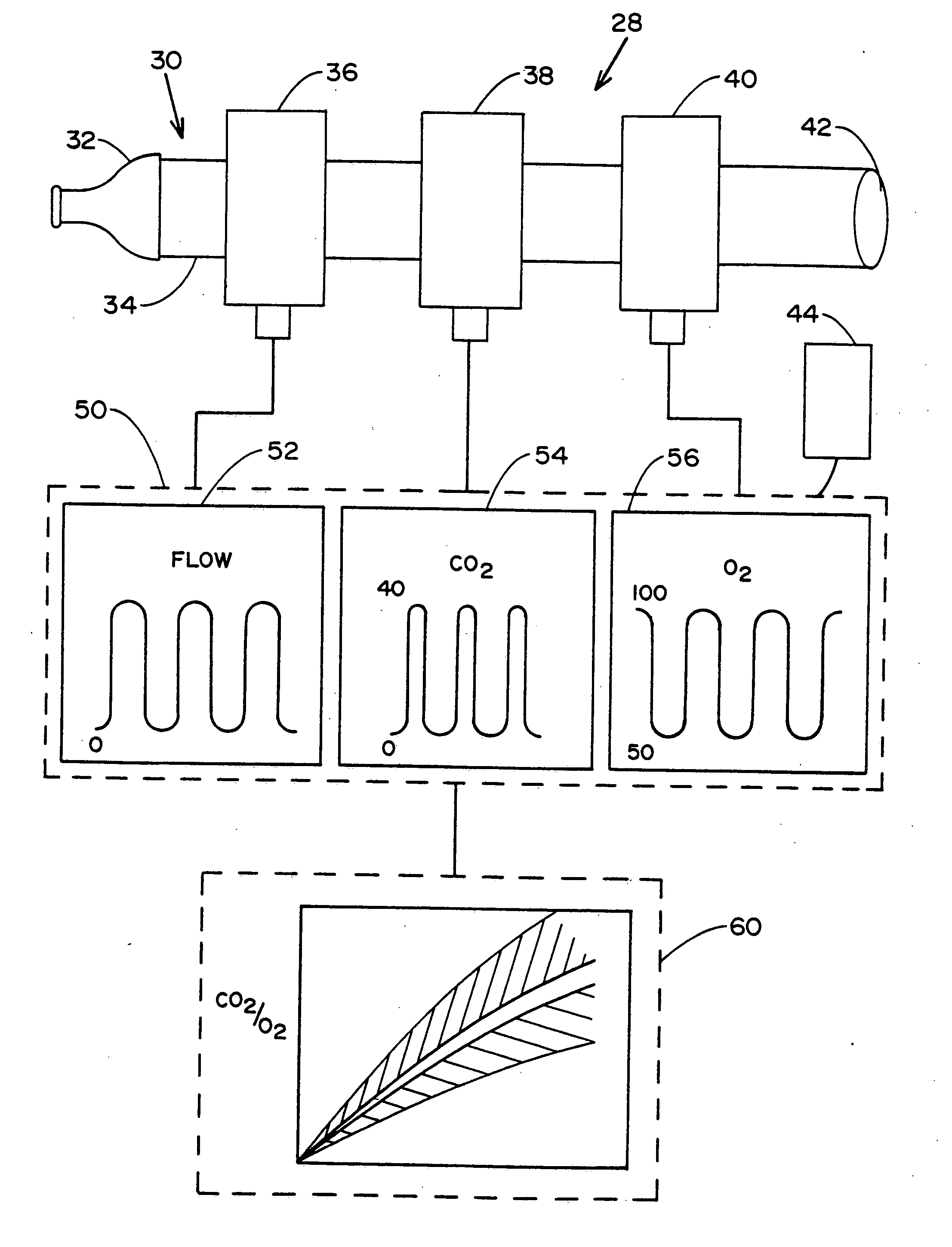
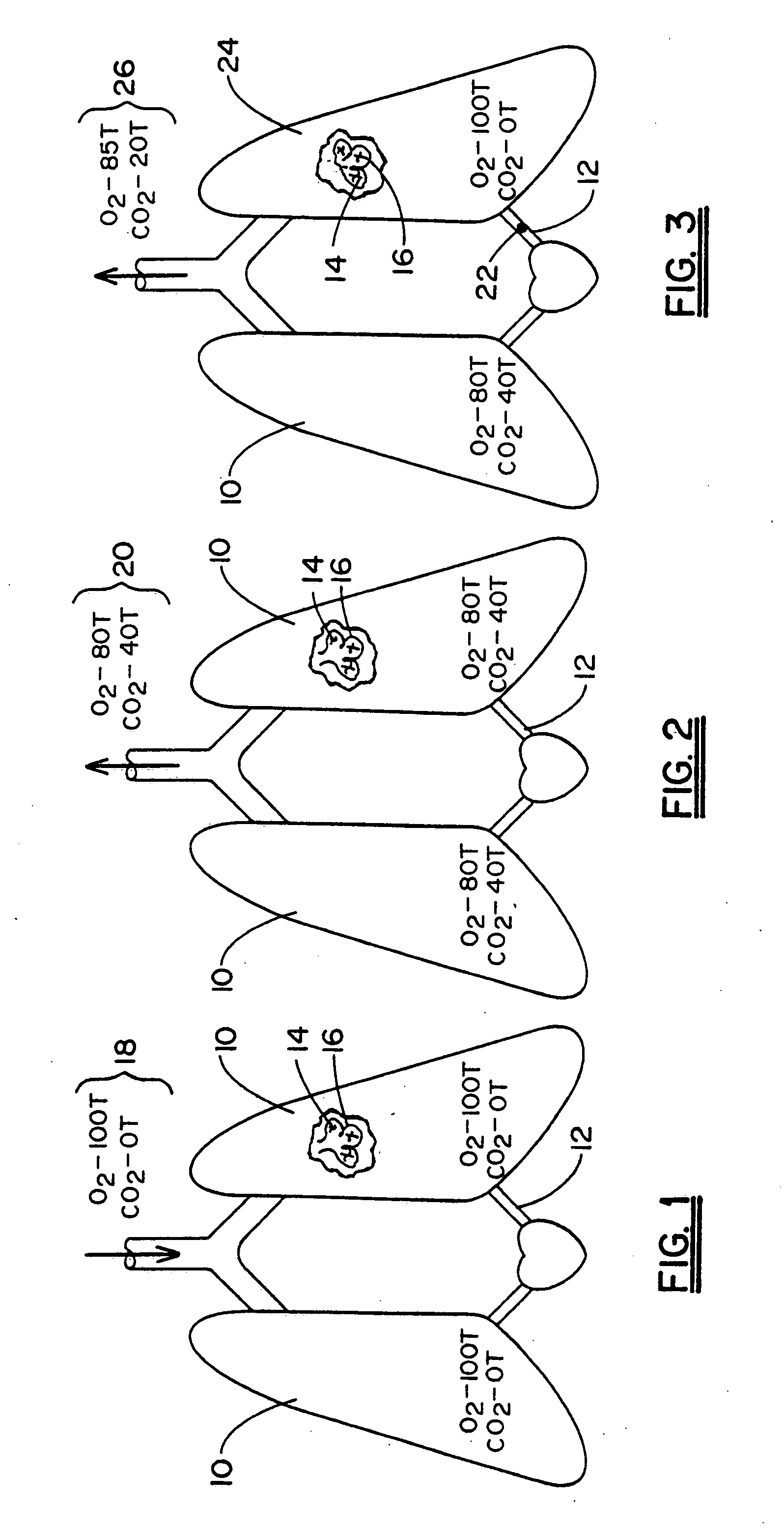
[0028] Referring now to the drawing in which like reference numerals refer to like parts throughout, there is seen in FIG. 1 a representation of lungs 10 free from any pulmonary occlusions. In healthy lungs 10, blood flows freely from the pulmonary arteries 12 into the capillaries 14 surrounding the individual alveoli 16 of the lungs 10. When inhaled air 18 is drawn into the lungs 10 and alveoli 16, oxygen is transferred from the inhaled air 18 to the blood stream and carbon dioxide is transferred out. Inhaled air 18 typically contains an oxygen partial pressure of approximately one hundred (100) torr and a carbon dioxide partial pressure of zero (0) torr.

[0029] Once the inhaled air 18 reaches the alveoli 16, the oxygen content decreases while the carbon dioxide content increases until an equilibrium with blood gas levels in the pulmonary arteries 12 is reached. The inhaled air 18 is then, as seen in FIG. 2, expired as exhaled air 20. Exhaled air 20 from properly functioning lungs ...


PUM

PropertyMeasurementUnit
partial pressureaaaaaaaaaa

Abstract

This invention involves a device and method for ascertaining the functioning of the respiratory system and determining whether a pulmonary embolism is present. The device comprises an apparatus containing sensors which measure the amount of a particular gas expired for a patient's lungs, including a non-ambient gas which is previously provided to the patent. From this data, a processor computes the concentration of expired gas as a function of expired volume and displays the results on a screen. By comparing the results to predetermined values from afflicted and healthy patient populations, an accurate determination can be made regarding the presence of a pulmonary embolism.

Description

CROSS-REFERENCE TO PRIOR APPLICATION [0001] The present application is divisional of U.S. patent application, Ser. No. 10 / 109,301 filed Mar. 27, 2002, now allowed, which is a continuation-in-part of U.S. patent application, Ser. No. 09 / 965,303 filed Sep. 27, 2001, U.S. Pat. No. 6,575,918.BACKGROUND OF THE INVENTION [0002] 1. Field of Invention [0003] The present invention relates generally to vascular occlusions of the respiratory system, and more particularly to non-invasive devices and methods for the diagnosis of a pulmonary embolism and related disorders. [0004] 2. Description of Prior Art [0005] A pulmonary embolism occurs when an embolus become lodged in lung arteries, thus blocking blood flow to lung tissue. An embolus is usually a blood clot, known as a thrombus, but may also comprise fat, amniotic fluid, bone marrow, tumor fragments, or even air bubbles that block a blood vessel. Unless treated promptly, a pulmonary embolism can be fatal. In the United States alone, around ...

Claims


Application Information

Patent Timeline
Patent Type & Authority Applications(United States)
IPC IPC(8): A61BA61B5/00A61B5/08A61B5/026A61B5/083A61B5/087A61B5/091A61B5/097A61B5/145A61B5/1455B32B5/02G01N1/22H02M5/458
CPCA61B5/083A61B5/417A61B5/091A61B5/087A61B5/08A61B5/082A61B5/0833A61B5/0836H02M5/4585
Inventor KLINE, JEFFREY A.
Owner CHARLOTTE MECKLENBURG HOSPITAL AUTHORITY
Why Eureka
Try Eureka