Time resolved contrast-enhanced MR projection imaging of the coronary arteries with intravenous contrast injection

Inactive Publication Date: 2005-11-24
NORTHWESTERN UNIV
View PDF6 Cites 6 Cited by
  • Summary
  • Abstract
  • Description
  • Claims
  • Application Information

AI Technical Summary

Benefits of technology

[0007] It is an object of the present invention to provide a magnetic resonance imaging method and apparatus for depicting coronary arteries as a

Problems solved by technology

Because of the strong signal produced by nearby cardiac chambers and the tortuous path of coronary arteri

Method used


Image

  • Time resolved contrast-enhanced MR projection imaging of the coronary arteries with intravenous contrast injection
  • Time resolved contrast-enhanced MR projection imaging of the coronary arteries with intravenous contrast injection
  • Time resolved contrast-enhanced MR projection imaging of the coronary arteries with intravenous contrast injection

Examples


Embodiment Construction

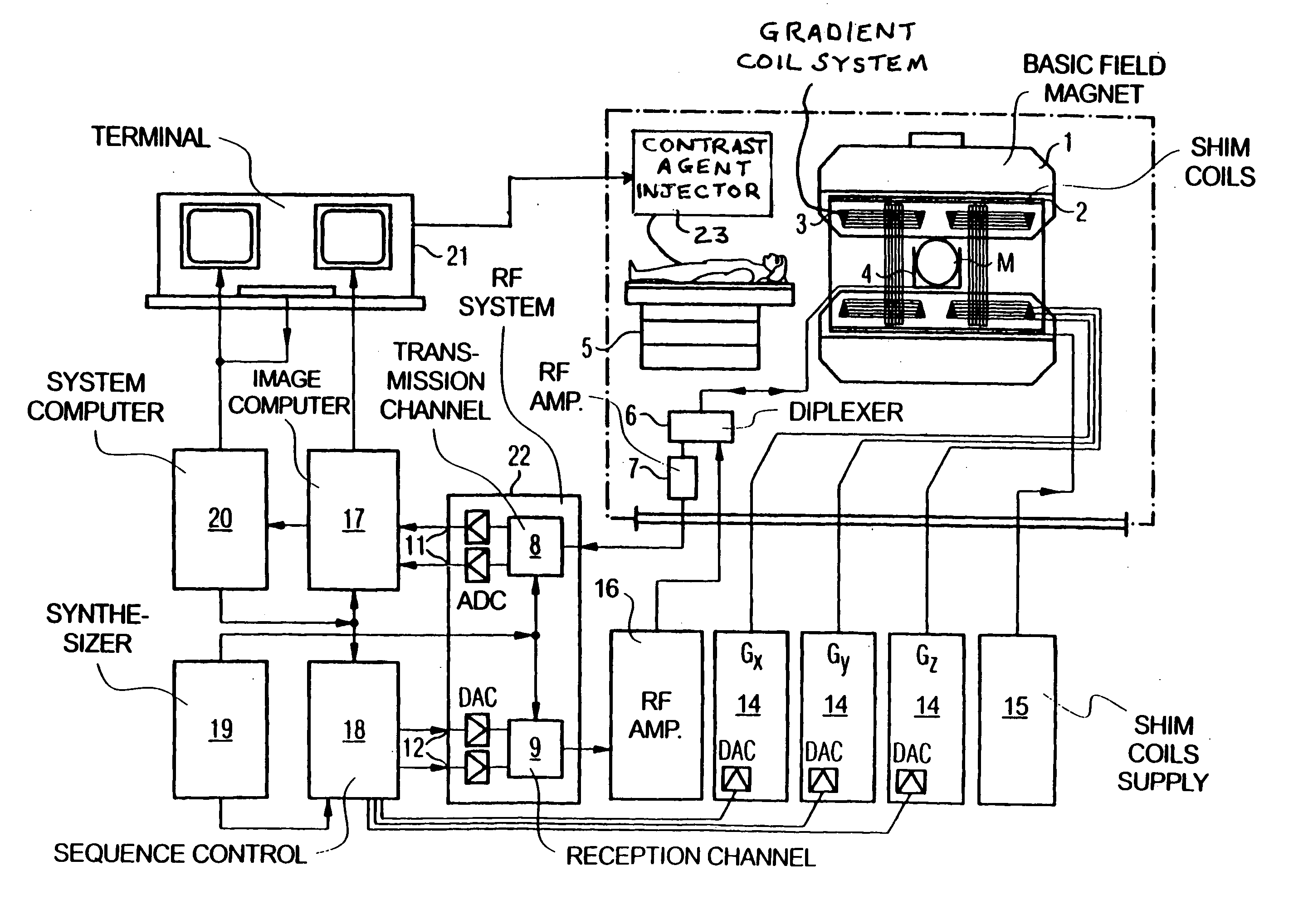
[0016]FIG. 6 schematically illustrates a magnetic resonance imaging (tomography) apparatus for generating a nuclear magnetic image of a subject according to the present invention. The components of the nuclear magnetic resonance tomography apparatus correspond to those of a conventional tomography apparatus, but it is controlled according to the invention. A basic field magnet 1 generates a time-constant, intense magnetic field for polarization (alignment) of the nuclear spins in the examination region of a subject such as, for example, a part of a human body to be examined. The high homogeneity of the basic magnetic field required for the nuclear magnetic resonance measurement is defined in a spherical measurement volume M in which the part of the human body to be examined is introduced. For supporting the homogeneity demands and, in particular, for eliminating time-invariable influences, shim plates of ferromagnetic material are attached at suitable locations. Time-variable influe...


PUM


Abstract

In a method and apparatus for contrast-enhanced magnetic resonance (MR) angiography of the right coronary artery of a patient, a bolus of MR contrast agent is selected to have a size that will cause the bolus, after injection into the patient, to wash out of right coronary chambers of the heart while still enhancing MR signals from the right coronary artery. The bolus of MR contrast agent is injected into the patient, and MR signals are generated in, and MR signals are obtained from, the patient in a time window after the bolus has washed out of the right coronary chambers and still enhances MR signals in the right coronary artery. An MR image of the right coronary artery is generated using only the MR signals obtained in the time window.

Description

FEDERAL FUNDING LEGEND [0001] This invention was produced in part using funds from the Federal government under NIH Grant Nos. HL38698 and HL70859. Accordingly, the Federal government has certain rights in this invention.BACKGROUND OF THE INVENTION [0002] 1. Field of the Invention [0003] The present invention relates to a method and apparatus for magnetic resonance imaging, and in particular to a method and apparatus for magnetic resonance imaging of the coronary arteries. [0004] 2. Description of the Prior Art [0005] A study by Kim et al. entitled “Coronary Magnetic Resonance Angiography for the Detection of Coronary Stenoses,” New England Journal of Medicine (2001), Vol. 345, No. 26, pages 1863-1869 showed that coronary artery magnetic resonance angiography (MRA) may be a viable diagnostic alternative to selective x-ray catheter-based angiography. The detection of coronary artery stenoses using MRA may be improved by viewing the entire artery as a whole, so as to prevent a torturo...

Claims


Application Information

Patent Timeline
IPC IPC(8): A61B5/05A61B5/055G01R33/28G01R33/561G01R33/563
CPCG01R33/281G01R33/56308G01R33/563A61B5/055
Inventor LI, DEBIAOGREEN, JORDIN D.
Owner NORTHWESTERN UNIV
Why Eureka
Try Eureka