Image guided interventions with interstitial or transmission ultrasound

a technology of interstitial or transmission ultrasound and image guided intervention, which is applied in the field of ultrasound imaging, can solve the problems of not being able to acquire and process pre-operative imagery in real-time setting, unsuitable for use in operation room setting, and subjecting patients to harmful radiation, so as to reduce the risk of radiation exposure, and assess the effect of intervention effectiveness

a technology of interstitial or transmission ultrasound and image guided intervention, which is applied in the field of ultrasound imaging, can solve the problems of not being able to acquire and process pre-operative imagery in real-time setting, unsuitable for use in operation room setting, and subjecting patients to harmful radiation, so as to reduce the risk of radiation exposure, and assess the effect of intervention effectiveness

US20050261591A1Inactive Publication Date: 2005-11-24THE JOHN HOPKINS UNIV SCHOOL OF MEDICINE

Image

  • Image guided interventions with interstitial or transmission ultrasound
  • Image guided interventions with interstitial or transmission ultrasound
  • Image guided interventions with interstitial or transmission ultrasound

Examples


Embodiment Construction

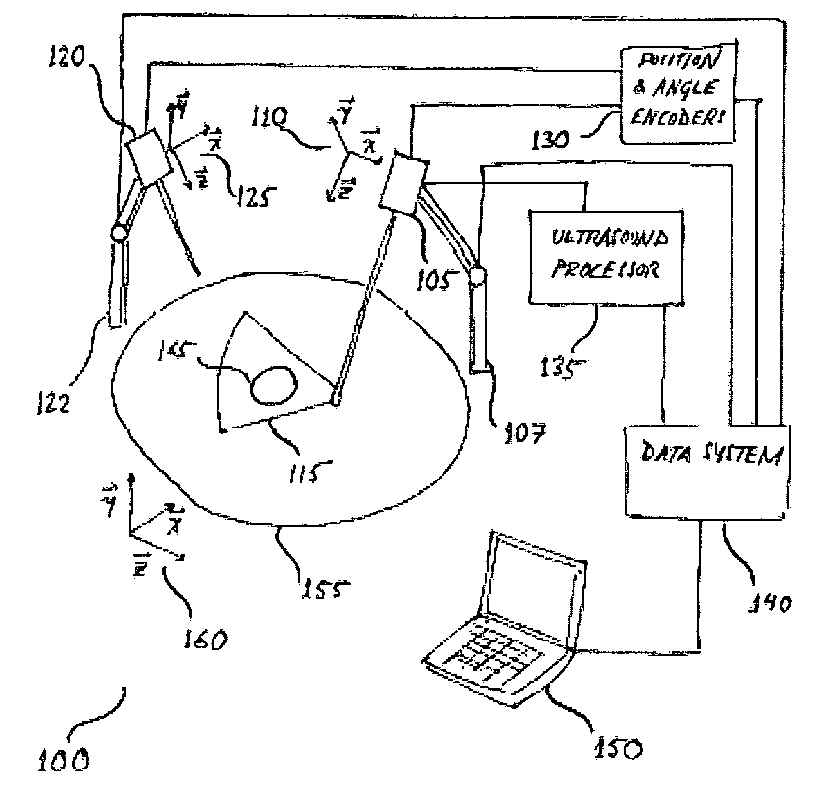
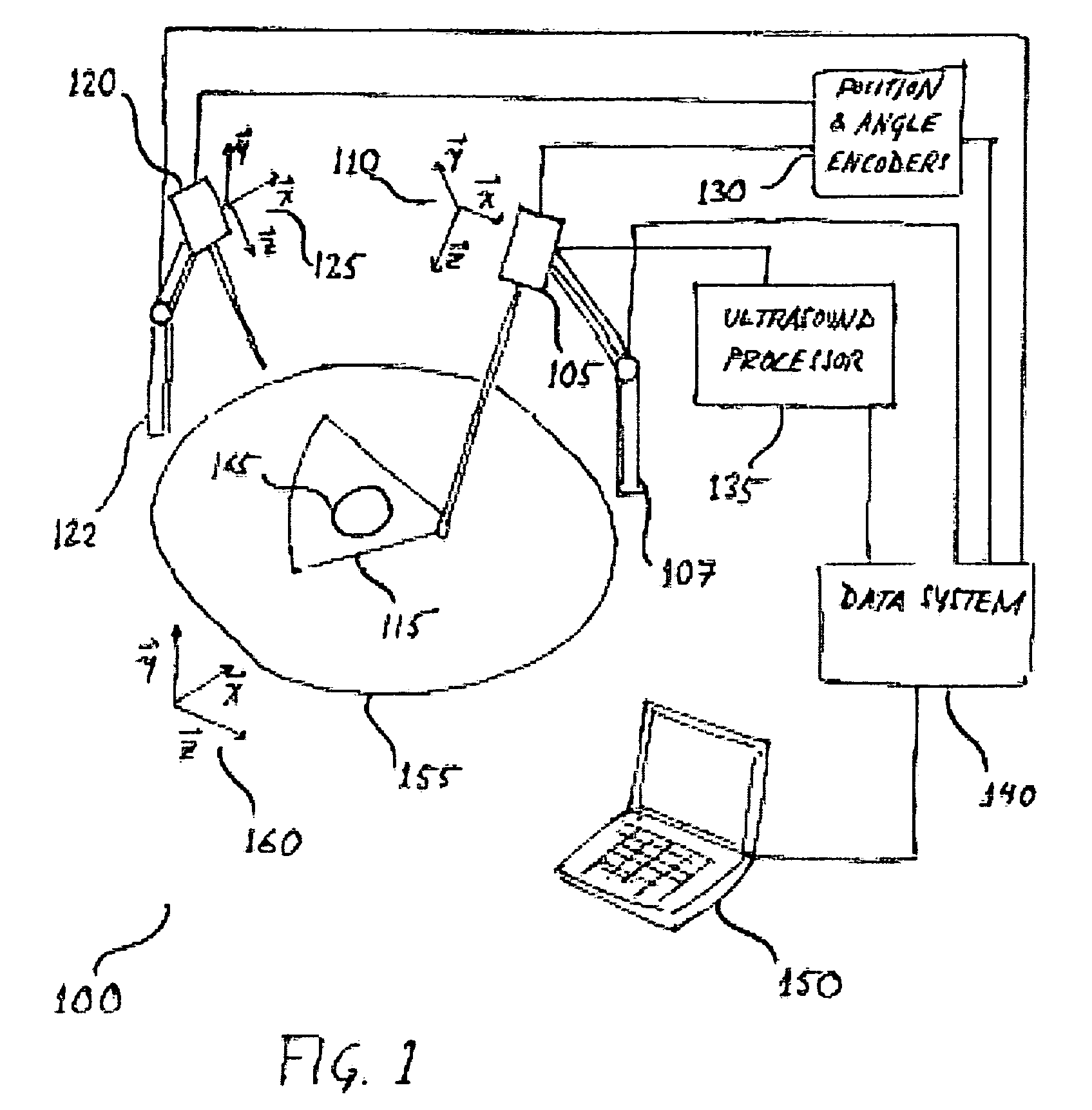
[0031]FIG. 1 illustrates an exemplary system 100 according to the present invention. The system 100 includes an interstitial ultrasound probe 105 having a probe reference frame 110 and a field of view 115; a surgical instrument 120 having an instrument reference frame 125; position and angle encoders 130 for measuring the position and orientation of the interstitial ultrasound probe 105 and the surgical instrument 120; an ultrasound processor 135 for providing signals to, and receiving signals from the interstitial ultrasound probe 105; a data system 140; and a user interface 150.

[0032] The system 100 may further include a probe mechanical arm 107 for controlling the position and orientation of the interstitial ultrasound probe 105. The probe mechanical arm 107 may be connected to the data system 140 for providing and receiving control signals and data. The position and angle encoders 130 corresponding to the interstitial ultrasound probe 105 may be attached to the probe mechanical...


PUM


Abstract

Disclosed is a system and method for providing image guidance for medical procedures in which an interstitial ultrasound probe is inserted into the tissue surrounding a tumor. The interstitial ultrasound probe is designed to โ€œrideโ€ in the surrounding tissue so that it may move in conjunction with the tumor and provide intra-operative imagery. A surgeon may guide a surgical instrument and perform interventions such as ablation while tracking the tumor in the ultrasound imagery. The position and orientation of the interstitial ultrasound probe and the surgical instrument are measured throughout the medical procedure to enable either the surgeon, or a robotic arm, to guide the surgical instrument such that it may move in conjunction with the tumor.

Description

[0001] This application claims the benefit of U.S. Provisional Patent Application No. 60 / 569,004, filed on May 7, 2004; U.S. Provisional Patent Application No. 60 / 577,788, filed on Jun. 8, 2004; and U.S. Non-provisional application Ser. No. 10 / 895,397 titled ROBOTIC 5D ULTRASOUND SYSTEM, which claims priority to U.S. Provisional Patent Application No. 60 / 488,941, filed Jul. 21, 2003, all of which are hereby incorporated by reference for all purposes as if fully set forth herein.[0002] The efforts associated with the subject matter of this patent application were supported by the National Science Foundation under grant no. EEC9731478.BACKGROUND OF THE INVENTION [0003] 1. Field of the Invention [0004] The present invention involves the field of ultrasound imagery. More particularly, the present invention involves a system and method for the use of ultrasound to provide real-time imaging for guiding surgical tools. [0005] 2. Discussion of the Related Art [0006] Computer Integrated Surg...

Claims


Application Information

Patent Timeline
24 Nov 2005
Publication
US20050261591A1
IPC
A61B8/08; A61B8/14
CPC
A61B8/4218; A61B8/08; A61B8/4245; A61B8/4254
Inventors
BOCTOR, EMAD; FICHTINGER, GABOR