Motion corrected multinuclear magnetic resonance imaging

a multi-nuclear magnetic resonance and imaging technology, applied in the field of acquiring magnetic resonance images, can solve the problems of low concentration of biomarkers, local change of image contrast, extra acquisition time, etc., and achieve the effect of increasing the good signal to noise ratio, and easy detection

Inactive Publication Date: 2010-03-04
KONINKLIJKE PHILIPS ELECTRONICS NV
View PDF8 Cites 12 Cited by
  • Summary
  • Abstract
  • Description
  • Claims
  • Application Information

AI Technical Summary

Benefits of technology

[0012]The present invention provides a method for acquiring MR images of an object, said object comprising at least first and second kinds of nuclei, the method comprising acquiring first MR image data of the object, wherein the first nuclei are excited. Concurrently or in the next step, second MR image data are acquired of the object, wherein the second nuclei are excited. The first MR image data are analyzed and motion parameters describing a motion of the object are determined based on said analysis. Finally a motion correction of the first and / or second MR image data is performed using said motion parameters. After the correction, multiple images resulting from the corrected second MR image data can be added to increase the signal to noise ratio. By performing the method according to the invention, second MR image data of the object can be acquired with a good signal to noise ratio.
[0013]This is especially important in the case of targeted contrast agents, which make this method especially suitable for the detection of certain diseases. For example, cancer cells can be detected by 19F labelled antibodies which specifically bind to said cancer cells. After injection into the human body, the fluorine labelled antibodies are more or less homogenously distributed in the human body. In the case of acquiring 19F MR image data, only a homogenous background (noise) signal originating from said homogenously distributed 19F labelled antibodies will be visible in the respective MR images. However, in the presence of respective cancer cells, the antibodies may bind to said cancer cells and accumulate at those cancer cell areas in the body. This leads to an accumulation of fluorine atoms at said locations, which can be easily detected in respective 19F MR images since due to the high concentration of fluorine atoms these areas light up as bright spots in the images. Using the image acquisition according to the invention, 19F MR image data acquisition can be performed over a long time scale, since any disturbing motion of the tissue is compensated.

Problems solved by technology

Some contrast agents possess permanent magnetic dipoles, which influence the relaxation process of the nearby water protons and so lead to a local change of the image contrast.
However, in general, the concentration of the biomarkers will be very low.
Averaging requires multiple acquisitions, hence extra acquisition time.
Additionally, isometric molecules like perfluorocarbons exhibit a large chemical shift, which must be corrected to obtain an optimal SNR and unambiguous results.
Therefore, chemical-shift corrected acquisition and related spectroscopic MRI methods have to be applied, which is additionally generally rather time consuming.
However, since acquisition of MR image data typically consists of multiple MRI measurements for the purpose of averaging MRI signals to increase the signal to noise ratio, it has to be ensured that during the measurements the image is not compromised by physiologic motion or deformation, since the set of measurements taken at different states of the object would not be immediately comparable.
Object motion during the acquisition of MR data produces image artifacts like blurring or ghosts in the phase encoded direction.
However, in case the tissue motion is irregular over time and complex throughout the volume, the traditional methods for motion estimation and correction fail.
Such irregular and complex motion typically takes place in the bowel.
Although, the colon itself exhibits peristaltic motion as well, its repeated local contractions appear in an irregular pattern, which are therefore unpredictable.
Chemical immobilization of the intestines, for example using an injection of Buscopan is only effective during a relatively short time span which is generally sufficient for CT yet insufficient for MRI.

Method used


Image

  • Motion corrected multinuclear magnetic resonance imaging
  • Motion corrected multinuclear magnetic resonance imaging
  • Motion corrected multinuclear magnetic resonance imaging

Examples


Embodiment Construction

[0041]In the following, similar elements are designated by the same reference numerals.

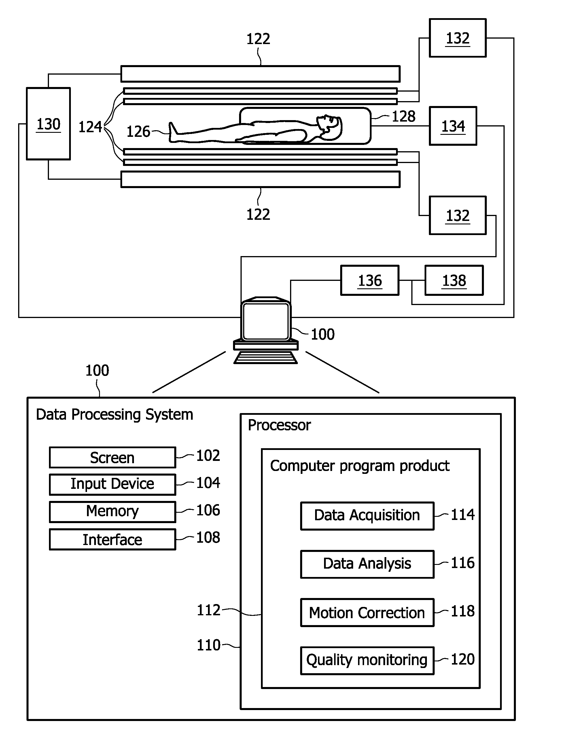
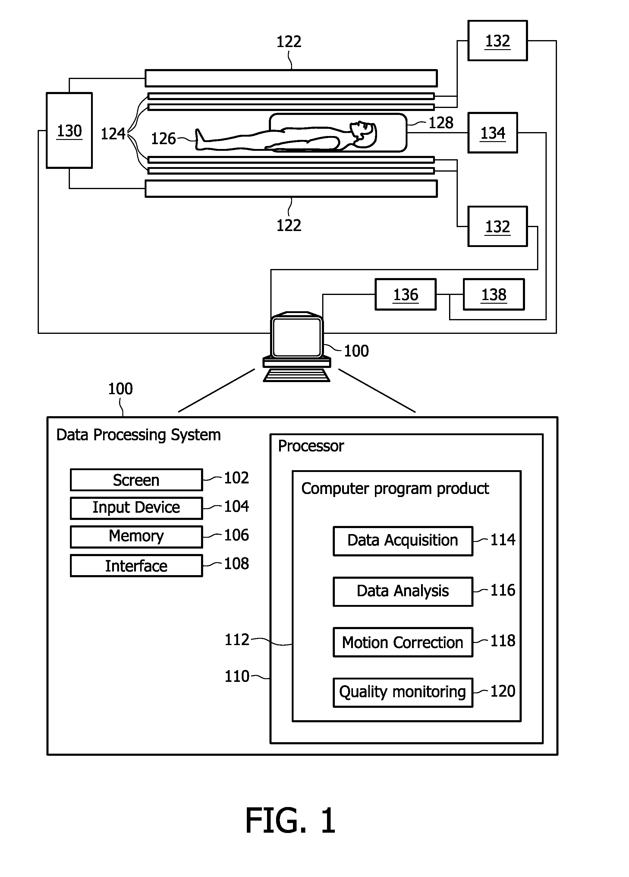
[0042]FIG. 1 is a block diagram of an embodiment of a magnetic resonance imaging apparatus. Thereby, only major components of a preferred MRI system which incorporates the present invention is shown in FIG. 1. The magnetic resonance imaging apparatus comprises a data processing system 100, whereby the data processing system 100 typically comprises a computer screen 102, an input device 104 which could for example be a keyboard and a mouse, as well as a memory 106 and an interface 108. Thereby, the interface 108 is adapted for communication and data exchange with typical MRI hardware components. These hardware components comprise for example a main field control unit 130 adapted for controlling the main field of the main magnet coils 122. The main magnets 122 may thereby be adapted as permanent super conducting magnets or being externally driven and switched on and off for each individual usage of ...


PUM


Abstract

The invention relates to a method for acquiring MR images (200-216) of an object, said object comprising at least first and second kinds of nuclei, the method comprising: acquiring (300; 304) first MR image data (200; 202; 204) of the object, wherein the first nuclei are excited, acquiring (302) second MR image data (206-216) of the object, wherein the second nuclei are excited, analyzing the first MR image data (200; 202; 204) determining motion parameters describing a motion of the object based on said analysis, motion correcting the first and/or second MR image data (206-216) using said motion parameters.

Description

TECHNICAL FIELD[0001]The invention relates to a method for acquiring Magnetic resonance (MR) images of an object, a magnetic resonance imaging apparatus for acquiring MR images of an object and a computer program product comprising computer executable instructions.BACKGROUND AND RELATED ART[0002]Magnetic resonance imaging (MRI) is one of the major imaging techniques in medicine. MRI is capable of generating detailed images of soft tissues. In MRI, specific properties of the various compounds found inside tissues are used to generate images, e.g., water is most commonly used for this purpose. When subjected to a strong external magnetic field, the protons 1H will align with this external field, resulting in a net magnetic moment. After excitation by radio frequency RF pulses, this magnetization will generate an RF signal that can be detected. This RF signal is characterized by a frequency that is related to the magnetic field strength. Therefore, magnetic field gradients are used to ...

Claims


Application Information

Patent Timeline
Patent Type & Authority Applications(United States)
IPC IPC(8): G06K9/00
CPCG01R33/281G01R33/446G01R33/5676G01R33/56509G01R33/4828
Inventor LAMERICHS, RUDOLF MATHIAS JOHANNES NICOLAASDE BRUIJN, FREDERIK JANGRUELL, HOLGERKEUPP, JOCHEN
Owner KONINKLIJKE PHILIPS ELECTRONICS NV
Why Eureka
Try Eureka