Methods and apparatus for scanning small sample volumes

a technology for optical probes and sample volumes, applied in the direction of fluorescence/phosphorescence, instruments, laboratory glassware, etc., can solve the problems of numerous technical challenges

Inactive Publication Date: 2005-12-01
BLUESHIFT BIOTECH
View PDF50 Cites 43 Cited by
  • Summary
  • Abstract
  • Description
  • Claims
  • Application Information

AI Technical Summary

Benefits of technology

[0009] Advantageous implementations can include one or more of the following features. Each sample well can be configured to hold a sample volume at most about 10 nanoliters. Each sample well can be configured to hold a sample volume smaller than one nanoliter. A substrate cover can be provided that seals edges of the individual sample wells and that is designed to minimize evaporation of the samples in the sample wells and to prevent cross-talk between individual sample wells. The substrate can have a substantially circular shape. The substrate can have a hole in its center, which allows the substrate to be placed on a rotating spindle. The circular substrate can further include a reference mark for referencing all sample well locations on the substrate. The substrate can be a glass substrate. The perimeter of a sample well can be substantially circular and the diameter of the sample well can be significantly larger than a depth of the sample well. The diameter of a sample well can be in the range of about 1-100 micrometers. The depth of the sample well can be in the range of about 10 nanometers to 10 micrometers.
[0010] A perimeter of each sample well can be surrounded by a lip for preventing overflow of the sample well and cross talk between the sample wells. The lip can extend to a height corresponding to about one-tenth to one-third of the depth of the respective sample wells. Each sample well can contain a sample that is unique with respect to the samples in the other sample wells on the substrate. The sample contained in a sample well can include at least one of: a biological sample and material derived from a biological sample. The centers of the sample wells can be separated by no more than about 100 micrometers. The substrate can have a surface height variation of at most about 10 micrometers over a region of the substrate that is readable by the apparatus, and the region that is readable by the apparatus can have a roughness of at most about than 10 nanometers. The substrate areas between the sample wells can be coated with a hydrophobic material and the sample wells can be hydrophilic in order to improve the confinement of the sample volumes into the sample wells.
[0012] Advantageous implementations can include one or more of the following features, some of which have been mentioned previously. A perimeter of each sample well can be surrounded by a lip for preventing overflow of the sample well and cross talk between the sample wells, and collecting light can include detecting when a focal point of a set of collection optical elements passes across an identifying feature on the substrate, and collecting light only when a focal point of a set of collection optical elements is located inside the perimeter defined by the lip. The identifying feature on the substrate can be a lip surrounding a sample well. The substrate can be substantially circular and collecting light can include rotating the circular substrate past a set of collection optical elements, and collecting light originating from within the sample well periodically each time the sample well rotates past the collection optical elements. The substrate can have a hole in the center and rotating can include rotating the circular substrate by means of a rotating spindle placed through the hole in the center of the substrate, past a set of collection optical elements.
[0013] In general, in one aspect, the invention provides a substrate for holding one or more biological samples in an apparatus for collecting optical data pertaining to one or more characteristics of the biological samples. The substrate includes a flat transparent glass substrate and a substrate cover. The glass slide has several sample wells formed therein and a set of fiducials against which the location of the sample wells can be determined by the apparatus. Each sample well holds a volume of a biological sample of at most about 50 nanoliters by one or more of: gravity, capillary action and surface tension and each sample well is surrounded by a lip arranged to prevent cross-talk between the sample wells. The substrate areas between the sample wells are coated with a hydrophobic material and the sample wells are hydrophilic in order to improve the confinement of the sample volumes into the sample wells. The substrate cover seals the edges of the individual sample wells and is designed to minimize evaporation of the biological samples in the sample wells.

Problems solved by technology

This presents numerous technical challenges.

Method used


Image

  • Methods and apparatus for scanning small sample volumes
  • Methods and apparatus for scanning small sample volumes
  • Methods and apparatus for scanning small sample volumes

Examples


Embodiment Construction

Overview of the Analysis System

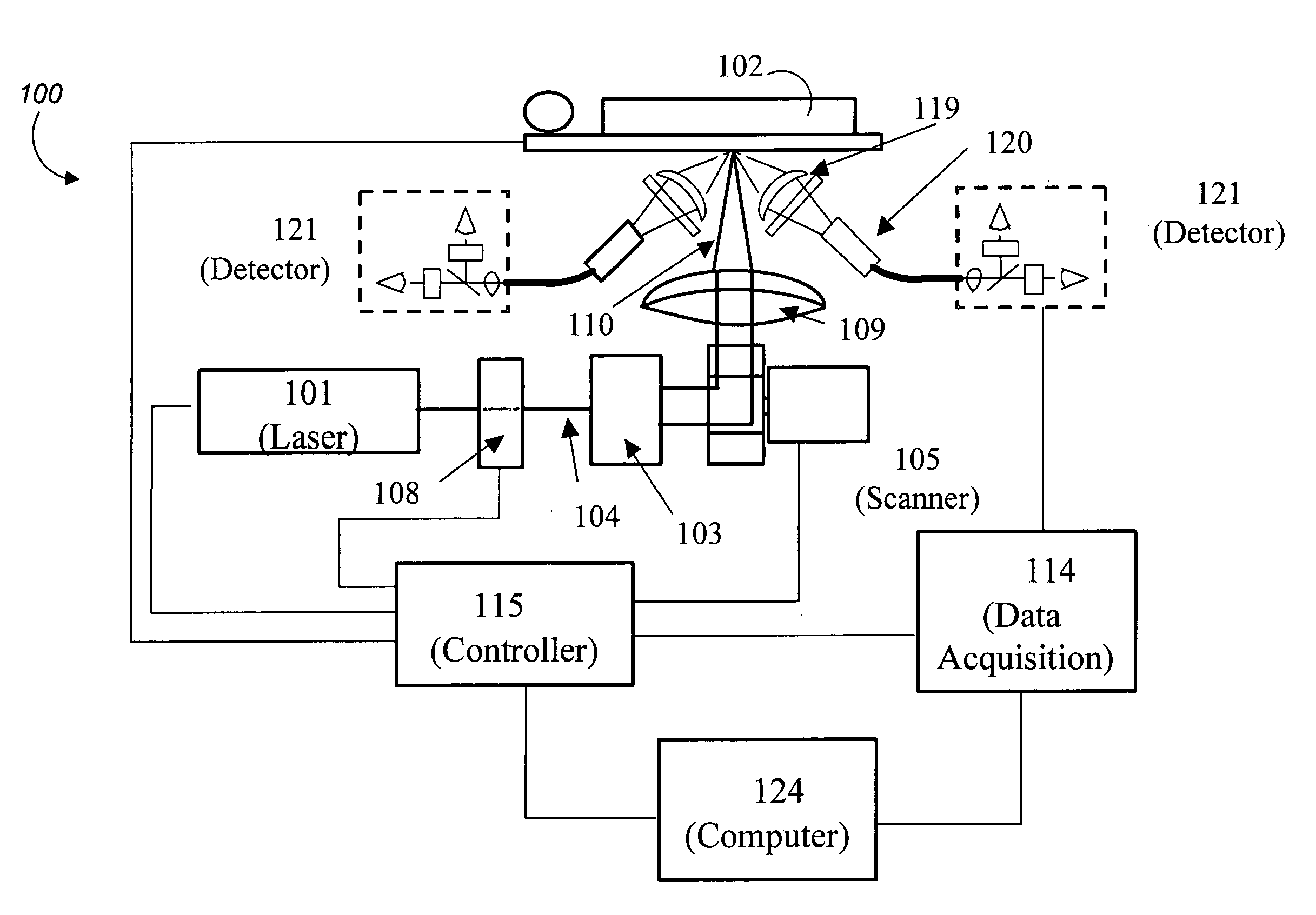
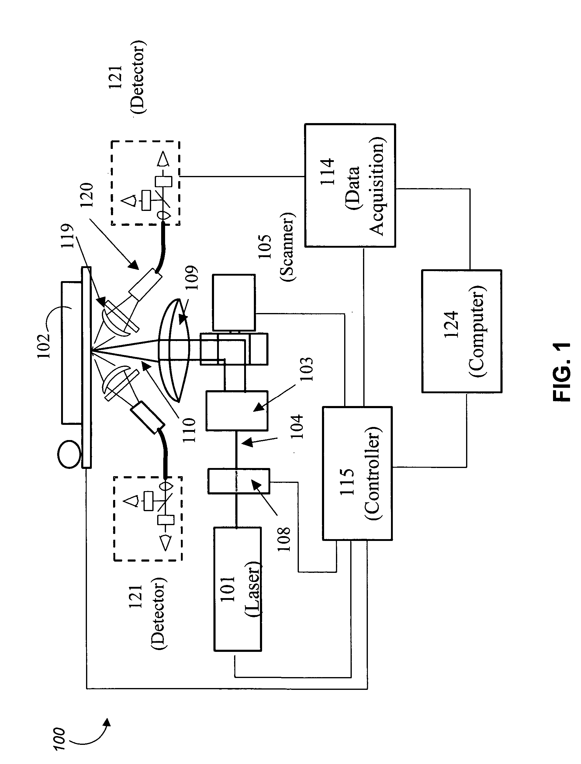
[0027] The invention provides an improved apparatus for interrogating and analyzing miniaturized samples, such as samples located in a number of small sample wells on a transparent substrate. An apparatus and methods suitable for analyzing such samples have been described in U.S. patent application Ser. No. 10 / 927,748 entitled “TIME DEPENDENT FLUORESCENCE MEASUREMENTS” and in U.S. patent application Ser. No. 10 / 928,484 entitled “MEASURING TIME DEPENDENT FLUORESCENCE,” the entire disclosures of both of which were incorporated herein by reference above.

[0028] In the described embodiment, the apparatus uses a scanning light source, which can be focused onto a substrate containing samples, with the ability to discriminate against background noise or signal, and makes use of image contrast mechanisms. The apparatus can be operated in several distinct modes or combinations thereof, depending on what type of sample data needs to be collected.

[0029] In a f...


PUM

PropertyMeasurementUnit
volumeaaaaaaaaaa
volumeaaaaaaaaaa
volumeaaaaaaaaaa

Abstract

Methods and apparatus for assaying biological materials employ multi-well substrates as described herein. The substrates include a plurality of wells, typically each of several nanoliters volume or smaller having consistent dimensions and formed in a rigid substrate such as a glass disk. Each well may be provided with a circumferential lip to minimize crosstalk between wells and / or facilitate optical location of the individual wells during interrogation. Samples are provided to the individual wells and assayed by an optical technique employing fluorescence, polarization, reflectance, or the like. A scanning laser system may be employed for this purpose. The substrate may rotate during the scan to allow consistent interrogation of the wells without stopping and starting the rotation. Multiple rotations may also be employed repeatedly interrogate the samples for use in a kinetic study, for example.

Description

CROSS-REFERENCE TO RELATED APPLICATIONS [0001] The present application claims priority under 35 U.S.C. §119(e) from U.S. Provisional Patent Application No. 60 / 543,409 entitled “SUBSTRATE FOR ULTRAFAST SCANNING OF MINIATURIZED ASSAYS” filed Feb. 9, 2004, the entire disclosure of which is incorporated herein by reference for all purposes. The present application is also related to U.S. patent application Ser. No. 10 / 927,748 entitled “TIME DEPENDENT FLUORESCENCE MEASUREMENTS” filed on Aug. 26, 2004, and to U.S. patent application Ser. No. 10 / 928,484 entitled “MEASURING TIME DEPENDENT FLUORESCENCE” filed on Aug. 26, 2004, the entire disclosures of both of which are incorporated herein by reference for all purposes. The present application is also related to U.S. Provisional Patent Application No. 60 / 497,764, entitled “TIME DEPENDENT FLUORESCENCE MEASUREMENTS” filed Aug. 26, 2003, and to U.S. Provisional Patent Application No. 60 / 497,803, filed Aug. 26, 2003, the entire disclosures of bo...

Claims


Application Information

Patent Timeline
Patent Type & Authority Applications(United States)
IPC IPC(8): B01L3/00G01N1/10G01N21/25G01N21/64G01N35/02
CPCB01L3/5085B01L2300/0806G01N21/253G01N35/028G01N21/6445G01N21/6452G01N21/6408
Inventor CROMWELL, EVAN F.MILLER, STEVEN C.SHUMATE, CHRISTOPHER B.COMITA, PAUL B.
Owner BLUESHIFT BIOTECH
Why Eureka
Try Eureka