Process for detecting PRPsc using an antibiotic from the family of aminoglycosides

a technology of aminoglycosides and enzymes, which is applied in the field of process for detecting prpsc using an antibiotic from the family of aminoglycosides, can solve the problems of slow development, significantly slowing the development of epidemiological models, and difficult detection of prpsc in infected animals

Inactive Publication Date: 2006-01-19
AGENCE FRE DE SECURITE SANITAIRE DES ALIMENT +3
View PDF5 Cites 6 Cited by
  • Summary
  • Abstract
  • Description
  • Claims
  • Application Information

AI Technical Summary

Problems solved by technology

The delayed appearance after an incubation period of 2 to 6 years and slow development of symptoms in cattle infected with ESB has considerably slowed the development of epidemiological models.
Detection of the protein pathogen PrPsc is difficult in infected animals healthy before the development of the disease and especially in the blood and urine of diseased animals.
However, prior methods have constantly come up against the difficulty of identifying PrPsc in a reliable manner when it is in a low quantity in a biological sample.

Method used


Image

  • Process for detecting PRPsc using an antibiotic from the family of aminoglycosides
  • Process for detecting PRPsc using an antibiotic from the family of aminoglycosides
  • Process for detecting PRPsc using an antibiotic from the family of aminoglycosides

Examples


example 1

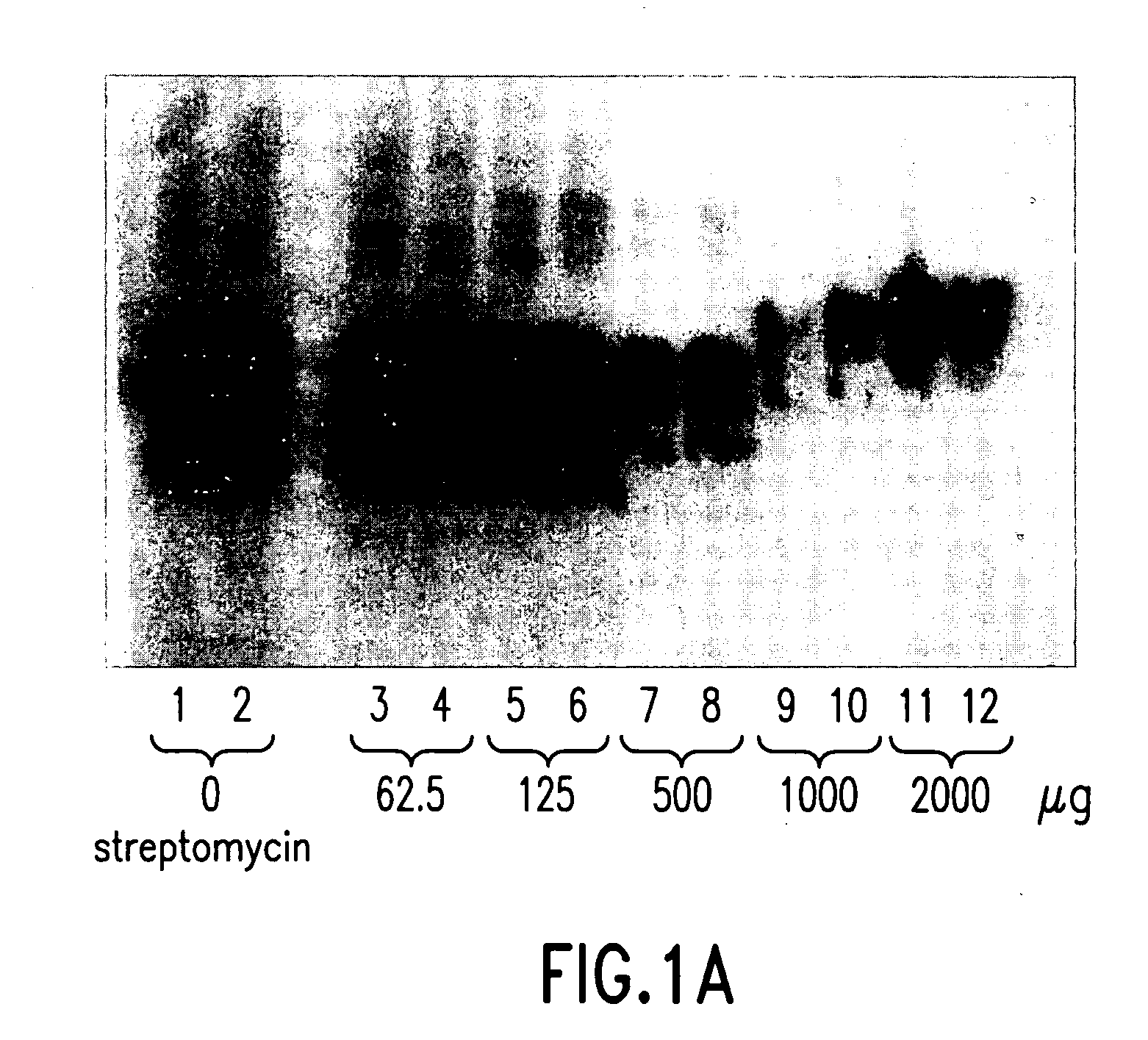
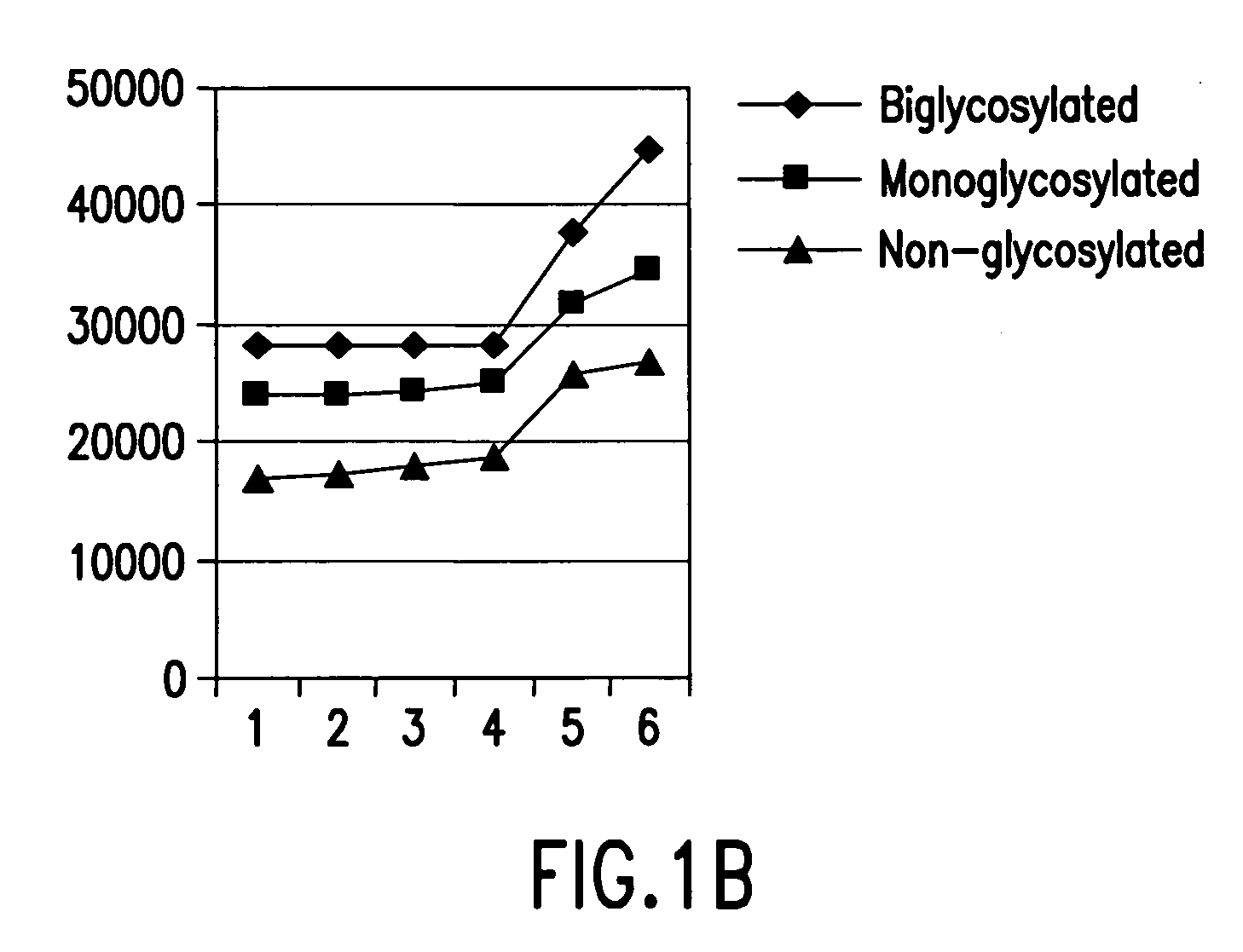
[0053] Example 1 relates to FIGS. 1A and 1B concerning trials in which increasing concentrations (0 μg, 62.5 μg, 125 μg, 500 μg, and 2000 μg) of streptomycin were added to constant quantities of PrPsc extracted from the equivalent of 920 μg of sheep brain afflicted with trembling. Then, the mixture was centrifuged. The supernatant was used for immunological detection by Western blot (FIG. 1A) and measurement of the average molecular mass of bands of PrPsc (FIG. 1B). The results show that the increase of the quantity of streptomycin, namely, an addition of 0, 62.5, 125, 500, 1000 and 2000 μg, permits the observation that at the lowest concentrations of streptomycin the band of the non-glycosylated protein is the first to show an increase of the apparent molecular mass. Then, the complexing concerning the band of monoglycosylated protein and, finally, the biglycosylated protein is complexed when the concentration of streptomycin is the greatest.

[0054] The number of streptomycin molec...

example 2

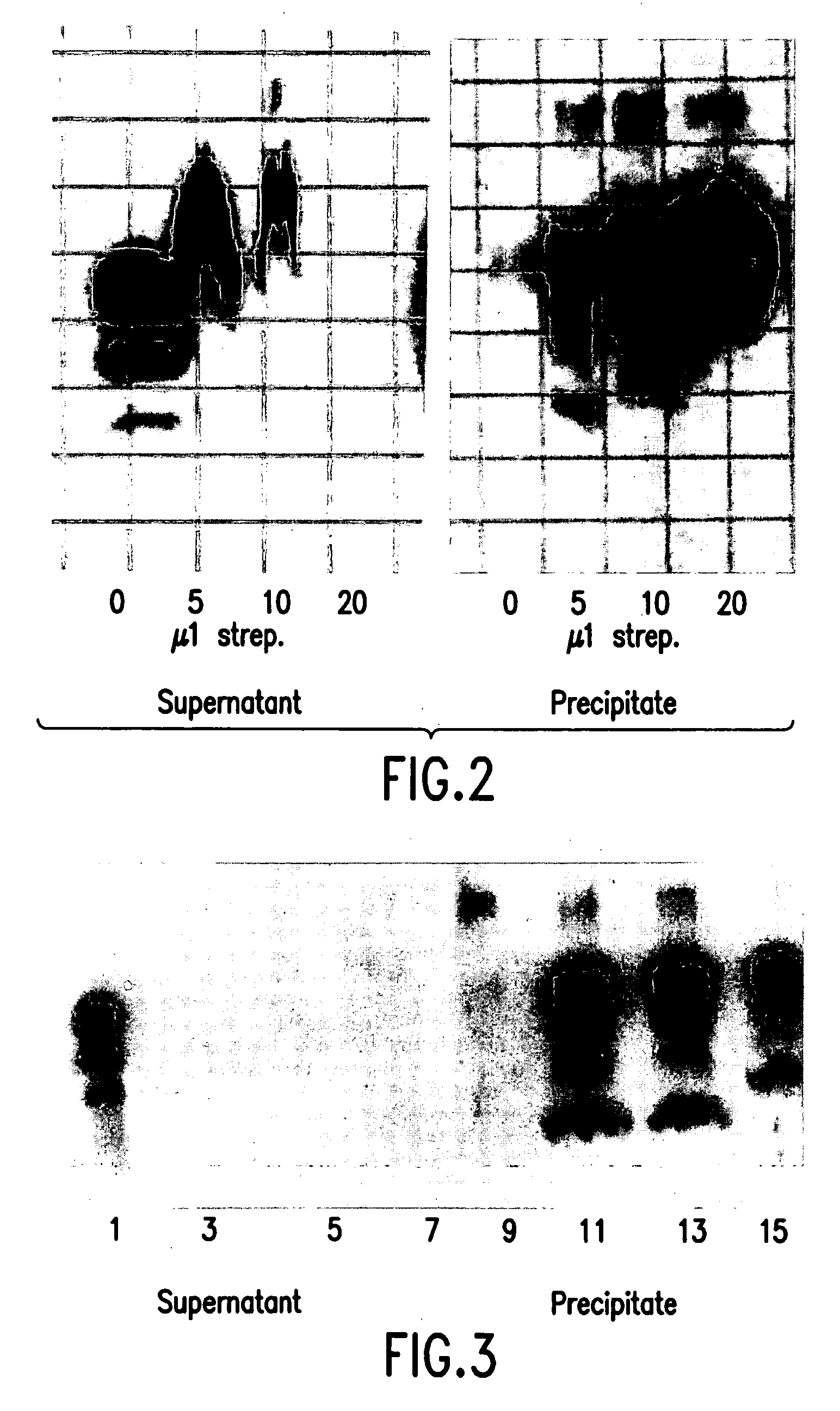
[0055] Example 2 relates to FIG. 2 concerning trials in which increasing concentrations (0 μg, 5 μg, 10 μg, and 20 μg) of streptomycin at 1 g / ml were added to constant quantities of the biological sample prepared as indicated above. In the absence of streptomycin, all the PrPsc bands are identified as being in the supernatant. They are detected progressively and simultaneously in the bottom and in the supernatant. The quantity of PrPsc present in the supernatant diminished progressively while the quantity increased progressively in the precipitates. The addition of 20 μl of streptomycin totally precipitated the PrPsc, that was then detected only in the precipitates.

example 3

[0056] Example 3 relates to FIG. 3.

[0057] The experiment of Example 2 was repeated, but with the same brain sample diluted to 1 / 25 relative to Example 2 and reducing the incubation period to one hour after the simultaneous addition of proteinase K and streptomycin to the brain suspensions. The result is that PrPsc bands were only detected in the supernatant in the absence of streptomycin and in the bottom in the presence of any concentration of streptomycin.


PUM

PropertyMeasurementUnit
detection thresholdaaaaaaaaaa
temperatureaaaaaaaaaa

Abstract

A process of concentration by precipitation of PrPsc for detecting or diagnosing PrPsc, wherein a tissue or biological fluid stemming from or obtained from an animal or human organism is brought together with an antibiotic from the family of aminoglycosides, preferably streptomycin or one of its derivatives. The use of such an antibiotic for eliminating PrPsc from a tissue or biological fluid and a kit for diagnosing pathologies associated with the presence of PrPsc is also disclosed.

Description

RELATED APPLICATION [0001] This is a continuation of International Application No. PCT / FR2003 / 003856, with an international filing date of Dec. 19, 2003 (WO 2004 / 059321 A1, published Jul. 15, 2004), which is based on French Patent Application No. 02 / 16382, filed Dec. 20, 2002. FIELD OF THE INVENTION [0002] This invention relates to a method of total precipitation of PrPsc by streptomycin sulfate and its use for the immunodetection or elimination of PrPsc from liquids or solutions. BACKGROUND [0003] The native or normal prion protein, designated “PrP” or “PrPc”, for the cellular prion protein is a glycoprotein broadly expressed in the lymphoid and neuronal cells of mammals. [0004] Conformational changes of PrPc result in the appearance and propagation of the protein pathogen PrPc that is resistant to the proteinase K. This protein pathogen can be indifferently called “PrPsc” or “PrPres.” Accumulation of PrPsc in the organs of animals is at the origin of numerous diseases and especial...

Claims


Application Information

Patent Timeline
Patent Type & Authority Applications(United States)
IPC IPC(8): G01N33/53G01N33/68
CPCG01N33/6896Y10T436/104998G01N2800/2828
Inventor MOUSSA, ALYCOLEMAN, ANTHONY WILLIAMBENCSIK-REYNIER, ANNASHAHGALDIAN, PATRICKPERRON, HERVEMARTIN, AMBROISE
Owner AGENCE FRE DE SECURITE SANITAIRE DES ALIMENT
Why Eureka
Try Eureka