
Apparatus and methods for facilitating hemostasis within a vascular puncture

a technology of vascular puncture and apparatus, applied in the field of apparatus and methods for sealing punctures, can solve the problems of time-consuming and expensive procedures, requiring as much as an hour of medical professionals' time, uncomfortable for patients, etc., and achieve the effect of enhancing hemostasis within the punctur

Active Publication Date: 2006-03-02
ACCESSCLOSURE
View PDF99 Cites 219 Cited by
  • Summary
  • Abstract
  • Description
  • Claims
  • Application Information

AI Technical Summary

Benefits of technology

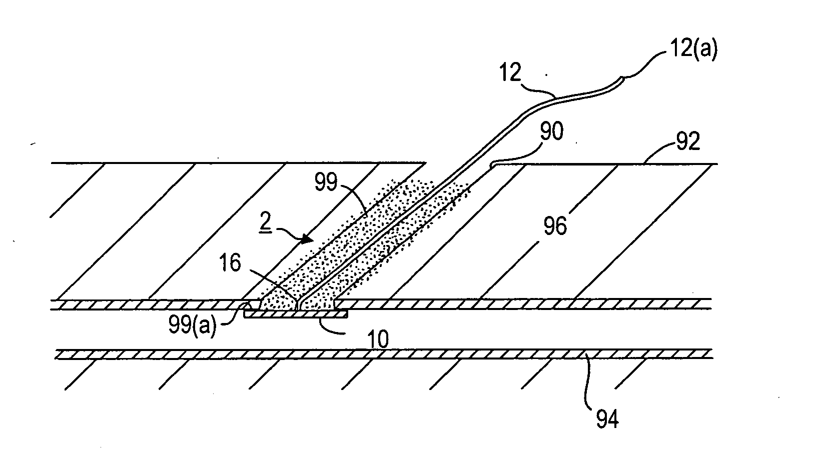
[0017] Optionally, sealing material, e.g., a plug and / or a single or multiple liquid sealing material, may be delivered into the puncture, e.g., around the retaining member, to further enhance hemostasis within the puncture. The sealing material may also be bioabsorbable, e.g., over a relatively longer time than the sealing member disposed within the body lumen.

Problems solved by technology

This procedure, however, may be time consuming and expensive, requiring as much as an hour of a medical professional's time.
It is also uncomfortable for the patient, and may require the patient to remain immobilized in the operating room, catheter lab, or holding area.
In addition, a risk of hematoma exists from bleeding before hemostasis occurs.
One of the disadvantages with these closure devices is that it may be difficult to position them properly with respect to the vessel, which may be significant since it is generally undesirable to expose collagen material to the bloodstream where it may float downstream and cause an embolism.
In addition, these closure devices take a relatively long period of time to resorb and / or dissolve within the body (i.e., ten days or more).
This is particularly undesirable for the portion of the device that is disposed within the blood vessel, because of the risk of a piece of the closure device breaking free and causing an embolism or other damage downstream of the puncture site.
Of course, if the device is absorbed too rapidly, it may adversely affect effective hemostasis of the puncture without risk of hematoma or other complications.

Method used


Image

  • Apparatus and methods for facilitating hemostasis within a vascular puncture
  • Apparatus and methods for facilitating hemostasis within a vascular puncture
  • Apparatus and methods for facilitating hemostasis within a vascular puncture

Examples


Embodiment Construction

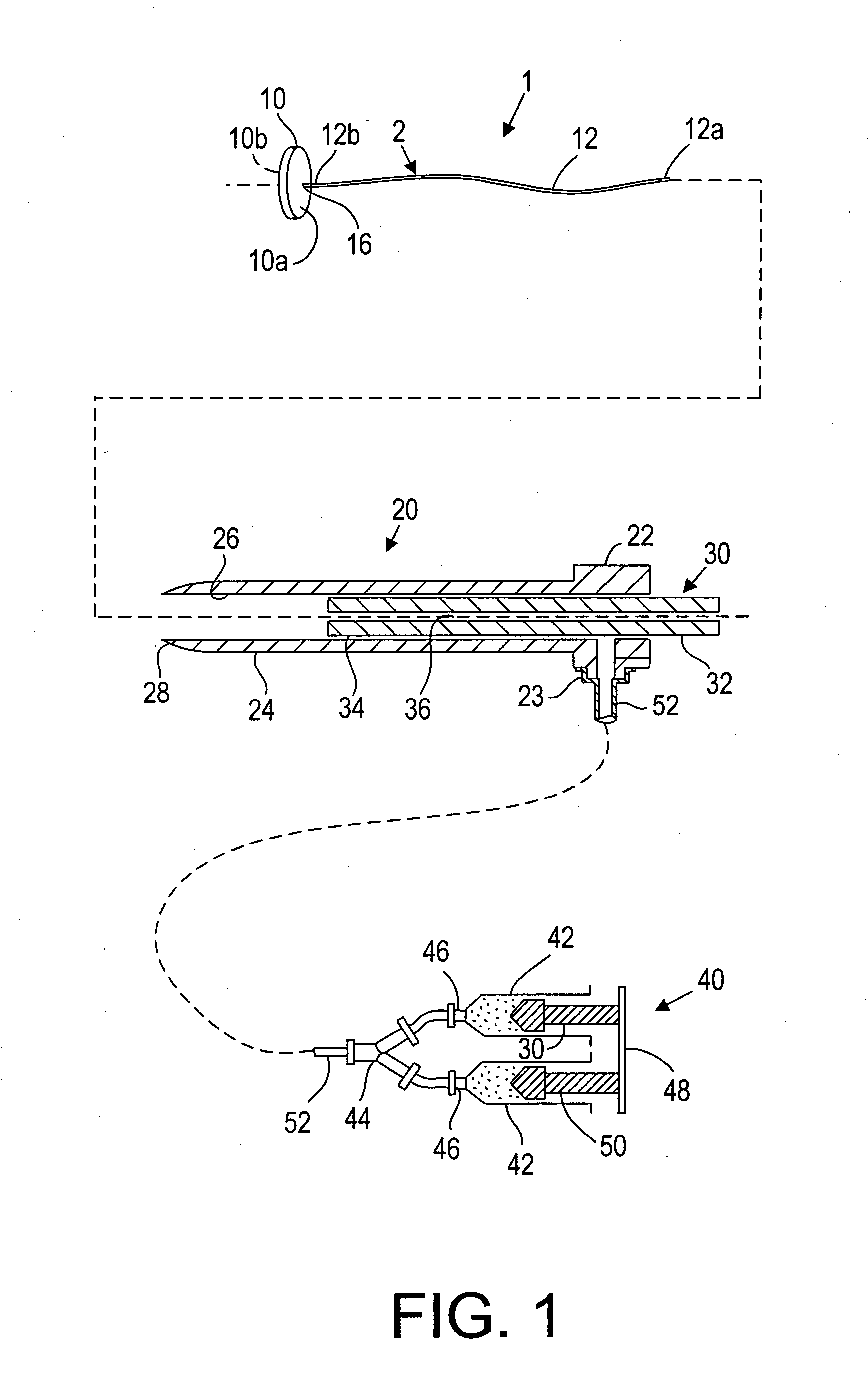
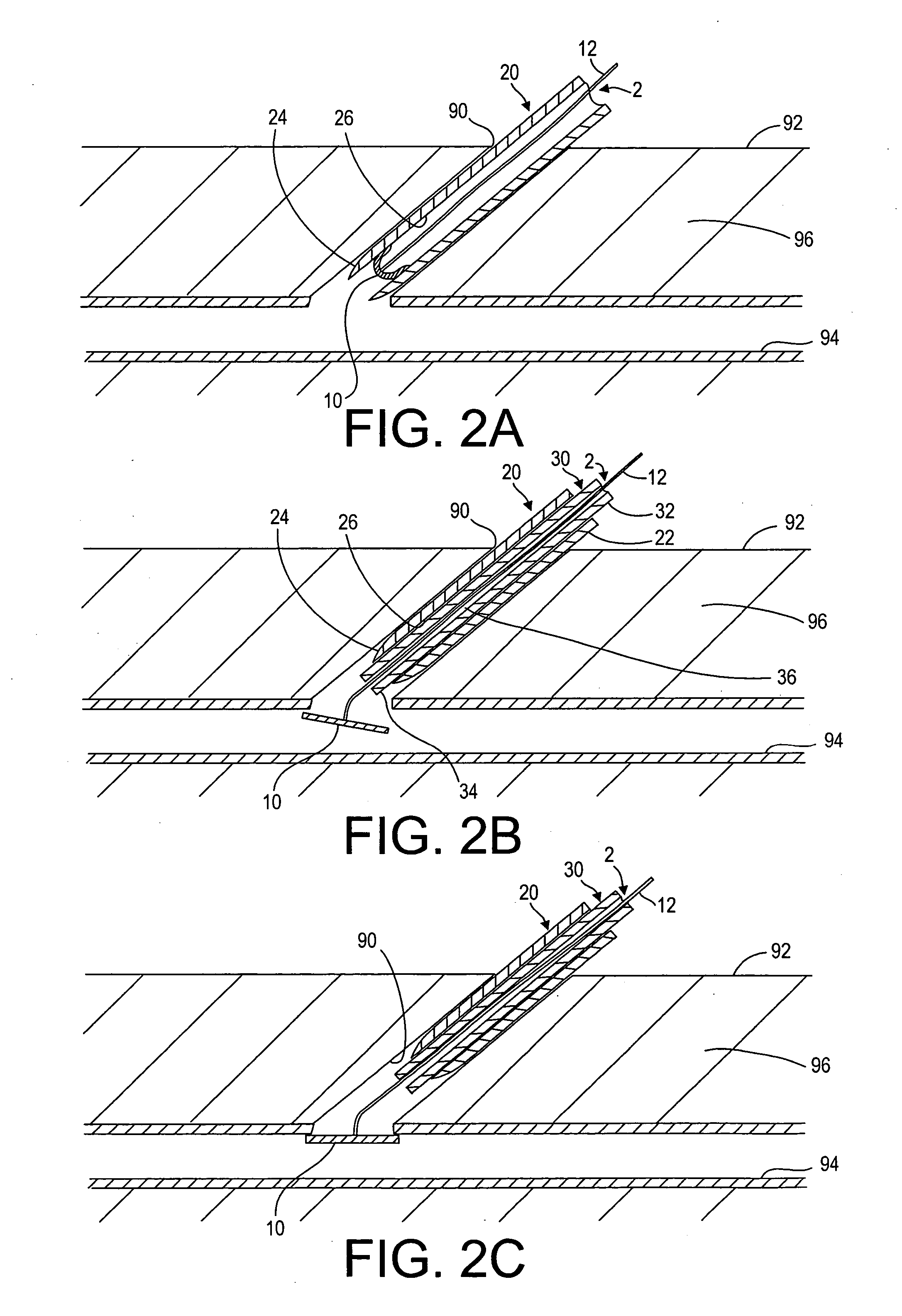
[0025] Turning to the drawings, FIG. 1 illustrates an exemplary embodiment of a closure device 2 and an apparatus 1 for facilitating temporary or permanent hemostasis of a puncture extending through tissue using the closure device 2. Generally, the apparatus 1 includes a delivery sheath 20, and a plunger, catheter, or other pusher member 30 for deploying the closure device 2 from the delivery sheath 20. Optionally, the apparatus 1 may include a source of sealing material 40, e.g., that may be delivered via the delivery sheath 20, as described further below.

[0026] The closure device 2 generally includes a filament or other retaining member 12 including a proximal end 12a and a distal end 12b, and a bioabsorbable sealing member 10 on the distal end 12b. The filament 12 may be a solid or hollow elongate body, e.g., a suture, string, wire, tube, and the like, e.g., having a diameter, thickness, or other cross-sectional dimension of not more than about 0.90 mm. In one embodiment, the fi...


PUM


Abstract

Apparatus for sealing a puncture communicating with a blood vessel includes a bioabsorbable sealing member secured to one end of a filament or other retaining member. The sealing member is delivered through the puncture into the vessel, and retracted against the wall of the vessel to provide temporary hemostasis. The sealing member is rapidly absorbed after exposure within the vessel, e.g., to an aqueous or heated physiological environment (e.g., exposure to blood or body temperature), immediately or shortly after completing a medical procedure via the puncture, e.g., within the time period that the patient is ambulatory optionally, extravascular sealing material is delivered into the puncture proximal to the sealing member. The retaining member and / or extravascular material may be bioabsorbable, being absorbed at a slower rate than the sealing member. Alternatively, the filament is removed from the puncture after hemostasis is established.

Description

FIELD OF THE INVENTION [0001] The present invention relates generally to apparatus and methods for sealing punctures in a body, and more particularly, to apparatus and methods for sealing a vascular puncture extending through tissue into a blood vessel, and to apparatus and methods for delivering a fast-dissolving sealing device into a puncture extending from a patient's skin to a blood vessel or other body lumen to provide temporary hemostasis. BACKGROUND [0002] Apparatus and methods are known for accessing a patient's vasculature percutaneously, e.g., to perform a procedure within the vasculature, and for sealing the puncture that results after completing the procedure. For example, a hollow needle may be inserted through a patient's skin and overlying tissue into a blood vessel. A guide wire may be passed through the needle lumen into the blood vessel, whereupon the needle may be removed. An introducer sheath may then be advanced over the guide wire into the vessel, e.g., in conj...

Claims


Application Information

Patent Timeline
IPC IPC(8): A61B17/04
CPCA61B17/0057A61B2017/00004A61B2017/00637A61B2017/0065A61B2017/00601A61B17/08A61B17/10A61B2017/00575A61B2017/00659
Inventor KHANNA, PUNEET K.KHOSRAVI, FARHAD
Owner ACCESSCLOSURE
Learn More