
Phacoemulsification device having rounded edges

a phacoemulsification device and rounded edge technology, applied in the field of surgical instruments, can solve the problems of cutting edge damage to the lens capsule and iris, damage to the incision, etc., and achieve the effects of reducing the risk of damage, and eliminating any sharp edges

Active Publication Date: 2006-03-09
DRT VENTURES
View PDF3 Cites 63 Cited by
  • Summary
  • Abstract
  • Description
  • Claims
  • Application Information

AI Technical Summary

Benefits of technology

[0018] A phacoemulsification needle according to the present invention has a hollow member with an inner surface defining a lumen, an outer surface, and a distal end tip. The distal end tip has an outer edge portion and an inner edge portion, both of which are rounded to eliminate any sharp edges. The needle can be used to deliver ultrasonic energy to a surgical site, such as a cataract, to emulsify unwanted tissue and to aspirate the tissue through the lumen of the hollow member without using any sharp surfaces for cutting. The rounded outer and inner edge portions of the distal end tip of the needle increase the safety of the surgical procedure by reducing the risk of damage to the capsule and other intraocular structures or tissues at or near the surgical site. This improvement in safety has allowed the development of new needle configurations to improve the efficiency of phacoemulsification, several of which are disclosed herein.
[0019] According to a broad aspect of the present invention, a phacoemulsification needle is provided, comprising: a hollow member having an inner surface, an outer surface, and a distal end tip; wherein the distal end tip has an outer edge portion and an inner edge portion, and wherein the outer and inner edge portions are rounded to eliminate any sharp edges.
[0020] According to another broad aspect of the present invention, a method of performing phacoemulsification is provided, comprising the steps of: providing a phacoemulsification needle comprising a hollow member having an inner surface, an outer surface, and a distal end tip, the distal end tip having outer and inner edge portions which are rounded to eliminate any sharp edges; and using the phacoemulsification needle to deliver ultrasonic energy to emulsify unwanted tissue at a surgical site and to aspirate the unwanted tissue through the hollow member without using a sharp surface to cut the unwanted tissue.

Problems solved by technology

Fundamentally, the sharp edge of the phacoemulsification probe enhances the cutting ability of the needle, but it poses several dangers.
The greatest danger is the ability of the cutting edge to tear intraocular structures while trying to remove the nuclear material.
This may result in damage to the lens capsule and iris.
Incidentally, the tip passing through the incision itself can cause damage to the incision.
Even when vacuum is turned off, an imbalance of the flow of fluid to stabilize the anterior chamber of the eye (infusion) and / or the process of the application of ultrasonic energy can create a vacuum-like effect to draw eye structures into the lumen of the phacoemulsification needle with or without intent.
Phacoemulsification needles with sharp edges on any surface can cause damage to ocular tissue.
Incidental contact of these sharp edges, either with the sharpened knife-like honing of the edge, or with burrs on the edge remaining from poor manufacturing techniques, will cause damage to the tissues these edges contact.
The iris is a readily distensible tissue that typically is not involved in intraocular surgery as the dilation is in most cases adequate to keep it out of the field of the active aspiration / emulsification occurring at the distal tip of the phacoemulsification needle.
Likewise, it becomes much more distensible as it becomes more exposed.
The anterior capsule is perhaps the least distensible of the tissues described, but can be damaged as well.
Damage to the capsule during this stage of surgery significantly compromises the likelihood of successfully removing the cataract without loss of vitreous, and can be shown to compromise the result of the cataract surgery by significantly increasing the intraoperative and postoperative complication rates.
The frequency of damage to the iris during cataract surgery is not documented, but can also result in significant compromise of the surgical result.
Such excess leakage can increase outflow and threaten chamber stability.
In addition, surgeon technique may not be sufficient to anticipate unwanted contact of these distensible tissues to the phacoemulsification tip.
Situations such as poor iris dilation, an unstable anterior chamber, or surgeon technique can result in an increased likelihood of a distensible eye tissue coming in contact with any of the sharpened or burred edges of a phacoemulsification needle, and this contact is likely to create damage to those eye tissues.
Another problem with prior art phacoemulsification needles is that active aspiration of a distensible ocular tissue can cut or damage that tissue by rubbing it against the sharp internal edge of the distal tip (i.e., the far distal inner edge of the lumen) or against small burrs that may exist despite honing this inner edge surface.
As previously discussed, these distensible tissues can be aspirated due to poor chamber stability (such as is seen when the incision sizes are not appropriate and allow excessive outflow), poor iris dilation, or poor surgeon technique as examples.
Contact of these tissues with the inner edge of the tip of the phacoemulsification needle (i.e., the outer edge of the inner lumen of the needle) can result in severe damage to these tissues, either from direct cutting of the sharp surface or through ripping or sawing as caused by the roughened burrs.
It is much more likely that the “vacuum” involved merely stabilizes the distensible capsule and allows it to be cut by either the sharpened, honed inner edge, or an incidental burr as a result of poor manufacturing.
This knuckle-tipped needle requires a new surgical technique to be used by cataract surgeons and a separate device to cut a cataract before removal, thereby reducing surgical efficiency.

Method used


Image

  • Phacoemulsification device having rounded edges
  • Phacoemulsification device having rounded edges
  • Phacoemulsification device having rounded edges

Examples


Embodiment Construction

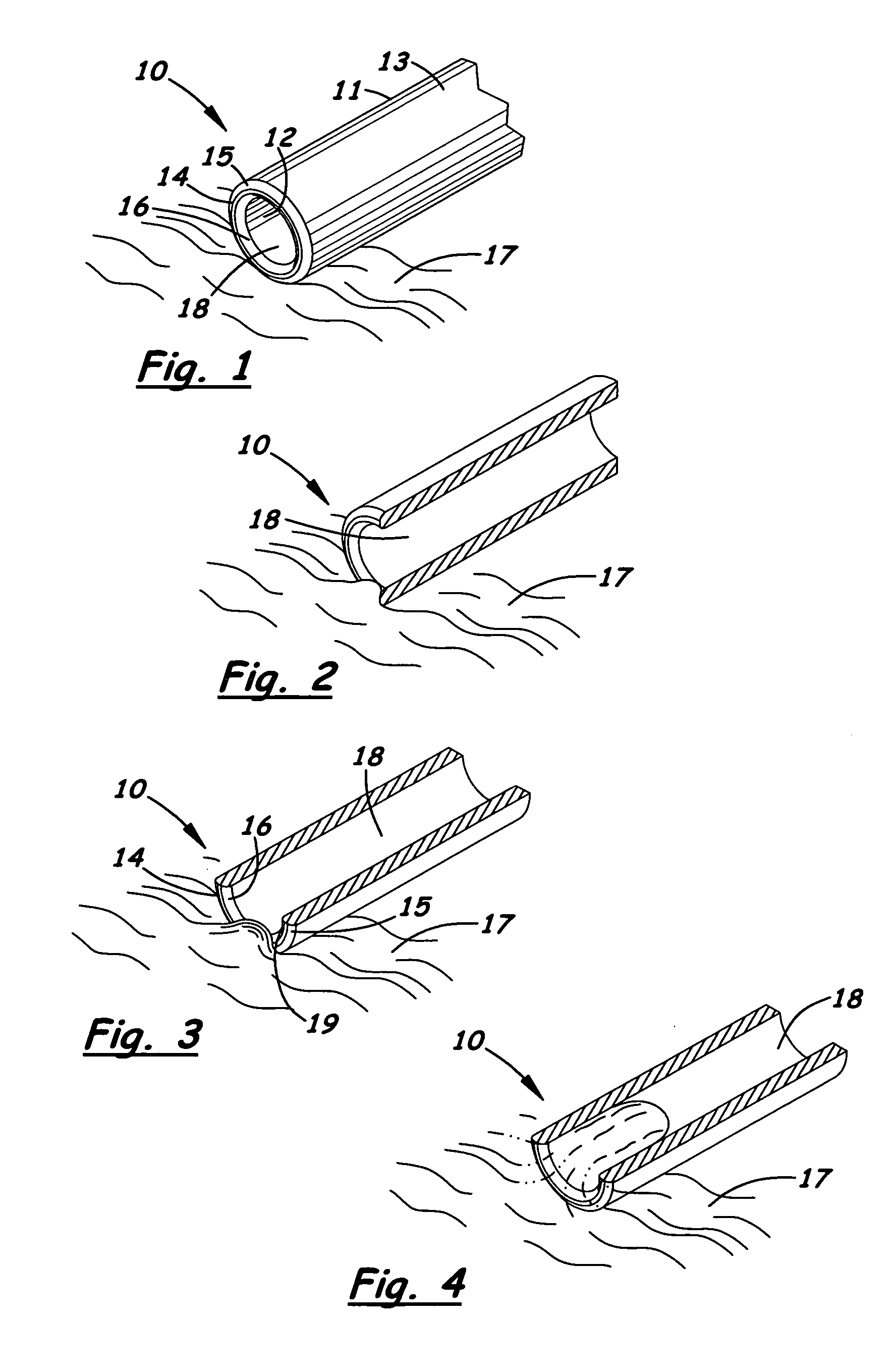
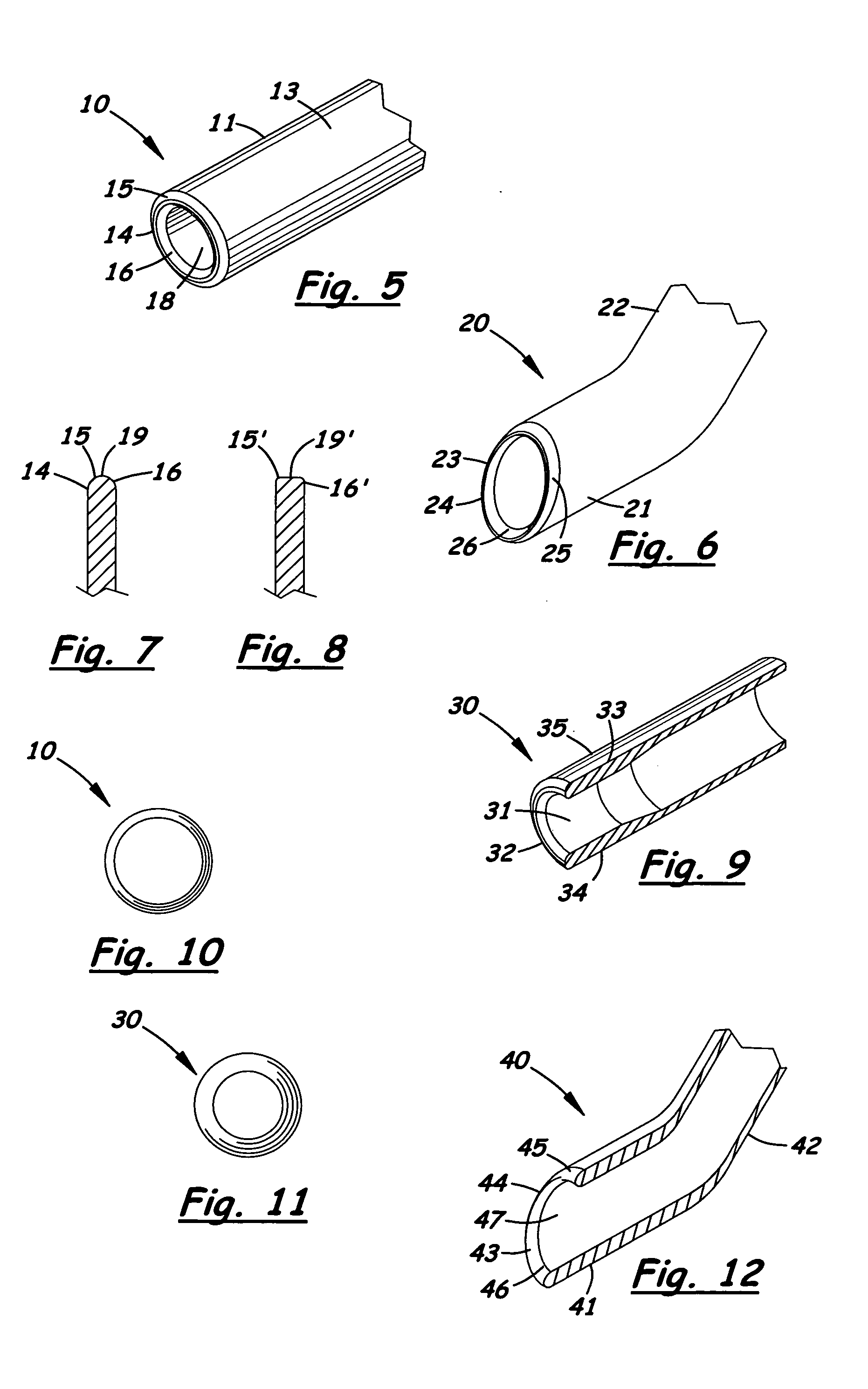
[0069] Several embodiments of phacoemulsification needles and device according to the present invention will now be described with reference to FIGS. 1 to 45 of the accompanying drawings.

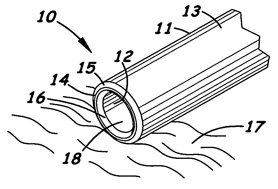
[0070] A phacoemulsification needle 10 according to a first embodiment of the present invention is shown in FIGS. 1 to 5. The needle 10 includes a hollow member 11 having an inner surface 12, a cylindrical outer surface 13, and a distal end tip 14. The distal end tip 14 has a rounded outer edge 15 and a rounded inner edge 16 and is carefully manufactured to avoid or eliminate any surfaces coming to a sharp point or sharp edge. Accordingly, the distal end tip 14 of the hollow needle 10 displays no surface coming to a sharp point or sharp edge.

[0071] The lens capsule 17, in its flaccid or resting state empty of contents, is essentially flat, depending on the fluidics in the eye at the time of observation. Typically it lies like a tablecloth draped over a bucket, provided the fluidics are at neutral....


PUM


Abstract

A phacoemulsification needle is disclosed having a hollow member with an inner surface, an outer surface, and a distal end tip. The distal end tip has an outer edge portion and an inner edge portion both of which are rounded to eliminate any sharp edges. The needle can be used to deliver ultrasonic energy to a surgical site, such as a cataract, to emulsify unwanted tissue and to aspirate the tissue through the lumen of the hollow member without using any sharp surfaces for cutting. The rounded outer and inner edge portions of the distal end tip of the needle increase the safety of the surgical procedure by reducing the risk of damage to the capsule or other intraocular structures or tissues near the surgical site. This improvement in safety has allowed the development of new needle configurations to improve the efficiency of phacoemulsification, several of which are disclosed herein.

Description

RELATED APPLICATIONS [0001] This application claims priority of U.S. Provisional Application No. 60 / 607,867 filed on Sep. 7, 2004, the content of which is incorporated herein by reference.BACKGROUND OF THE INVENTION [0002] 1. Field of the Invention [0003] The present invention relates generally to surgical instruments. In particular, the present invention relates to improved phacoemulsification needles used in ophthalmic surgery. [0004] 2. Description of the Related Art [0005] Phacoemulsification needles are used for breaking apart and removing unwanted tissue and material, particularly cataracts. It should be understood that phacoemulsification procedures can also be used to remove the soft lens of an eye before the soft lens becomes a cataract, and that the procedures described herein for removing cataracts also encompasses such soft lens removal and other common procedures utilizing phacoemulsification. A phacoemulsification needle is typically attached to a handpiece that vibrat...

Claims


Application Information

Patent Timeline
IPC IPC(8): A61M5/32
CPCA61F9/00745
Inventor DEWEY, STEVEN H.
Owner DRT VENTURES
Learn More