
System and method for displaying images on a pacs workstation based on level of significance

a workstation and significance technology, applied in the field of display of information in a healthcare environment, can solve problems such as interference with job performance, health care personnel may encounter many difficulties or obstacles in their workflow, and interrupt medical personnel

Active Publication Date: 2006-06-29
GENERAL ELECTRIC CO
View PDF18 Cites 41 Cited by
  • Summary
  • Abstract
  • Description
  • Claims
  • Application Information

AI Technical Summary

Problems solved by technology

Healthcare personnel may encounter many difficulties or obstacles in their workflow.
A variety of distractions in a clinical environment may frequently interrupt medical personnel or interfere with their job performance.
Furthermore, workspaces, such as a radiology workspace, may become cluttered with a variety of monitors, data input devices, data storage devices, and communication device, for example.
Cluttered workspaces may result in efficient workflow and service to clients, which may impact a patient's health and safety or result in liability for a healthcare facility.
Data entry and access is also complicated in a typical healthcare facility.
Thus, management of multiple and disparate devices, positioned within an already crowded environment, that are used to perform daily tasks is difficult for medical or healthcare personnel.
Additionally, a lack of interoperability between the devices increases delay and inconvenience associated with the use of multiple devices in a healthcare workflow.
In a healthcare environment involving extensive interaction with a plurality of devices, such as keyboards, computer mousing devices, imaging probes, and surgical equipment, repetitive motion disorders often occur.
During a medical procedure or at other times in a medical workflow, physical use of a keyboard, mouse or similar device may be impractical (e.g., in a different room) and / or unsanitary (i.e., a violation of the integrity of an individual's sterile field).
Re-sterilizing after using a local computer terminal is often impractical for medical personnel in an operating room, for example, and may discourage medical personnel from accessing medical information systems.
Imaging systems are complicated to configure and to operate.
In many situations, an operator of an imaging system may experience difficulty when scanning a patient or other object using an imaging system console.
For example, using an imaging system, such as an ultrasound imaging system, for upper and lower extremity exams, compression exams, carotid exams, neo-natal head exams, and portable exams may be difficult with a typical system control console.
An operator may not be able to physically reach both the console and a location to be scanned.
Additionally, an operator may not be able to adjust a patient being scanned and operate the system at the console simultaneously.
An operator may be unable to reach a telephone or a computer terminal to access information or order tests or consultation.
Providing an additional operator or assistant to assist with examination may increase cost of the examination and may produce errors or unusable data due to miscommunication between the operator and the assistant.
Hospitals and other healthcare environments currently have many disparate enterprise information systems that are not integrated, networked or in communication with each other.
Such a process may be time consuming.
This slows down the practitioner and introduces a potential for errors due to the sheer volume of paper.
In the second case, electronic information systems often do not communicate well across different systems.
Such a tedious task results in significant delays and potential errors.
Additionally, even if systems are integrated using mechanisms such as Clinical Context Object Workgroup (CCOW) to provide a practitioner with a uniform patient context in several systems, the practitioner is still provided with too much information to browse through.
Too much information from different applications is provided at the same time and slows down the reading and analysis process.
Currently, PACS and other medical information systems lack tools to extract interpretation behavior of healthcare practitioners to customize workflow.
That is, workflow, such as a radiology or cardiology workflow, is designed by PACS or other medical information system developers and is not customized for particular practitioners or types of practitioners.
The PACS or other system does not adapt to interpretation patterns of the user, such as a radiologist or cardiologist.
Thus, a practitioner's efficiency at using a PACS workstation or other system workstation may no improve over time.
In some healthcare settings, such as an OR, ten or more displays may clutter the room and cause great difficulty for practitioners trying to locate key information without scanning each display.
Head movement, particularly during a medical procedure, is neither ergonomically correct nor comfortable for the practitioner.
Additionally, such head movement may be very repetitive throughout an examination or procedure.
In addition, multiple displays results in an overabundance of information presented simultaneously to a healthcare practitioner.
Thus, too much information on too many displays creates difficulty for a practitioner attempting to locate relevant information.
Further difficulties may arise from having too many displays in a healthcare environment, such as an OR.
For example, multiple displays are not cost effective from an information technology perspective.
Purchasing multiple displays for one or more rooms represents a significant expense for a healthcare provider.
Excess heat contributes to higher electric bills and may pose a health hazard to patients, practitioners, and equipment in a healthcare environment.
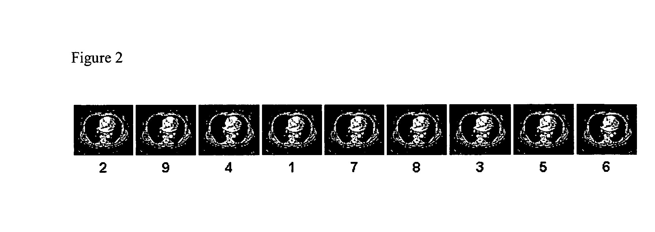
Once the study is read by the radiologist, the PACS workstations does not currently allow the radiologist to quickly review the images on which the radiologist spent most of the time reading and analyzing.
However, users are not currently allowed to determine how significant are images.
Currently, users are unable to prioritize of the images based on the level of significance.

Method used


Image

  • System and method for displaying images on a pacs workstation based on level of significance
  • System and method for displaying images on a pacs workstation based on level of significance
  • System and method for displaying images on a pacs workstation based on level of significance

Examples


Embodiment Construction

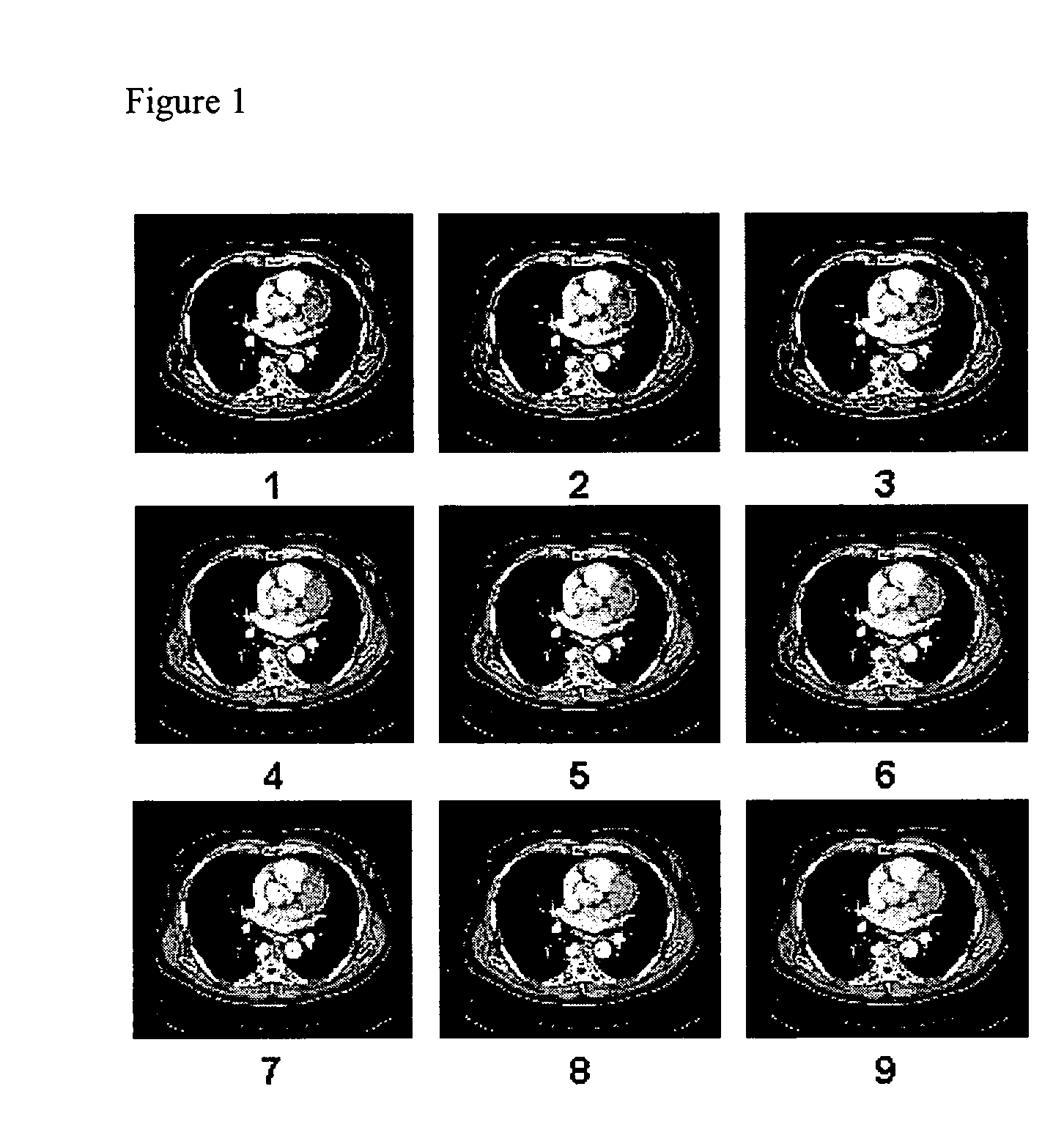
[0042] Image review and analysis may be a significant component of a healthcare workflow. For example, in a radiology reading, a radiologist spends a significant portion of his or her time reading and analyzing clinically relevant images. Clinically relevant images are the images that may be used for diagnosing a patient's medical condition, for example. In order to read images, a radiologist may use a variety of tools, such as window level, zoom / pan, rotate, image filter(s), process, cine, annotate, etc. For example, a user may dynamically modify a display window level to read images. Different window level settings may allow a user to better view an image or anatomy within an image, for example. A user may zoom and / or pan images for details, for example. A user may rotate and / or flip an image, for example. A user may apply one or more filters to an image to read specific areas of a body part or anatomy of interest. A user may apply image processing tools to image data, for example...


PUM


Abstract

Certain embodiments of the present invention provide methods and systems for determine a level of significance of an image. Certain embodiments include selecting one or more parameters for use in determining a level of significance for an image, assigning a value to each of the one or more parameters, and determining the level of significance for the image based on the parameters and the values associated with each of the one or more parameters. One or more parameters may be weighted to affect determination of the level of significance. An offset may be added to the determination of the level of significance for the image. Parameters may include clinical relevance, diagnosis pattern, segmented area, number of pixels, and / or reading time for the image, for example.

Description

RELATED APPLICATIONS [0001] The present application is a continuation-in-part of, and claims priority from, U.S. patent application Ser. No. 11 / 077,299, filed on Mar. 10, 2005, which claims priority as a continuation-in-part of U.S. patent application Ser. No. 11 / 039,152, filed on Jan. 19, 2005, and entitled “System and Method for Presentation of Enterprise, Clinical, and Decision Support Information Utilizing Eye Tracking Navigation”, which claims priority from, U.S. Provisional Application No. 60 / 631,045, filed on Nov. 24, 2004.FEDERALLY SPONSORED RESEARCH OR DEVELOPMENT [0002] [Not Applicable]MICROFICHE / COPYRIGHT REFERENCE [0003] [Not Applicable]BACKGROUND OF THE INVENTION [0004] The present invention generally relates to display of information in a healthcare environment. In particular, the present invention relates to use of visual tracking technology to improve selection and display of significant images in a healthcare environment. [0005] A clinical or healthcare environment ...

Claims


Application Information

Patent Timeline
Patent Type & Authority Applications(United States)
IPC IPC(8): G09G5/00
CPCG06F3/013G06F19/321G06F19/3406A61B2017/00216G16H30/20G16H40/63G16H50/20
Inventor KARIATHUNGAL, MURALI KUMARANMAHESH, PRAKASHMORITA, MARK M.
Owner GENERAL ELECTRIC CO
Learn More