X-ray diagnostic apparatus

a diagnostic apparatus and x-ray technology, applied in the field of x-ray diagnostic apparatus, can solve the problems of large installation area, difficult operation, high cost, etc., and achieve the effect of improving operability of moving a c-arm

Active Publication Date: 2006-09-28
TOSHIBA MEDICAL SYST CORP
View PDF2 Cites 33 Cited by
  • Summary
  • Abstract
  • Description
  • Claims
  • Application Information

AI Technical Summary

Benefits of technology

[0013] It is an object of the present invention to improve operability in moving a C-arm and performing imaging operation in an X-ray diagnostic apparatus.

Problems solved by technology

In the present circumstances, although an X-ray diagnostic apparatus of the biplane system requires no complicated operation for the movement of the C-arms because observations from two positions can be simultaneously performed by using the two C-arms, the apparatus demands a large installation area and is expensive, and hence its installation is limited.
Such operations are cumbersome and difficult for an operator who manually operates a device such as a catheter or guide wire during diagnosis or medical treatment.

Method used


Image

  • X-ray diagnostic apparatus
  • X-ray diagnostic apparatus
  • X-ray diagnostic apparatus

Examples


Embodiment Construction

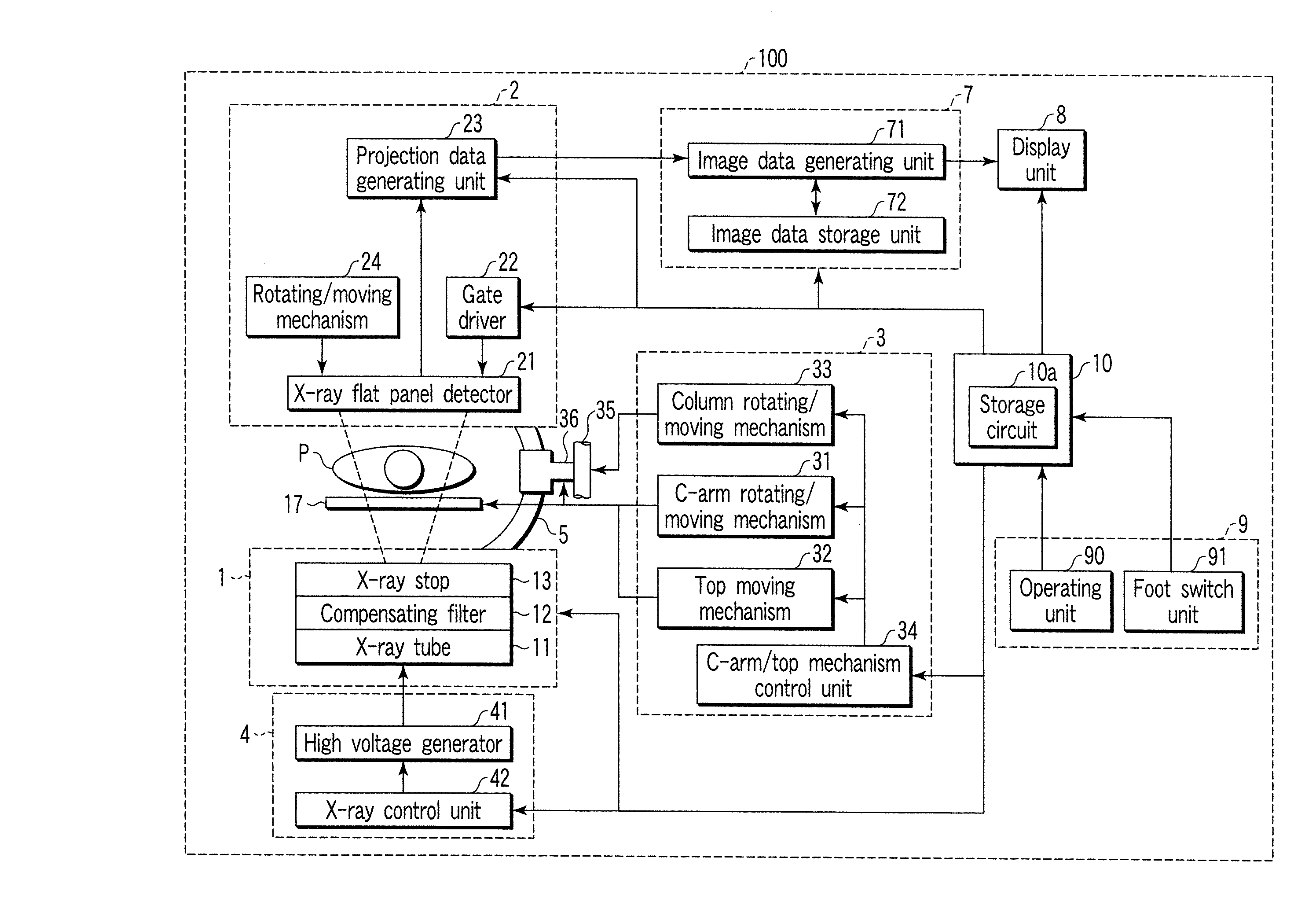
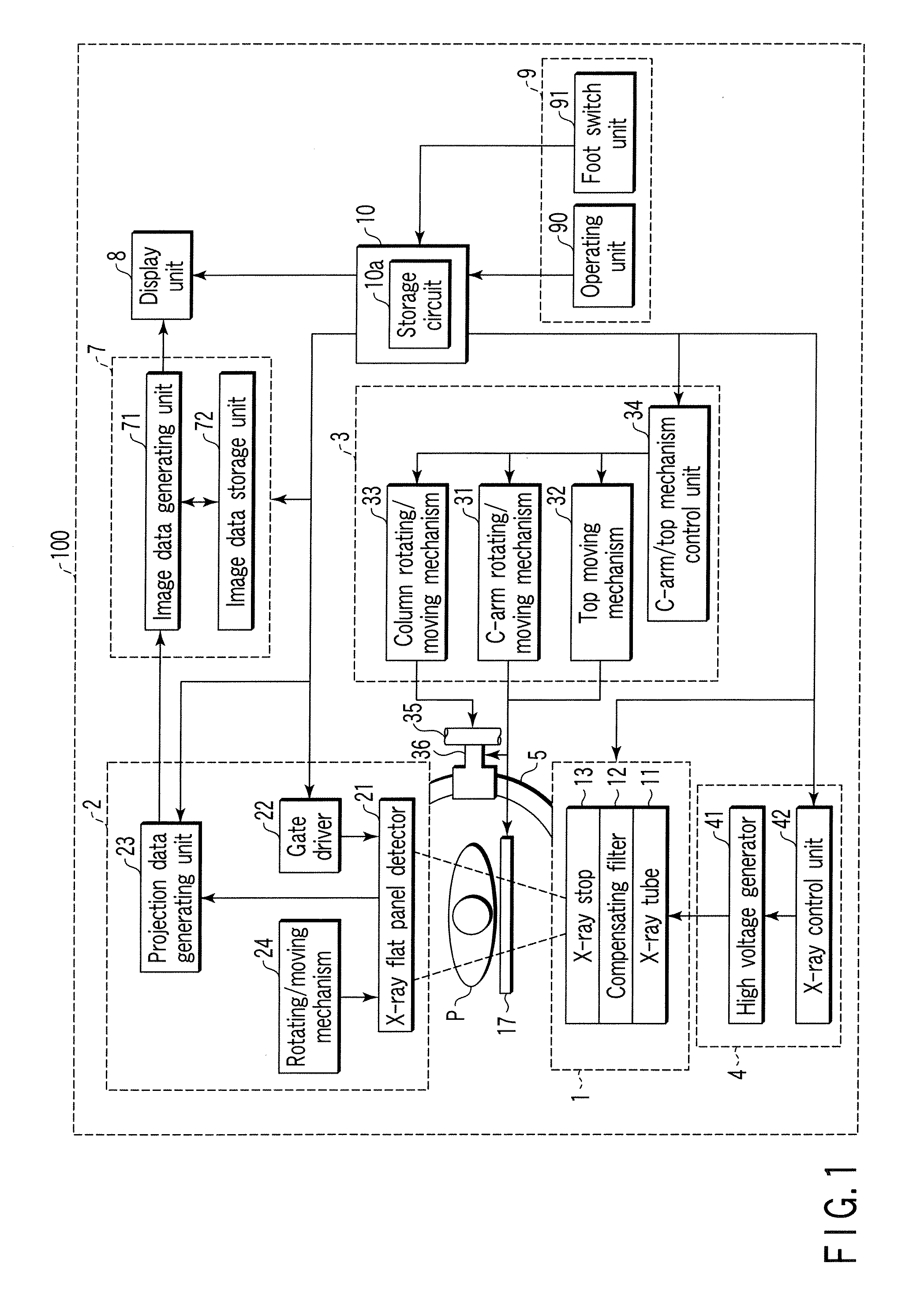
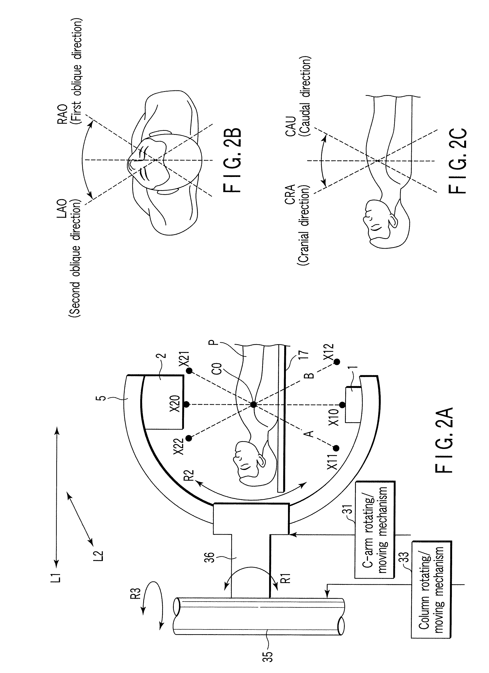
[0034] An X-ray diagnostic apparatus according to an embodiment of the present invention will be described with reference to the views of the accompanying drawing. FIG. 1 is a block diagram showing the arrangement of an X-ray diagnostic apparatus according to this embodiment. An X-ray diagnostic apparatus 100 comprises an X-ray generating unit 1 which generates X-rays, an X-ray detecting unit 2 which detects X-rays transmitted through a subject P, a C-arm 5 on which an X-ray generating unit 1 and an X-ray detecting unit 2 are mounted, a top 17 on which the subject P is placed, and a high voltage control unit 4 which generates a high voltage for generating X-rays from the X-ray generating unit 1.

[0035] The X-ray diagnostic apparatus 100 comprises a mechanical unit 3 which moves the C-arm 5, the top 17, and the like, an image data processing unit 7 which generates and stores various kinds of image data from X-ray projection data generated by the X-ray detecting unit 2, and a display ...


PUM


Abstract

An X-ray diagnostic apparatus includes an X-ray generating unit which generates X-rays, an X-ray detecting unit which detects X-rays transmitted through a subject, a C-arm on which the X-ray generating unit and the X-ray detecting unit are mounted, a support mechanism which movably supports the C-arm, a high voltage generating unit which generates a high voltage for generating X-rays from the X-ray generating unit, a first foot switch to input a first user instruction associated with generation of the X-rays, a second foot switch to input a second user instruction associated with movement of the C-arm, and a control unit which controls the high voltage generating unit in accordance with the input of the first user instruction and controls the support mechanism in accordance with the input of the second user instruction.

Description

CROSS-REFERENCE TO RELATED APPLICATIONS [0001] This application is based upon and claims the benefit of priority from prior Japanese Patent Application No. 2005-081989, filed Mar. 22, 2005, the entire contents of which are incorporated herein by reference. BACKGROUND OF THE INVENTION [0002] 1. Field of the Invention [0003] The present invention relates to an X-ray diagnostic apparatus having a C-arm. [0004] 2. Description of the Related Art [0005] In diagnosis or medical treatment based on angiography, a device such as a guide wire or catheter is inserted into a blood vessel. An operator advances a device to a morbid region of a subject in diagnosis of the morbid region while referring to fluoroscopic image data obtained by fluoroscopy and reference image data obtained by imaging at the same position as that of the fluoroscopic image data upon injecting a contrast medium in advance by using an X-ray diagnostic apparatus for circulatory organs. [0006] In addition, in many cases, an o...

Claims


Application Information

Patent Timeline
Patent Type & Authority Applications(United States)
IPC IPC(8): H05G1/56
CPCA61B6/4441A61B6/481A61B6/503A61B6/504
Inventor WATANABE, IZUMI
Owner TOSHIBA MEDICAL SYST CORP
Why Eureka
Try Eureka