
Cytotoxicity mediation of cells evidencing surface expression of CD63

Inactive Publication Date: 2006-11-30
F HOFFMANN LA ROCHE & CO AG
View PDF30 Cites 6 Cited by
  • Summary
  • Abstract
  • Description
  • Claims
  • Application Information

AI Technical Summary

Benefits of technology

This patent describes a method for creating patient-specific anti-cancer antibodies that can be used for diagnosis, staging, and treatment of cancer. These antibodies can target molecules and pathways not previously known to be involved in cancer growth, making them ideal for personalized treatment. The antibodies can be used alone or in combination with traditional cancer therapies, and can also be conjugated to red blood cells or other hematogenous cells for targeted treatment of metastases. The patent also discusses the various mechanisms by which anti-cancer antibodies can kill cancer cells, including inducing an immune response, targeting growth receptors, and directly killing cells. Overall, this patent provides a technical solution for creating effective, personalized cancer treatments.

Problems solved by technology

There have been few effective treatments for metastatic cancer and metastases usually portend a poor outcome resulting in death.

Method used


Image

  • Cytotoxicity mediation of cells evidencing surface expression of CD63
  • Cytotoxicity mediation of cells evidencing surface expression of CD63
  • Cytotoxicity mediation of cells evidencing surface expression of CD63

Examples


example 1

Human Liver Tumor Tissue Staining

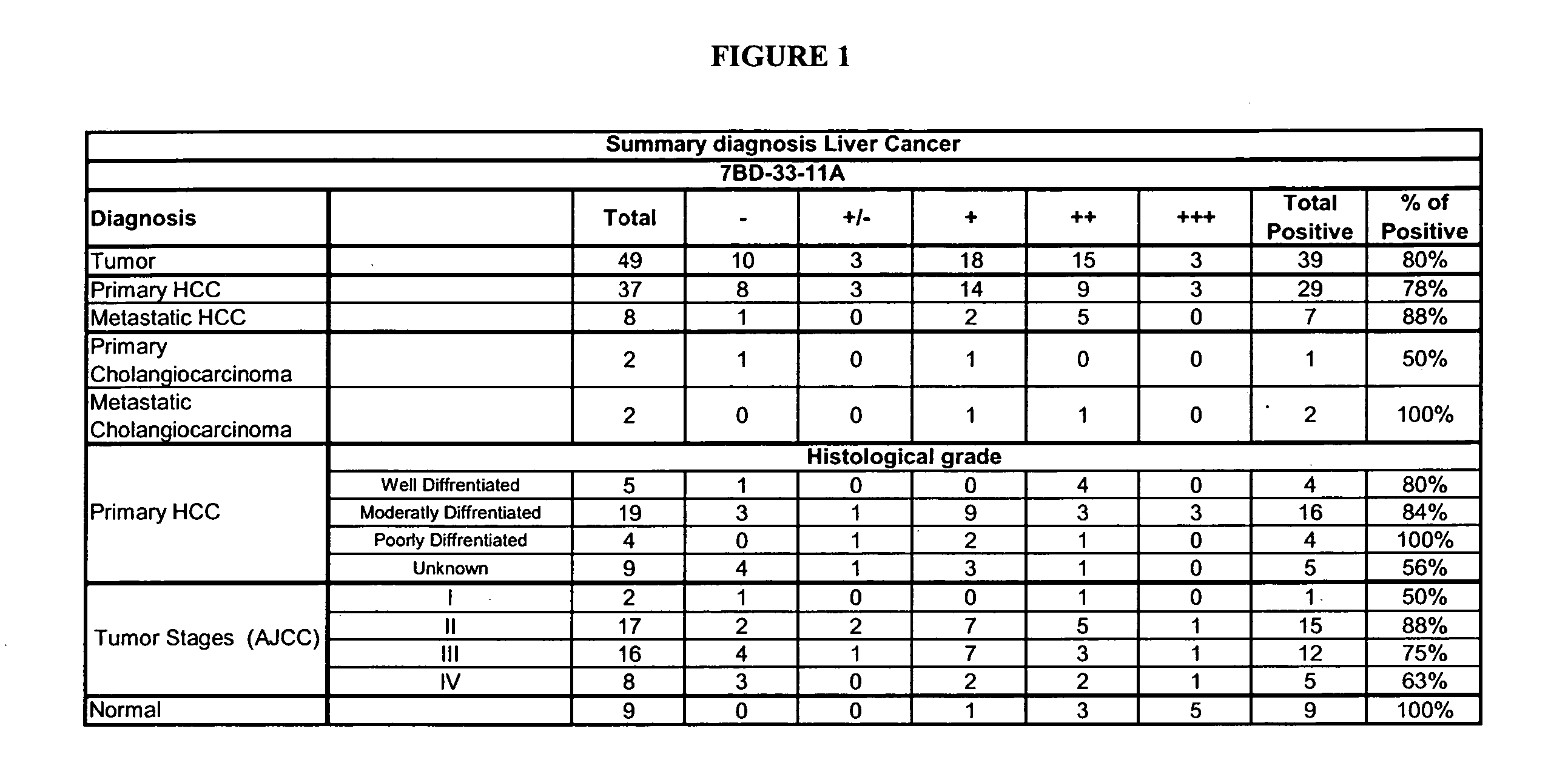
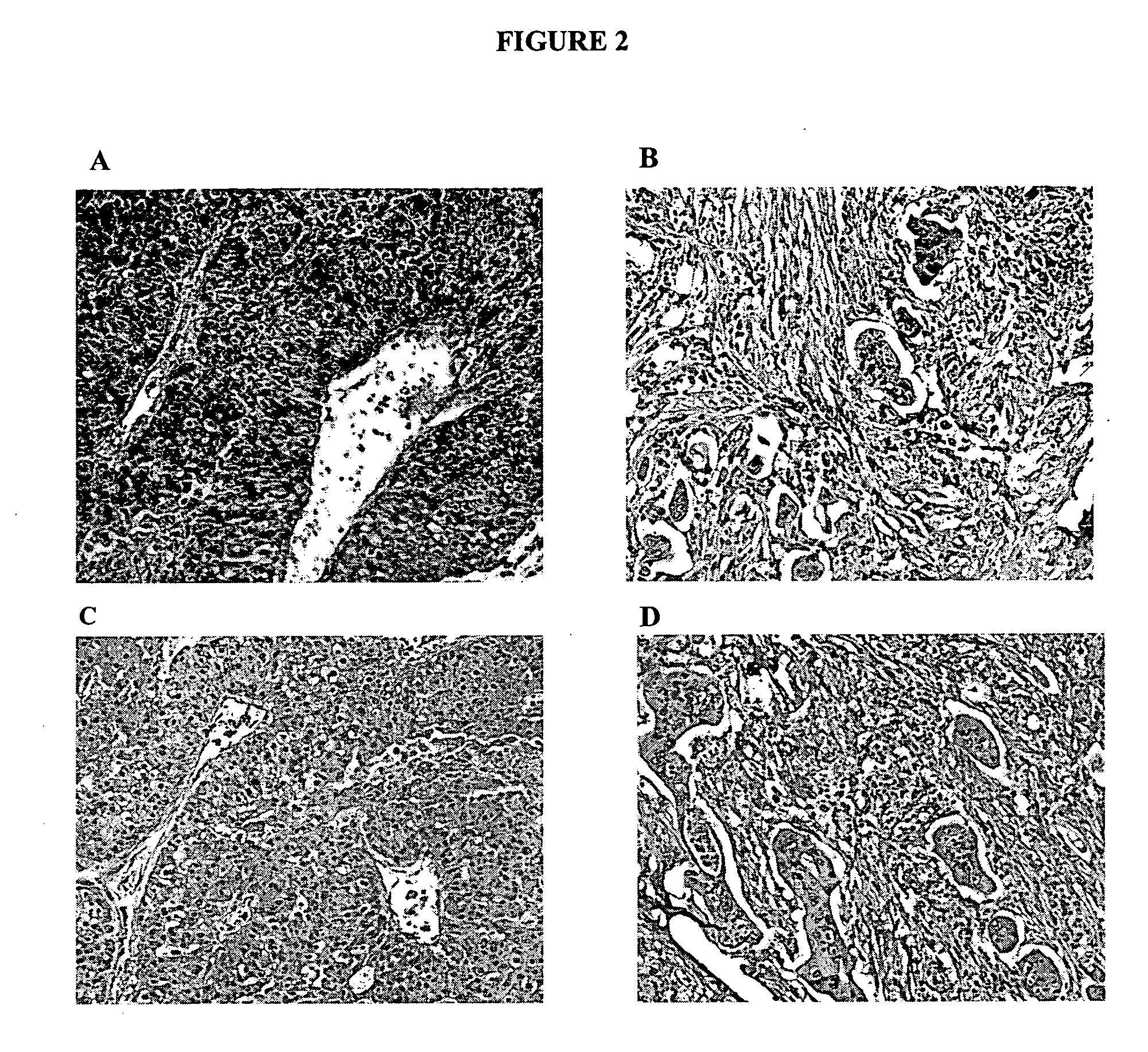
[0163] IHC studies were conducted to evaluate the binding of 7BD-33-11A to human liver tumor tissue. IHC optimization studies were performed previously in order to determine the conditions for further experiments.

[0164] Tissue sections were deparaffinized by drying in an oven at 58° C. for 1 hour and dewaxed by immersing in xylene 5 times for 4 minutes each. Following treatment through a series of graded ethanol washes (100 percent-75 percent) the sections were re-hydrated in water. The slides were immersed in 10 mM citrate buffer at pH 6 (Dako, Toronto, Ontario) then microwaved at high, medium, and low power settings for 5 minutes each, then left 15 minutes at room temperature and washed with PBS 3 times for minutes each. Slides were then immersed in 3 percent hydrogen peroxide solution for 10 minutes, washed with PBS three times for 5 minutes each, then incubated with Universal blocking solution (Dako, Toronto, Ontario) for 5 minutes at room tem...

example 2

Immunostaining on Hepatocellular Carcinoma Tissue Microarray

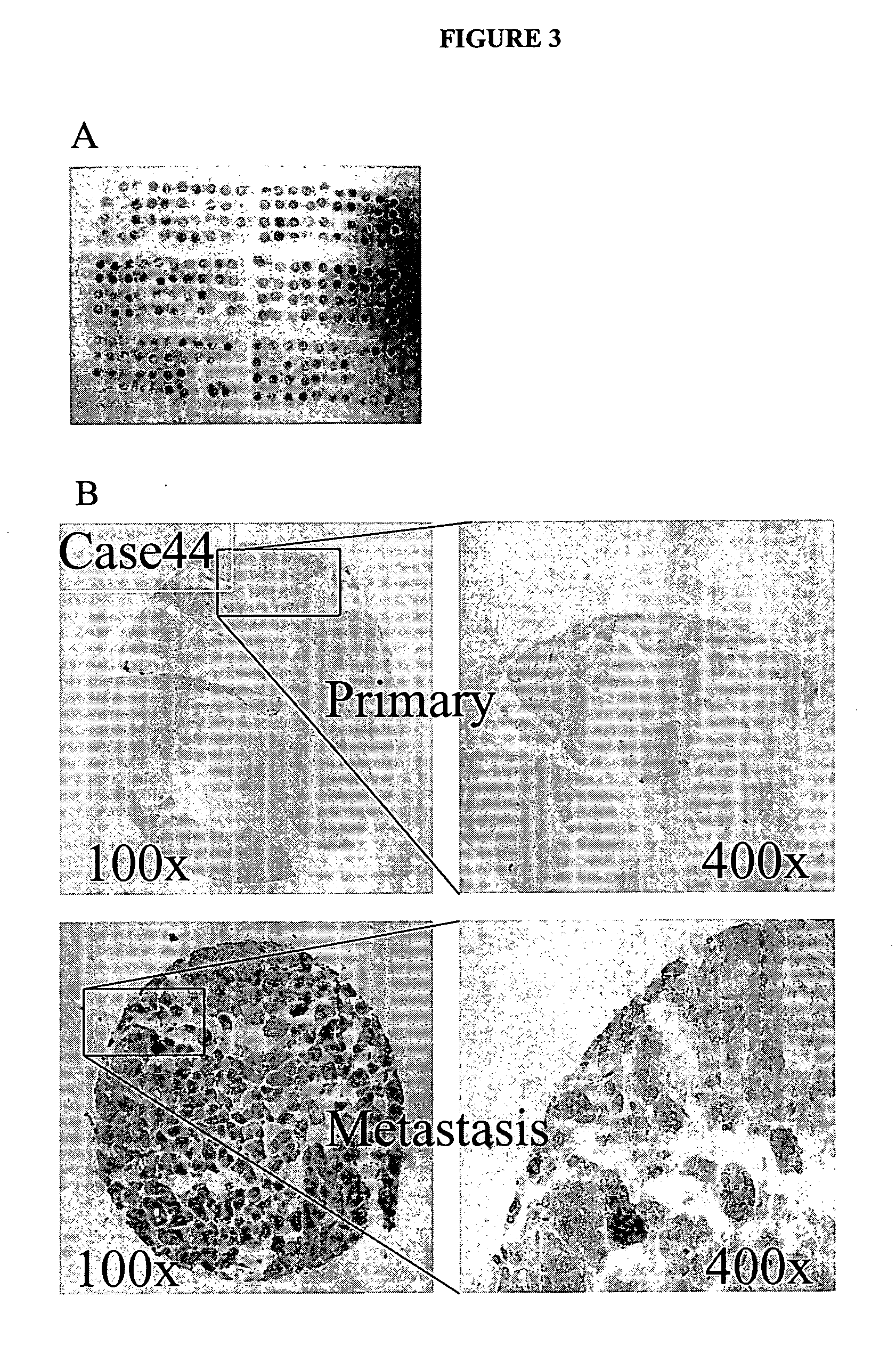
[0169] To further study the results obtained in Example 1, 7BD-33-11A binding was directly compared to human primary and mestastatic hepatocellular carcinoma tissue. With reference to FIG. 3, the human hepatocellular carcinoma (HCC) tissue microarray was constructed as outlined below. Briefly, tissue samples were obtained from Eastern Hepato-biliary Surgery Hospital (Shanghai, China) from 1995 to 1999 and were embedded in paraffin using standard protocols. The embedded tissue samples were freshly sectioned and stained with hematoxylin and eosin. The representative regions of lesion were reviewed carefully and defined by two pathologists. Based on the clinicopathological information, specimens were grouped in tissue cylinders and a diameter of 0.6 mm was taken from the selected regions of the donor block and then punched precisely into a recipient paraffin block using a tissue array instrument (Beecher Instruments, Silver ...

example 3

Immunostaining on 50 Cases of Paraffin-Embedded HCC Tumor Tissues

[0171] To determine the correlation of CD63 expression with several clinical pathological features, immunostaining was performed on 50 cases of paraffin-embedded human HCC clinical samples, obtained during 1999 to 2001 from the Department of Surgery, University of Hong Kong Medical Centre, Queen Mary Hospital, Hong Kong, with the 7BD-33-11A antibody (FIG. 5).

[0172] Cytoplasmic expression of CD63 was determined by two independent observers who visually assessed the percentage of stained tumor cells as well as staining intensity. The percentage of positive cells was rated as follows: 2 points, 11-50 percent positive tumor cells; 3 points, 51-80 percent positive cells; and 4 points, >81 percent positive cells. Staining intensity was rated as follows: 1 point, weak intensity; 2 points, moderate intensity; and 3 points, strong intensity. Points for expression and percentage of positive cells were added, and specimens wer...


PUM

PropertyMeasurementUnit
molecular weightaaaaaaaaaa
median timeaaaaaaaaaa

Abstract

This invention relates to the diagnosis and treatment of cancerous diseases, particularly to the mediation of cytotoxicity of primary and metastatic human tumor cells; and most particularly to the use of an isolated monoclonal antibody or cancerous disease modifying antibodies (CDMAB) thereof, optionally in combination with one or more chemotherapeutic agents, as a means for initiating the cytotoxic response in such human tumors, e.g. any primary or metastatic tumor sites which arise from hepatocytes. The invention further relates to binding assays which utilize the CDMAB of the instant invention.

Description

REFERENCE TO RELATED APPLICATIONS [0001] This application is a continuation-in-part to U.S. patent application Ser. No. 11 / 321,624, filed on Dec. 29, 2005, and is a continuation-in-part to U.S. patent application Ser. No. 10 / 810,751, filed Mar. 26, 2004, which is a continuation-in-part to U.S. patent application Ser. No. 10 / 603,006, filed Jun. 23, 2003, which is a continuation-in-part to U.S. patent application Ser. No. 10 / 348,231, filed Jan. 21, 2003 (including U.S. divisional application Ser. No. 10 / 891,866, filed Jul. 15, 2004), and is a continuation-in-part to U.S. patent application Ser. No. 09 / 727,361 now U.S. Pat. No. 6,657,048 issued Dec. 2, 2003 (including U.S. divisional application Ser. No. 10 / 713,642, filed Nov. 13, 2003), the contents of each of which are herein incorporated by reference.FIELD OF THE INVENTION [0002] This invention relates to the diagnosis and treatment of cancerous diseases, particularly to the mediation of cytotoxicity of tumor cells; and most particu...

Claims


Application Information

Patent Timeline
Patent Type & Authority Applications(United States)
IPC IPC(8): A61K51/00G01N33/574A61K39/395
CPCA61K51/1045A61K51/1051A61K51/1057A61K51/1072G01N2500/00C07K16/2896C07K16/303G01N33/57438A61K2039/505A61P1/16A61P35/00A61P35/04A61K39/395A61K51/00
Inventor YOUNG, DAVID S.F.HAHN, SUSAN E.FINDLAY, HELEN P.DACRUZ, LUIS A. G.SAYEGH, DAADFAN, SHEUNG TATPOON, RONNIE TUNG PINGLEE, TERENCE KIN WAH
Owner F HOFFMANN LA ROCHE & CO AG
Learn More