Cytotoxicity mediation of cells evidencing surface expression of CD44

Inactive Publication Date: 2007-10-25
F HOFFMANN LA ROCHE INC
View PDF41 Cites 5 Cited by
  • Summary
  • Abstract
  • Description
  • Claims
  • Application Information

AI Technical Summary

Benefits of technology

[0110]FIG. 4 demonstrates the effect of (ch)ARH460-16-2-IgG1 on HCC tumor growth and metastasis in an established orthotopic HCC tumor model. Data points represent the mean +/−SEM.
[0111]FIG. 5 demonstrates the quantitative effect of (ch)ARH460-16-2-IgG1 on tumor growth in an established orthotopic HCC tumor model. The bar graph su

Problems solved by technology

There have been few effective treatments for metastatic cancer a

Method used


Image

  • Cytotoxicity mediation of cells evidencing surface expression of CD44
  • Cytotoxicity mediation of cells evidencing surface expression of CD44
  • Cytotoxicity mediation of cells evidencing surface expression of CD44

Examples


example 1

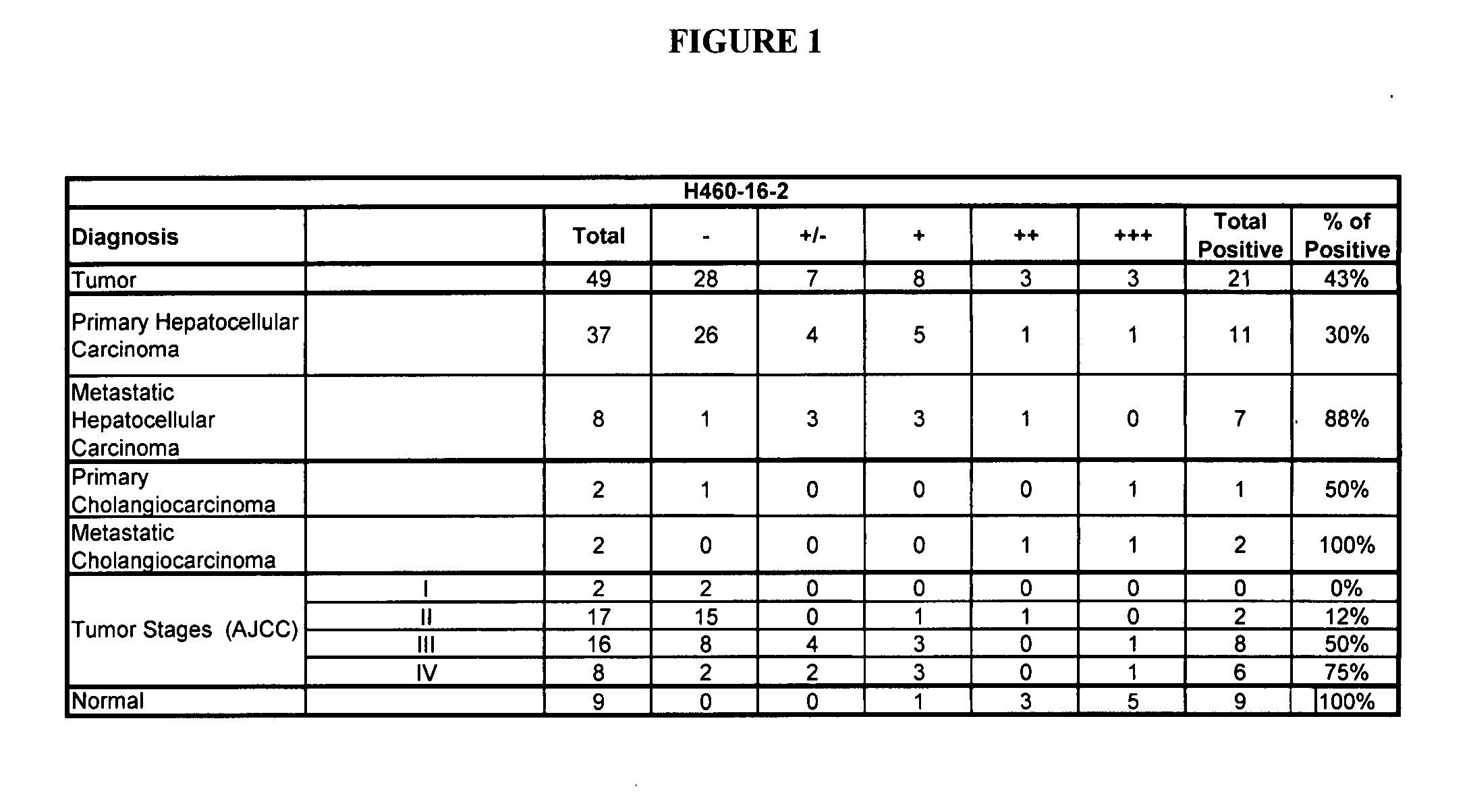
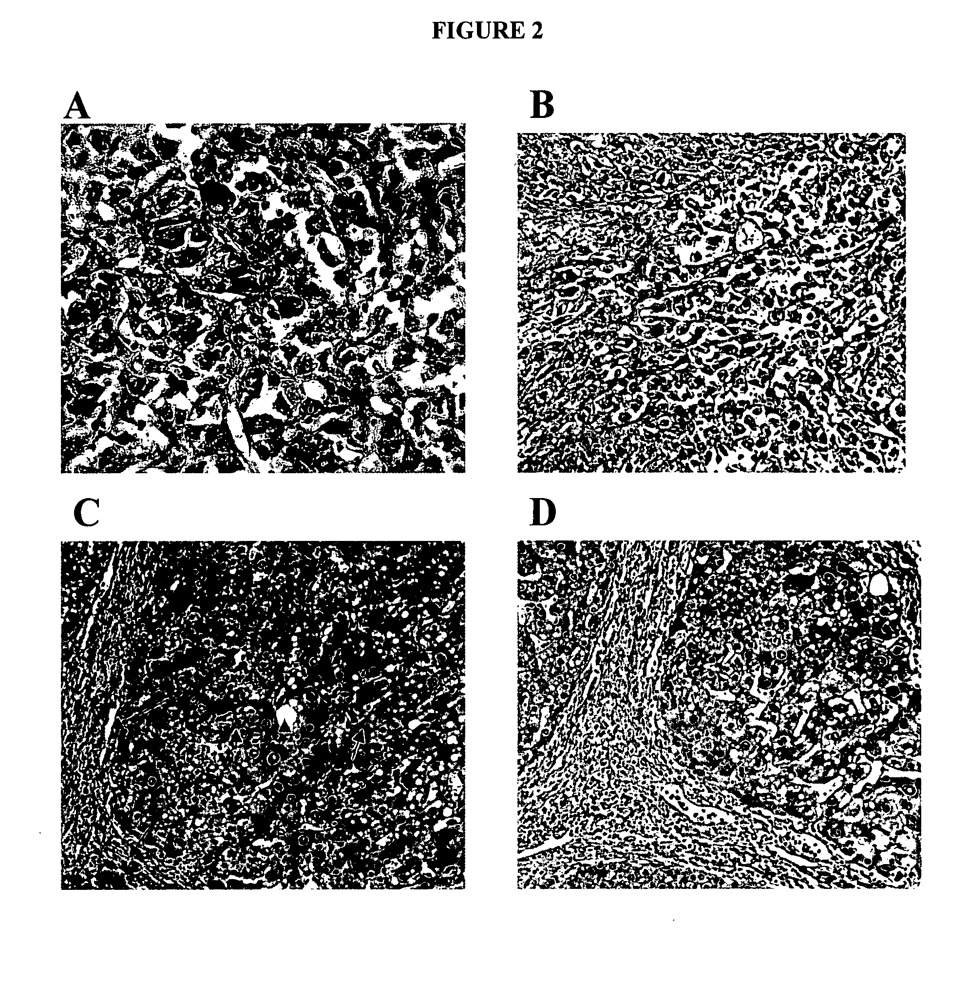
Human Liver Tumor Tissue Staining

[0185] IHC studies were conducted to further evaluate the binding of H460-16-2 to human liver tumor tissue. IHC optimization studies were performed previously in order to determine the conditions for further experiments. H460-16-2 monoclonal antibodies were produced and purified as previously disclosed in Ser. No. 10 / 603,000.

[0186] Binding of antibodies to 49 human liver tumor and 9 normal liver tissues was performed using a human, liver normal and tumor tissue microarray (Imgenex, San Diego, Calif.). The following information was provided for each patient: age, sex, organ and diagnosis. Tissue sections were deparaffinized by drying in an oven at 58° C. for 1 hour and dewaxed by immersing in xylene 5 times for 4 minutes each in Coplin jars. Following treatment through a series of graded ethanol washes (100 percent-75 percent) the sections were re-hydrated in water. The slides were immersed in 10 mM citrate buffer at pH 6 (Dako, Toronto, Ontario) t...

example 2

Correlation of CD44 with Metastatic Potential of Various HCC Cell Lines

[0189] To further evaluate this correlation in vitro, six heptaocellular carcinoma (HCC) cell lines with various metastatic potential (Hep3B (American Type Culture Collection, Manassas, Va.), Huh-7 (a gift from Dr. H. Nakabayashi, Hokkaido University School of Medicine, Sapporo, Japan), PLC (Japanese Cancer Research Bank, Tokyo, Japan), MHCC-97L, MHCC-97H and HCCLM3 (Liver Cancer Institute, Fudan University, Shanghai, China)) were evaluated for CD44 expression by flow cytometry using (ch)ARH460-16-2-IgG1 antibody. To detect expression of CD44 in various HCC cell lines, cells were stained for 1 hour with (ch)ARH460-16-2-IgG1 or isotype control antibody (10 micrograms / mL) and 30 minutes with the appropriate secondary antibody and analyzed by FACS.

[0190] By flow cytometry, CD44 expression levels were found to be higher in the metastatic HCC cell lines (MHCC-97L, HCCLM3 and MHCC-97H) when compared with the primary...

example 3

Orthotopic HCC Tumor Model with HCCLM3 Cells

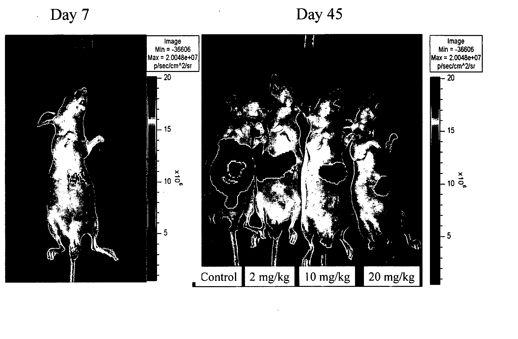
Luciferase Labeling of Cells

[0191] To further evaluate this correlation in vivo, (ch)ARH460-16-2-IgG1 was tested in an orthotopic HCC tumor model. For the luciferase labelling of HCCLM3 (a metastatic HCC cell line Yang et al., Cancer Genet. Cytogenet. 158(2):180-183 2005) cells, lentiviral vector harboring the luciferase gene was constructed and transfected into the cells using the method described previously (Cheung et al., Cancer Res. 66(8):4357-4367 2006). Stable transfectants were generated from a pool of greater than 20 positive clones (which were selected with blasticidin at a concentration of 2 micrograms / mL).

Animal CCD Experiments

[0192] One million HCCLM3 luciferase labeled cells were sub-cutaneously injected into the right flank of nude mice, which were then observed daily for signs of tumor development. Once the tumors reached 1 to 1.5 cm in diameter, it was removed and cut into about 1- to 2-mm cubes, which were then subs...


PUM

PropertyMeasurementUnit
Fractionaaaaaaaaaa
Fractionaaaaaaaaaa
Fractionaaaaaaaaaa

Abstract

This invention relates to the diagnosis and treatment of cancerous diseases, particularly to the mediation of cytotoxicity of primary and metastatic human tumor cells; and most particularly to the use of an isolated monoclonal antibody or cancerous disease modifying antibodies (CDMAB) thereof, optionally in combination with one or more chemotherapeutic agents, as a means for initiating the cytotoxic response in such human tumors, e.g. any primary or metastatic tumor sites which arise from hepatocytes. The invention further relates to binding assays which utilize the CDMAB of the instant invention.

Description

REFERENCE TO RELATED APPLICATIONS [0001] This application is a continuation-in-part to U.S. patent application Ser. No. 11 / 716,216, filed on Mar. 9, 2007, which is a continuation-in-part to U.S. patent application Ser. No. 11 / 364,013, filed on Feb. 28, 2006, which is a continuation-in-part to U.S. patent application Ser. No. 10 / 810,165, filed Mar. 26, 2004, now abandoned, which is a continuation-in-part to U.S. patent application Ser. No. 10 / 647,818, filed Aug. 22, 2003, which is a continuation-in-part to U.S. patent application Ser. No. 10 / 603,000, filed Jun. 23, 2003, which is a continuation-in-part to U.S. patent application Ser. No. 09 / 727,361 now U.S. Pat. No. 6,657,048, issued Dec. 2, 2003, which is a continuation-in-part to U.S. patent application Ser. No. 09 / 415,278 now U.S. Pat. No. 6,180,357, issued Jan. 30, 2001, the contents of each of which are herein incorporated by reference.FIELD OF THE INVENTION [0002] This invention relates to the isolation and production of cancer...

Claims


Application Information

Patent Timeline
IPC IPC(8): A61K51/10A61K39/395A61P35/04G01N33/574A61P43/00A61K39/44
CPCA61K47/48561A61K49/0013A61K2039/505C07K16/2884C07K16/30G01N33/574G01N33/57492G01N2333/70585C07K2317/24A61K47/6849A61P1/16A61P35/00A61P35/02A61P35/04A61P37/04A61P43/00
Inventor YOUNG, DAVID S. F.SAYEGH, DAADFAN, SHEUNG TATPOON, RONNIE TUNG PINGLEE, TERENCE KIN WAH
Owner F HOFFMANN LA ROCHE INC
Why Eureka
Try Eureka