Anti-inflammatory agents and methods of their use
- Summary
- Abstract
- Description
- Claims
- Application Information
AI Technical Summary
Benefits of technology
Problems solved by technology
Method used
Image
Examples
example 1
Differential Regulation of BMP4 Gene by LS and OS in Endothelial Cells
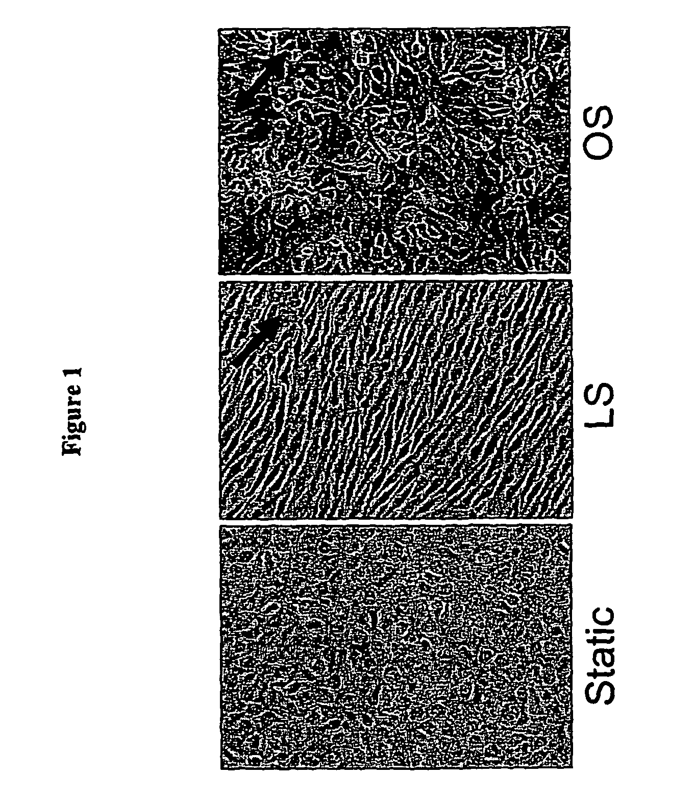
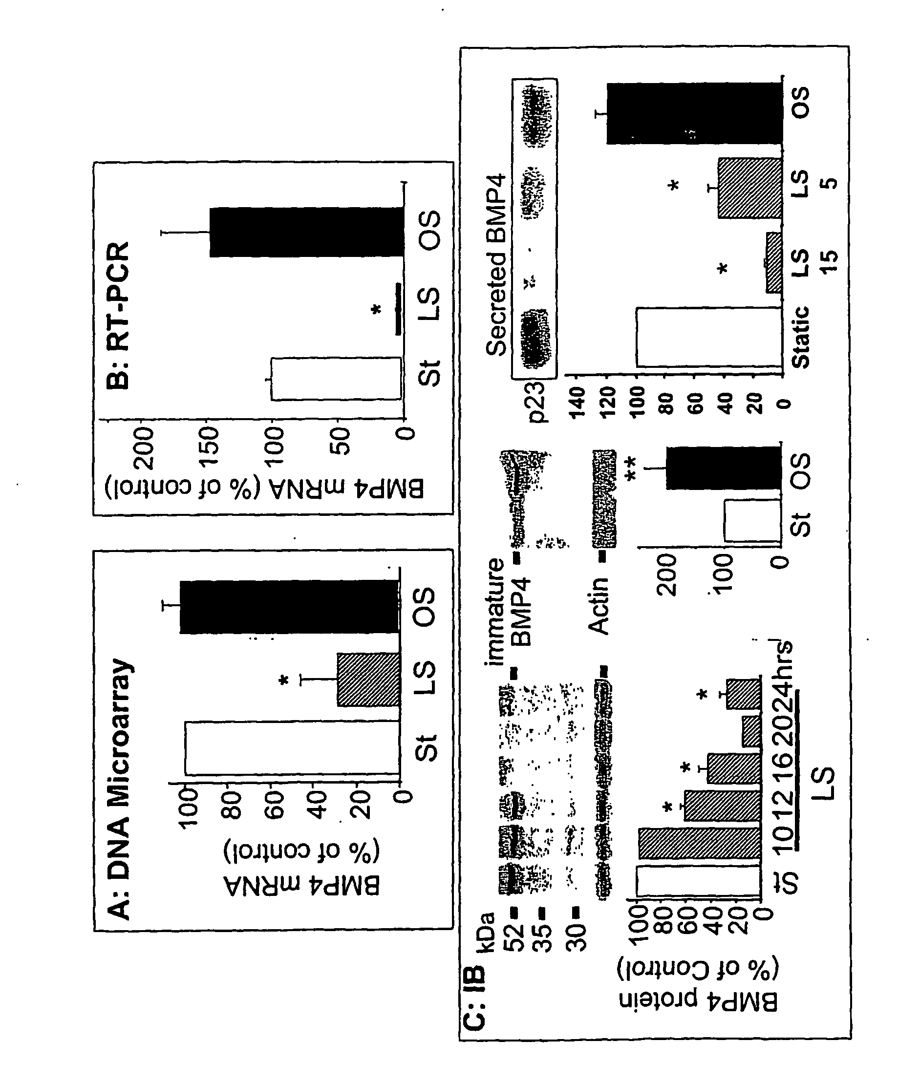
[0166] To identify the genes that may be responsible for the atheroprotective and pro-atherogenic effects of LS and OS, respectively, DNA microarray studies were performed using cultured MAEC. Exposing MAEC to LS, but not OS, for 1 day using the modified “cone-and-plate” device, induced a cell shape alignment to the direction of the flow from a typical polygonal “cobblestone shape” found in static cultured cells (FIG. 1). FIG. 1 shows confluent monolayers of MAEC were exposed to static condition (St), LS (15 dynes / cm2) or OS (±5 dyn / cm2, 1 Hz cycle) for 24 his using the cone-and-plate apparatus. Following shear exposure, cell morphology was determined by light microscopy. Arrows indicate the direction of imposed shear stress.
[0167] The total RNAs prepared from these cells were used to determine mRNA expression profiles by using Affymetrix and / or Motorola DNA chips according to the manufacturers′ protocols. The a...
example 2
BMP4 Expression in the Selective Patches of Endothelial Cells Over Foam-Cell Lesions in Human Coronary Arteries
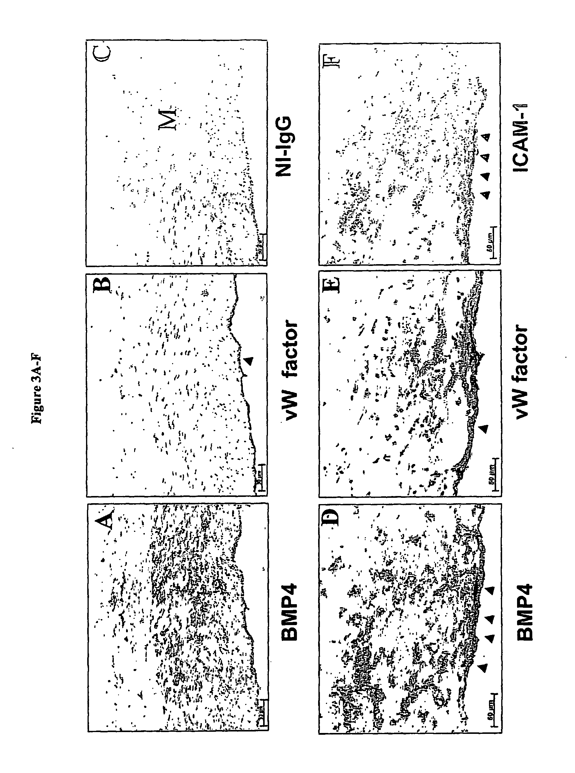
[0175] Next, the presence of BMP4 protein in endothelial cells of human atherosclerotic lesions from human coronary arteries was determined. The coronary arteries exhibiting a spectrum of atherosclerotic lesion complexity were obtained from patients undergoing heart transplantations and examined by immunohistochemical staining (Sorescu, D., et al. (2002) Circulation 105, 1429-1435). BMP4 protein expression was not apparent in the intimal endothelial cells in relatively normal, “minimally diseased” human coronary arteries (FIG. 3A) as well as advanced lesions (data not shown).
[0176]FIG. 3A shows human coronary arteries stained with antibodies specific to BMP4 (FIGS. 3A and 3D), von Willebrand factor (FIGS. 3B and 3E), ICAM-1 (FIGS. 3F), and non-immune mouse IgG (NI-IgG) (FIGS. 3C). FIGS. 3A, 3B and 3C are serial sections obtained from minimally diseased (normal) arterial s...
example 3
BMP4 produced in endothelial cells by OS stimulates monocyte adhesion.
[0178] The role of BMP4 in the inflammatory responses observed in lesion-prone areas was investigated. To begin to test the hypothesis, MAEC were treated with increasing amounts of BMP4 for 24 hr, and then monocyte adhesion to endothelium was determined. As a positive control, some cells were treated with a well-known inflammatory cytokine TNF-α (100 U / ml). BMP4 stimulated monocyte binding in a concentration dependent manner with a maximum activation of 4 to 7-fold over control (FIG. 4A, p<0.05). In FIG. 4A to MAEC that were treated with increasing concentrations of BMP4 overnight, BCECF-labeled THP-1 monocytes were added to determine monocyte adhesion. The Bar graph represents mean numbers of bound monocytes per 10× objective field (6-12 different fields per dish) mean±SEM (n=4-6). As a positive control, monocyte binding was determined using MAEC treated with TNF-α (100 U / ml for 2 hrs). As low as 0.1 ng / ml of BM...
PUM
| Property | Measurement | Unit |
|---|---|---|
| Time | aaaaa | aaaaa |
| Adhesion strength | aaaaa | aaaaa |
| Therapeutic | aaaaa | aaaaa |
Abstract
Description
Claims
Application Information
 - R&D
- Intellectual Property
- Life Sciences
- Materials
- Tech Scout
- Unparalleled Data Quality
- Higher Quality Content
- 60% Fewer Hallucinations
Browse by: Latest US Patents, China's latest patents, Technical Efficacy Thesaurus, Application Domain, Technology Topic, Popular Technical Reports.
© 2025 PatSnap. All rights reserved.Legal|Privacy policy|Modern Slavery Act Transparency Statement|Sitemap|About US| Contact US: help@patsnap.com


