Ultrasound diagnostic system and method of automatically controlling brightness and contrast of a three-dimensional ultrasound image

a diagnostic system and ultrasound image technology, applied in the field of ultrasound imaging systems, can solve the problems of complicated manual adjustment required to optimize the 3d ultrasound image, and achieve the effect of convenient and efficien

Inactive Publication Date: 2007-02-15
MEDISON CO LTD
View PDF13 Cites 8 Cited by
  • Summary
  • Abstract
  • Description
  • Claims
  • Application Information

AI Technical Summary

Benefits of technology

[0004] The present provides an ultrasound diagnostic system and method for automatically controlling the brightness and contrast of a three-dimensional (3D) ultrasound image to optimize the 3D ultrasound image and to reduce diagnostic time by minimizing the operations of a system user.
[0007] According to the present invention, the 3D ultrasound image can be optimized by automatically controlling the brightness and contrast of the 3D ultrasound image. Thus, a system user can conduct a diagnosis in a convenient and efficient manner.

Problems solved by technology

However, in the conventional ultrasound diagnostic system, the image parameters are adjusted manually (not automatically).
That is, a complicated manual adjustment is required to optimize the 3D ultrasound image.

Method used


Image

  • Ultrasound diagnostic system and method of automatically controlling brightness and contrast of a three-dimensional ultrasound image
  • Ultrasound diagnostic system and method of automatically controlling brightness and contrast of a three-dimensional ultrasound image
  • Ultrasound diagnostic system and method of automatically controlling brightness and contrast of a three-dimensional ultrasound image

Examples


Embodiment Construction

[0021] Hereinafter, a preferred embodiment of the present invention will be described with reference to FIGS. 1 to 11B.

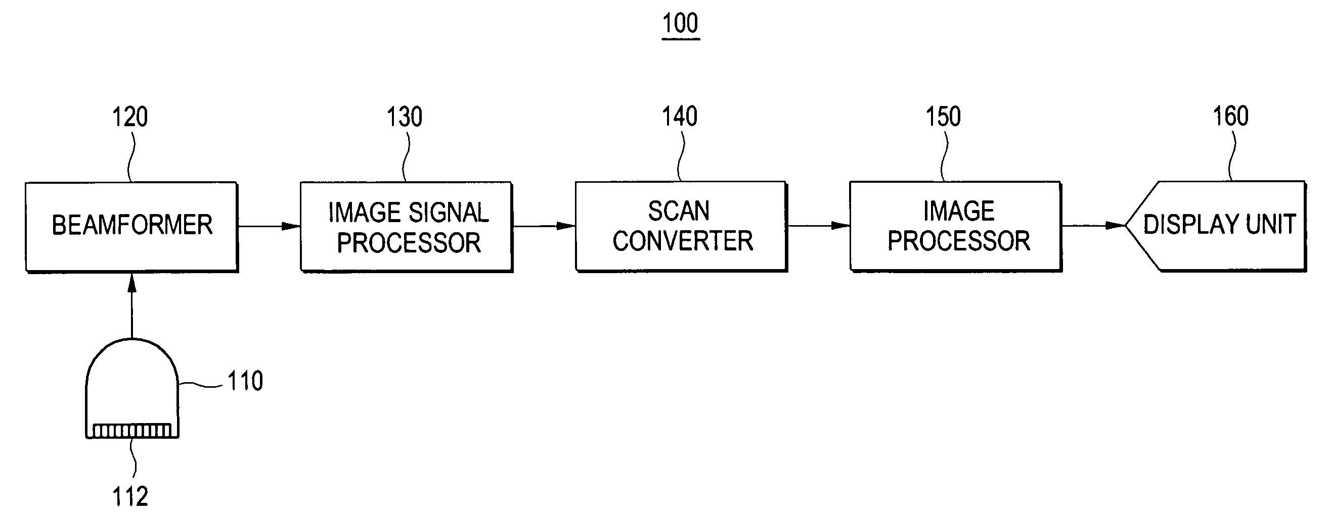
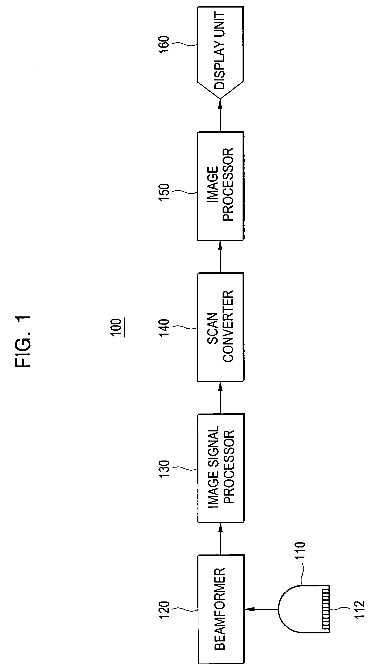
[0022]FIG. 1 is a block diagram showing an ultrasound diagnostic system constructed in accordance with a preferred embodiment of the present invention. As shown in FIG. 1, an ultrasound diagnostic system 100 includes a probe 110, a beamformer 120, an image signal processor 130, a scan converter 140, an image processor 150 and a display unit 160. The image signal processor 130 and image processor 150 may be implemented by using a single processor.

[0023] The probe 110 includes a one-dimensional or two-dimensional (2D) transducer array 112 having a plurality of transducer elements. By properly delaying the pulses applied to the transducer elements, the probe 110 transmits a focused ultrasound beam to a target object (not shown) along a transmit scan line. Ultrasound echo signals reflected from a focal point (not shown) on the transmit scan line are received by the tr...


PUM


Abstract

The present invention relates to an ultrasound diagnostic system and method for automatically controlling the brightness and contrast of a three-dimensional (3D) ultrasound image. The method for automatically controlling the brightness and contrast of a 3D ultrasound image includes the steps of: creating 3D ultrasound image data based on ultrasound echo signals; setting a critical value for rendering the 3D ultrasound image data; rendering the 3D ultrasound image data by using the critical value to form a 3D ultrasound image; analyzing a histogram of the 3D ultrasound image to set image parameters for the 3D ultrasound image; and adjusting the brightness and contrast of the 3D ultrasound image based on the image parameters.

Description

FIELD OF THE INVENTION [0001] The present invention generally relates to an ultrasound imaging system, and more particularly to an ultrasound diagnostic system and method for automatically controlling the brightness and contrast of a three-dimensional ultrasound image. BACKGROUND OF THE INVENTION [0002] An ultrasound diagnostic system has become an important and popular diagnostic tool due to its wide range of applications. Specifically, due to its non-invasive and non-destructive nature, the ultrasound diagnostic system has been extensively used in the medical profession. Modem high-performance ultrasound diagnostic systems and techniques are commonly used to produce two or three-dimensional diagnostic images of internal features of an object (e.g., organs of a human patient). The ultrasound diagnostic system generally uses a wide bandwidth transducer to transmit and receive ultrasound signals. The ultrasound diagnostic system forms images of the internal tissues of a human body by...

Claims


Application Information

Patent Timeline
Patent Type & Authority Applications(United States)
IPC IPC(8): A61B8/00
CPCG01S7/52026G01S15/8993G06T2207/30004G06T5/40G06T2207/10132G06T5/009A61B8/00
Inventor KIM, JAE GYOUNGSONG, YOUNG SEUKCHOI, DO YOUNG
Owner MEDISON CO LTD
Why Eureka
Try Eureka