
Medical device guide locator

a technology for medical devices and locators, applied in tomography, ultrasonic/sonic/infrasonic diagnostics, applications, etc., can solve problems such as unneeded guides, unsuitable guides appended to ultrasound transducers, and unsuitable placement of medical devices

Active Publication Date: 2007-03-01
FUJIFILM SONOSITE
View PDF16 Cites 140 Cited by
  • Summary
  • Abstract
  • Description
  • Claims
  • Application Information

AI Technical Summary

Benefits of technology

The present invention provides systems and methods for providing feedback on the proper placement of a medical device guide without adding any distortions to the surface of the device used in medical procedures. The invention uses sensors, such as Hall effect sensors or other types of sensors, placed in the device and a magnet or magnets placed in the guide bracket to detect when the guide is properly located on the device. The guide bracket is designed to be minimally obtrusive with respect to the operator of the device. The sensors can provide feedback to the user through audible and / or visual signals when the guide is properly located or improperly located. The invention can also control the operation of the device based on the feedback from the sensors. Overall, the invention ensures accurate placement of the medical device guide and improves the overall efficiency and accuracy of medical procedures.

Problems solved by technology

Likewise, such a growth may be found within or near other tissue structure which is subject to damage by misplaced or misguided medical devices, such a needle used in a biopsy procedure.
This is difficult at best and sometimes results in improper angles of attack and could result in improper placement of the medical device.
Moreover, it is often necessary for the physician to manipulate a plurality of instruments, such as an ultrasound transducer and the needle, while looking at the screen, thereby adding to the difficulty of the procedure.
However, procedures benefiting from such guides often account for only a small portion (e.g., 10%) of the procedures which use such ultrasound imaging devices.
Accordingly, such guides appended to an ultrasound transducer may be unneeded, and thus undesired (or perhaps even interfering), much of the time the ultrasound imaging device is used.
Surface protuberances on devices used in medical procedures, such as ultrasound transducers, have been found to be undesirable to the users thereof.
For example, such devices are often handheld and surface protuberances present irregularities in the surface, thereby resulting in stress points experienced by the users.
Where such devices are used more commonly without optional structure, such as the aforementioned guide bracket, being disposed on the device, discomfort associated with such stress points can be particularly problematic.

Method used


Image

  • Medical device guide locator
  • Medical device guide locator
  • Medical device guide locator

Examples


Embodiment Construction

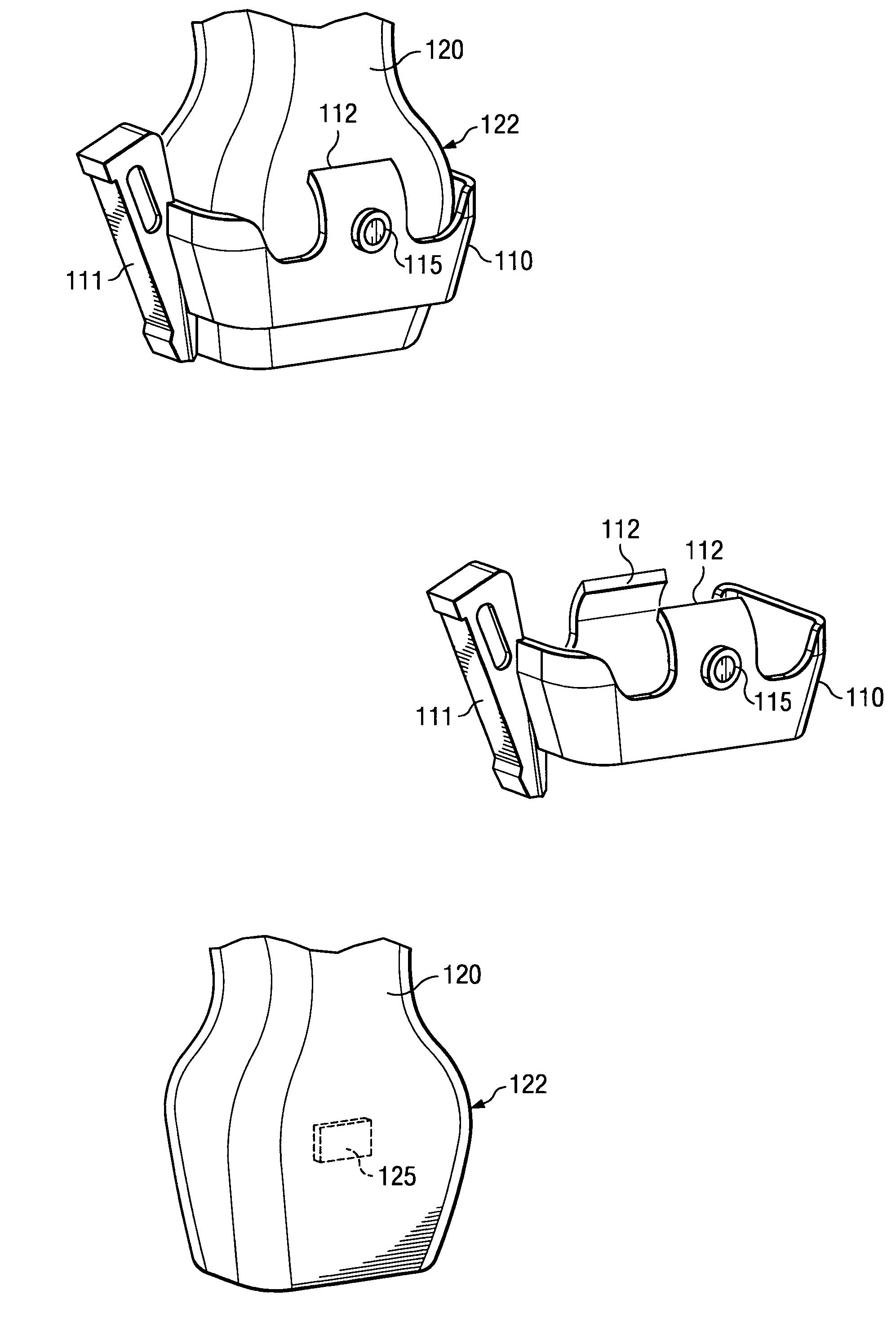
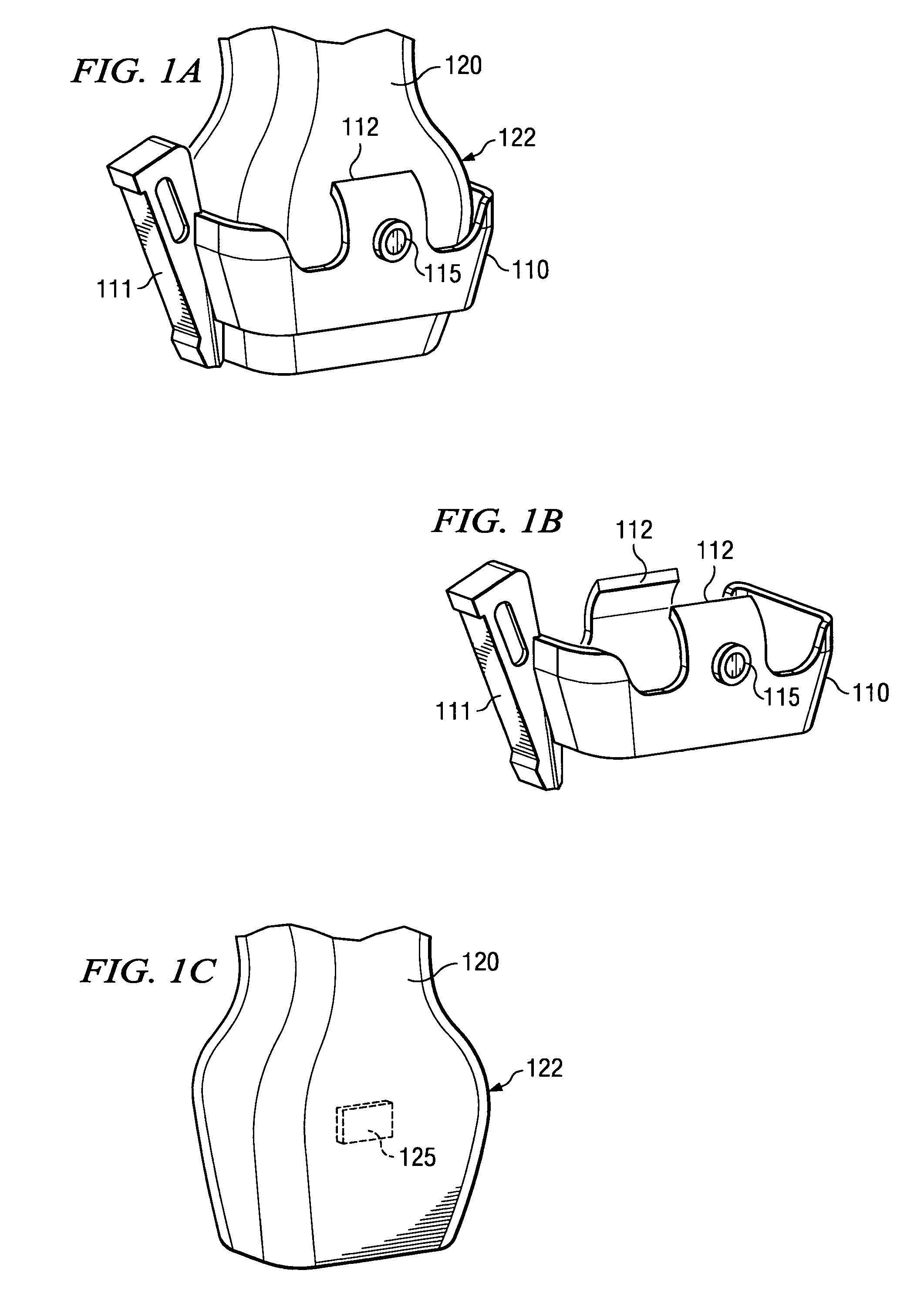
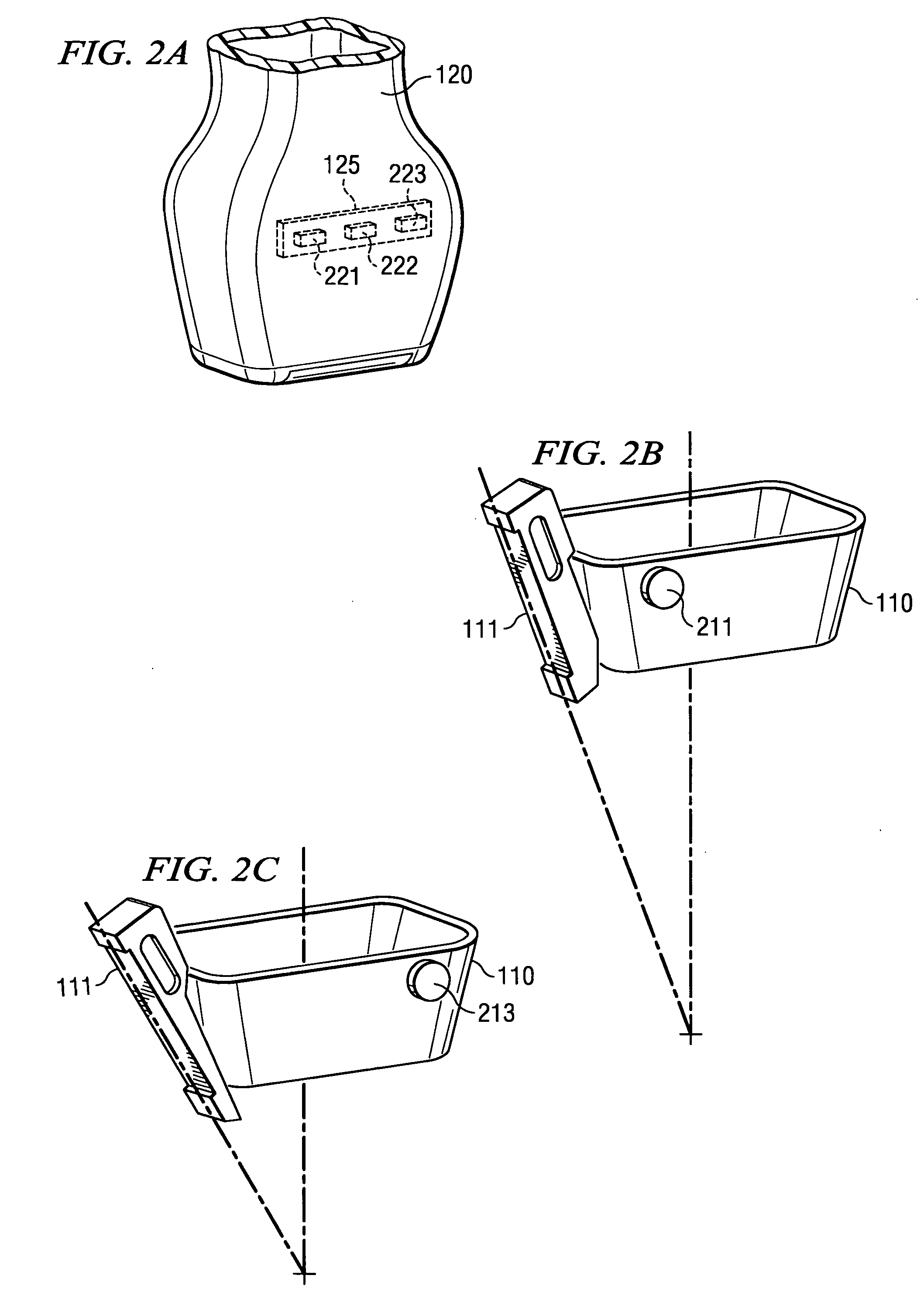
[0018]FIGS. 1A-1C show a device used in medical procedures and a corresponding medical device guide bracket adapted according to an embodiment of the present invention. Specifically, FIGS. 1A-1C illustrate an embodiment wherein medical device guide bracket 110, shown here as a biopsy needle guide bracket, and assembly 120, shown here as an ultrasound transducer assembly 120, are adapted to provide feedback with respect to the desired or proper placement of medical device guide 111 disposed on medical device guide bracket 110 without introducing protuberances or other structure to the surface of assembly 120.

[0019] It should be appreciated that, although the illustrated embodiment shows a biopsy needle guide and ultrasound transducer, embodiments of the present invention may be utilized with respect to any number of medical devices. For example, embodiments of the present invention may be utilized with respect to needles, catheters, drills, saws, scalpels, stints, and / or the like. L...


PUM


Abstract

Systems and methods are shown which provide feedback with respect to the desired or proper placement of a medical device guide without introducing protuberances or other perturbations to the surface of devices used in medical procedures. Embodiments provide a biopsy needle guide bracket for use with an ultrasound transducer assembly, wherein the biopsy needle guide bracket and ultrasound transducer assembly are adapted to detect when the biopsy needle guide bracket is properly located on the ultrasound transducer assembly using one or more sensors. Embodiments may implement various sensor technology, such as Hall effect sensors, optical sensors, capacitive coupled sensors, inductive coupled sensors, and / or the like to provide feedback with respect to placement of a medical device guide.

Description

TECHNICAL FIELD [0001] The invention relates generally to positioning of medical devices and more particularly to systems and methods for determining if a medical device guide is properly and / or improperly located. BACKGROUND OF THE INVENTION [0002] Proper movement and placement (referred to collectively as positioning) of medical devices, such as needles, catheters, drills, saws and even scalpels, is often critical in the proper performance of certain medical procedures. For example, a physician may wish to obtain a tissue sample from a small tumor or cyst in order to determine if the growth is malignant. Accordingly, a needle biopsy procedure may be performed in which the physician percutaneously extracts a tissue sample. It is critical, however, that the physician actually extract a tissue sample of the growth rather than nearby tissue. Likewise, such a growth may be found within or near other tissue structure which is subject to damage by misplaced or misguided medical devices, ...

Claims


Application Information

Patent Timeline
Patent Type & Authority Applications(United States)
IPC IPC(8): A61B8/00
CPCA61B8/00A61B17/3403A61B2019/4857A61B2017/00482A61B2017/3413A61B2017/00477A61B2090/0811
Inventor BUNCE, STEVEN M.LITTLE, BLAKE W.
Owner FUJIFILM SONOSITE
Learn More