Nerve surveillance cannulae systems

a technology of cannulae and surveillance system, which is applied in the direction of diagnostic recording/measuring, applications, and catheters. it can solve the problems of inadvertent contact or damage of paraspinal nerves, danger of pinching or damaging spinal nerves when accessing an intervertebral space, and limiting the methods and devices used in minimally invasive spinal surgery

Inactive Publication Date: 2007-03-01
NUVASIVE
View PDF18 Cites 102 Cited by
  • Summary
  • Abstract
  • Description
  • Claims
  • Application Information

AI Technical Summary

Benefits of technology

[0004] Optionally, the present nerve surveillance expandable tip cannula may also be adapted to selectively electrically induce cauterization of severed blood vessels when the cannula or other surgical instruments sever small blood vessels when they are inserted percutaneously into the patient and are advanced along a path into the patient's intervertebral space. An additional advantage of the present cannula system therefore is that, prior to piercing the annulus of an intervertebral disc, vessels on the surface of the disc may be cauterized to assure clear vision inside the disc after surgical entry is made.
[0005] In one embodiment, the present expandable tip nerve surveillance cannula preferably comprises a hollow tubular body with a expandable tip portion mounted at its distal end. In a preferred aspect of the invention, the expandable tip portion comprises a plurality of generally triangular shaped petals which are held together in a radially-inwardly tapering arrangement by breakable seals disposed between adjacent petals. Since the expandable tip portion of the cannula tapers to a narrow blunt end, the cannula can be easily pushed through the patient's facia and spinal musculature using blunt dissection, while minimizing the amount of cutting and tearing of such structures.
[0007] An obturator shaft which is slidably received within the hollow tubular cannula body provides support for the cannula, giving the cannula sufficient strength such that the cannula can be inserted percutaneously through the patient's facia and para-spinal musculature. Preferably, the obturator has a large solid handle which allows the surgeon to grasp and push the cannula through the resistance of the facia and para-spinal musculature.

Problems solved by technology

A significant danger of performing intervertebral operations or accessing an intervertebral space during spine surgery is that of inadvertently contacting or damaging the para-spinal nerves, including the exiting nerve roots, traversing nerves and the nerves of the cauda equina.
Moreover, intervertebral spaces in the spine have other sensitive nerves disposed at locations which are not entirely predictable prior to insertion of the surgical tool into the intervertebral area.
Accordingly, the danger of pinching or damaging spinal nerves when accessing an intervertebral space has proven to be quite limiting to the methods and devices used during minimally invasive spinal surgery.

Method used


Image

  • Nerve surveillance cannulae systems
  • Nerve surveillance cannulae systems
  • Nerve surveillance cannulae systems

Examples


Embodiment Construction

[0045] As will be set forth herein, the present invention encompasses both nerve surveillance probes which are received through cannulae, and various expandable tip cannulae comprising nerve surveillance probes at their distal ends.

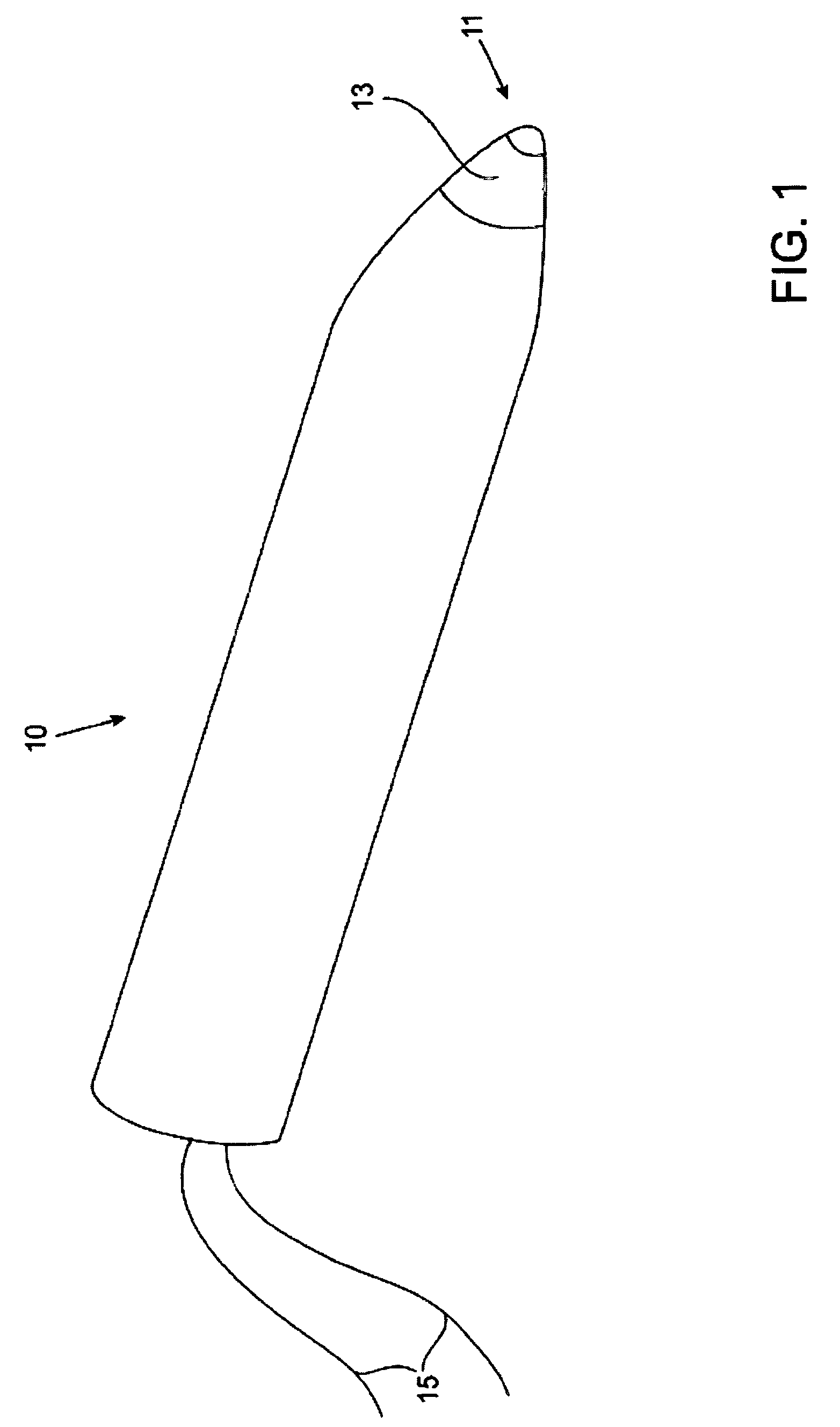
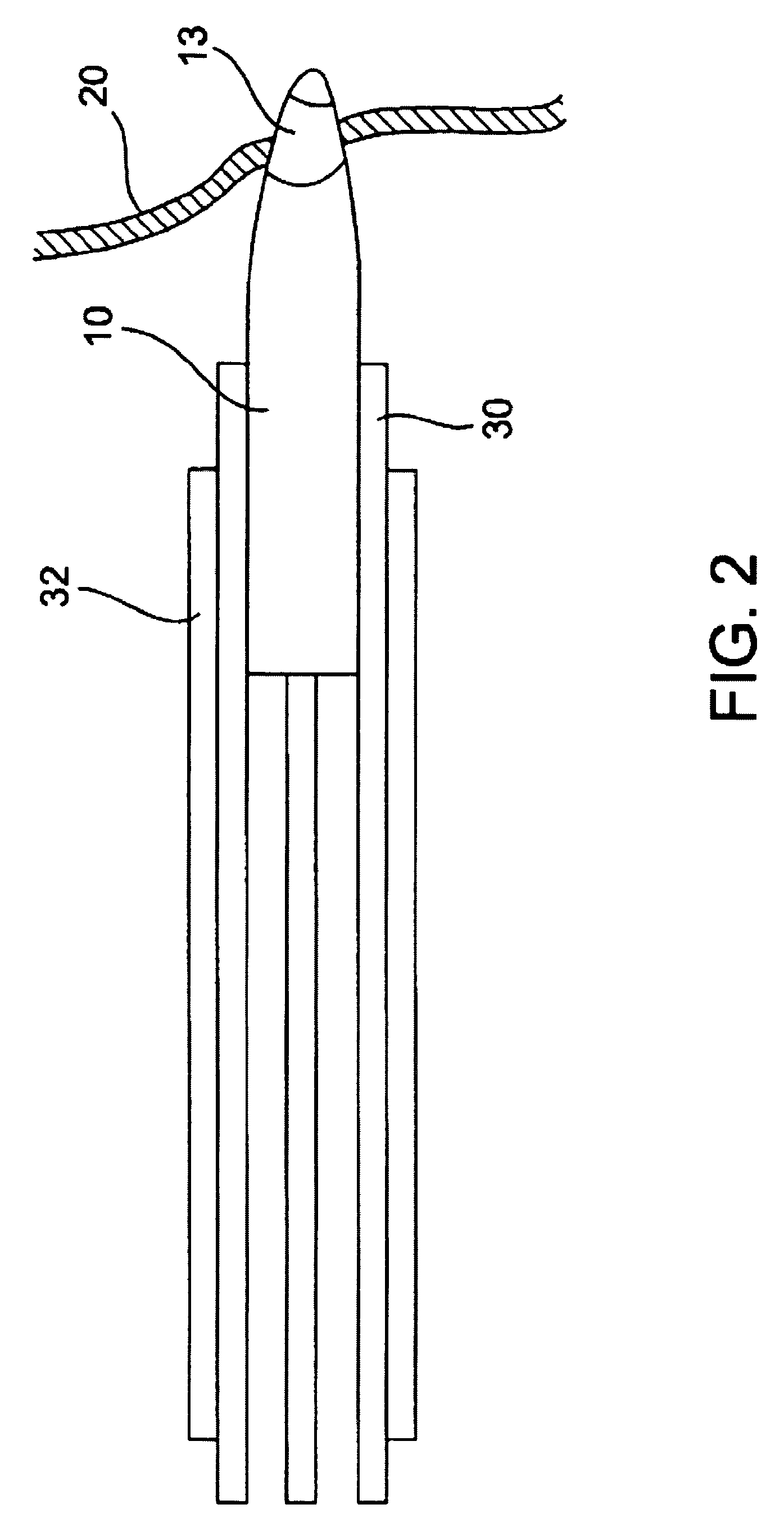
[0046] In a first preferred embodiment, as is seen in FIG. 1, an electromyography nerve surveillance probe 10 having a blunt end 11 is provided. Electrode 13 is disposed at the distal end of probe 10 and is charged by electrical contacts 15. As electrode 13 approaches nerve 20 (as seen in FIG. 2), the minimal threshold depolarization value elicited by the electrode will result in corresponding electromyography activity, such that the presence of nerve 20 can be sensed by standard electromyographic techniques, thus indicating the presence of the nerve. Specifically, using standard electromyographic techniques, the presence of nerve 20 will be sensed by appropriate needles or patches attached to the appropriate muscle as electrode 13 stimulates, and thereb...


PUM


Abstract

An expandable tip cannula system, comprising: a hollow cannula shaft having a proximal end and a distal end; and an expandable tip mounted at the distal end of the hollow cannula shaft, the expandable tip comprising a plurality of generally-triangular shaped petals held together in a radially-inwardly tapered arrangement between adjacent petals, each petal comprising a nerve sensing electrode disposed therein.

Description

CROSS-REFERENCES TO RELATED APPLICATIONS [0001] The present application is a division of commonly owned and co-pending U.S. patent application Ser. No. 10 / 431,619, filed on May 7, 2003, which is a divisional application of commonly owned and co-pending U.S. patent application Ser. No. 09 / 325,998, filed on Jun. 4, 1999 and issued as U.S. Pat. No. 6,564,078, the complete disclosure of which is hereby incorporated by reference in its entirety for all purposes. Additionally, the present application claims benefit under 35 U.S.C. §119(e) from U.S. Provisional Patent Applications Serial No. 60 / 113,651 filed Dec. 23, 1998; U.S. Provisional Patent Application Serial No. 60 / 120,663 filed Feb. 12, 1999; and U.S. Provisional Patent Application Serial No. 60 / 123,268 filed Mar. 8, 1999; the complete disclosures of which are hereby incorporated herein by reference in their entirety for all purposes.TECHNICAL FIELD [0002] The present invention relates to nerve surveillance systems and to cannulae ...

Claims


Application Information

Patent Timeline
Patent Type & Authority Applications(United States)
IPC IPC(8): A61M29/00A61B5/296A61B17/00A61B17/17A61B17/34A61B17/56A61B18/04
CPCA61B5/04012A61B5/0492A61B5/4893A61B17/1757A61B17/3417A61B17/3439A61B5/04001A61B18/1487A61B2017/3484A61B2018/00589A61B2018/00595A61N1/0551A61B18/148A61B5/4029A61B5/4041A61B5/316A61B5/296A61B5/24A61B17/3423
Inventor MARINO, JAMES F.AHIGREN, DAN K.
Owner NUVASIVE
Why Eureka
Try Eureka