Method for acquiring cardiac image data

a cardiac image and data acquisition technology, applied in the field of cardiac image acquisition, can solve the problems of inability to repeat the measurement of all slice orientations, and the exposure time is too long

Inactive Publication Date: 2007-04-12
SIEMENS AG
View PDF9 Cites 5 Cited by
  • Summary
  • Abstract
  • Description
  • Claims
  • Application Information

AI Technical Summary

Benefits of technology

[0007] An object of the present invention is to provide a method that is improved in this respect for acquir...

Problems solved by technology

Conventionally, the images are subsequently inspected, usually by a physician, whereupon measurements of individual slice orientations are repeated using a second acquiring techn...

Method used


Image

  • Method for acquiring cardiac image data
  • Method for acquiring cardiac image data
  • Method for acquiring cardiac image data

Examples


Embodiment Construction

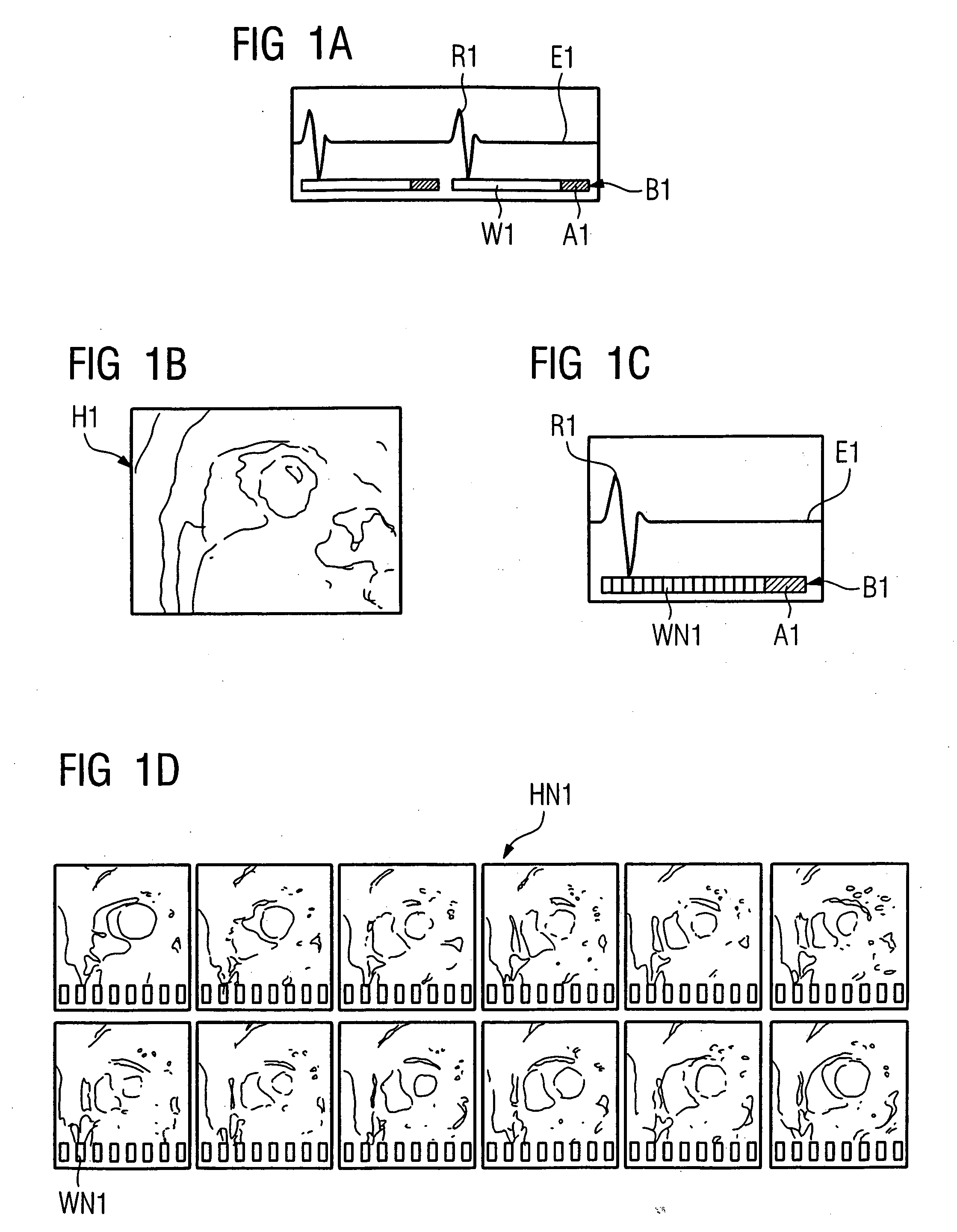
[0034]FIG. 1A shows a sketch of an electrocardiogram E1; two cycles are shown with their respective R-spikes R1. Below electrocardiogram E1, time bars B1 are shown that symbolize the elapsing of time and that each have a long waiting time W1 in which no image data are acquired, followed by, in the end diastole, a relatively short acquiring time A1. Waiting time W1 makes up by far the longest part of the cardiac cycle, which accordingly remains unused for acquiring data. FIG. 1B shows a sketch of a heart exposure H1 corresponding to FIG. 1A that was created from image data acquired in the end diastolic phase. A limitation to the first image acquiring technique having the short exposure time window A1 thus yields only a limited amount of data that can be used to create a diagnosis.

[0035]FIG. 1C shows a sketch for the use of waiting time W1 of FIG. 1A, using a second image acquiring technique according to the method of the present invention. A sketch of electrocardiogram E1 is again s...


PUM


Abstract

In a method and apparatus for acquiring cardiac image data of a subject of examination, in particular in the context of magnetic resonance tomography, a first image acquiring technique is implemented, in which image data acquiring takes place only in a part of the cardiac cycle, and/or not in each cardiac cycle, and a second image acquiring technique is alternatingly implemented, such that the image data acquiring using the second image acquiring technique takes place at times in which image data acquiring using the first image acquiring technique is not occurring.

Description

BACKGROUND OF THE INVENTION [0001] 1. Field of the Invention [0002] The present invention relates to a method for acquiring cardiac image data obtained from an examination subject, in particular in the context of magnetic resonance tomography (MRT). [0003] 2. Description of the Prior Art [0004] For acquiring cardiac image data of a human or animal subject, various image acquiring techniques are used in which data are actually acquired only in a small time region of the cardiac cycle. The remaining time is unused during the acquiring, because this time is merely spent waiting for the heart to, for example, return to the desired segment of the cardiac cycle, or for preparation pulses (in the case of MRT) to achieve their effect on the resulting image, or for the tissue to subsequently relax again. [0005] If, for example, images of the myocardium are produced in the context of a morphological imaging, this often takes place only in the end diastolic phase. In addition, there are other ...

Claims


Application Information

Patent Timeline
IPC IPC(8): A61B5/05A61B5/352
CPCG01R33/5607G01R33/56325G01R33/5673
Inventor SPRUNG, KATRIN CHRISTEL
Owner SIEMENS AG
Why Eureka
Try Eureka