Programmable phase velocity in an ultrasonic imaging system

a phase velocity and ultrasonic imaging technology, applied in the field of medical imaging process system and method, can solve the problems of inability to consistently determine the functional relationship between the hardness profile measurement, and the limitations of the application of ultrasonic imaging technology to accurately detect biological tissues of different hardness

Inactive Publication Date: 2007-04-12
SONOWISE
View PDF44 Cites 39 Cited by
  • Summary
  • Abstract
  • Description
  • Claims
  • Application Information

AI Technical Summary

Benefits of technology

[0013] In yet another aspect, the present invention further provides an ultrasonic imaging scanning system that is implemented to allow an operator's adjustment for the best image focusing estimation controlled by the operator. Additional flexibility may also provide that enables a computer's algorithm for automatic best image searching.

Problems solved by technology

Even though the hardness of a biological tissue is an important parameter for detection of abnormal tissue growth to enable an early detection of diseases occurring to a biological organ and greatly improve the curing rates, there are still technical difficulties and limitations faced by those applying ultrasonic imaging technologies for accurately detect biological tissues of different hardness.
However, several of these measurements can only obtain hardness profile measurement with limited accuracies.
However, there are no consistently determinable functional relationships between the attenuation of the ultrasonic transmission through the biological tissues of different values of tissue hardness.
Additionally, there are operator's errors such as the Time Gain Compensation (TGC), Focus point and Aperture size settings, etc. can also affect the attenuation, which discounts the accountability of the tissue hardness characterization.
For these reasons, the measurements of attenuations do not usually provide reliable indications of the tissue hardness and the hardness profile measurements therefore have only limited usefulness.
However, as discussed above, the functional relationship between the hardness profile and the attenuation are changed under different measurement circumstances including but not limited to operators' skills and possible errors, the accuracies of measurements based on attenuation measurements are therefore degraded and hasn't been successful in the commercial available ultrasound imaging system.
The conventional ultrasound system assumes the phase velocity in the soft tissue is constant at 1540 m / s, however in the soft organ, liver for instance, when the homogeneous tissue becomes harder, e.g. cirrhosis, the phase velocity in the liver tissue changes, therefore, the assuming of 1540 m / s is no longer valid.
The consequences of such assumptions lead to blurred images due to the inaccurate of the beam focusing and the deviations of the geometry distance measurements.
The degradation of the image quality might not be considered as significant in the past due to the facts that the conventional image scanning system did not require such image quality and furthermore, it was not expected that the scanned images can achieve consistent quality of sharp and clear images anyway.
The ultrasound phase velocity measurement is easier done with the tissue cut out from the body, but extremely difficult in the live organ.
As discussed above, the measurement accuracies are limited due to this single set of characteristics projected from a single light source operated without controllable input to change the output from the light source.
Such methodology may be useful for some image analyses applications but would not provide practical solution to overcome the difficulties of insufficient accuracy in measuring and mapping the hardness profile.
However, such disclosures do not provide effective methods for a person of ordinary skill to over the problem that accuracy of hardness measurements cannot be easily improved by applying convention techniques as now available.

Method used


Image

  • Programmable phase velocity in an ultrasonic imaging system
  • Programmable phase velocity in an ultrasonic imaging system
  • Programmable phase velocity in an ultrasonic imaging system

Examples


Embodiment Construction

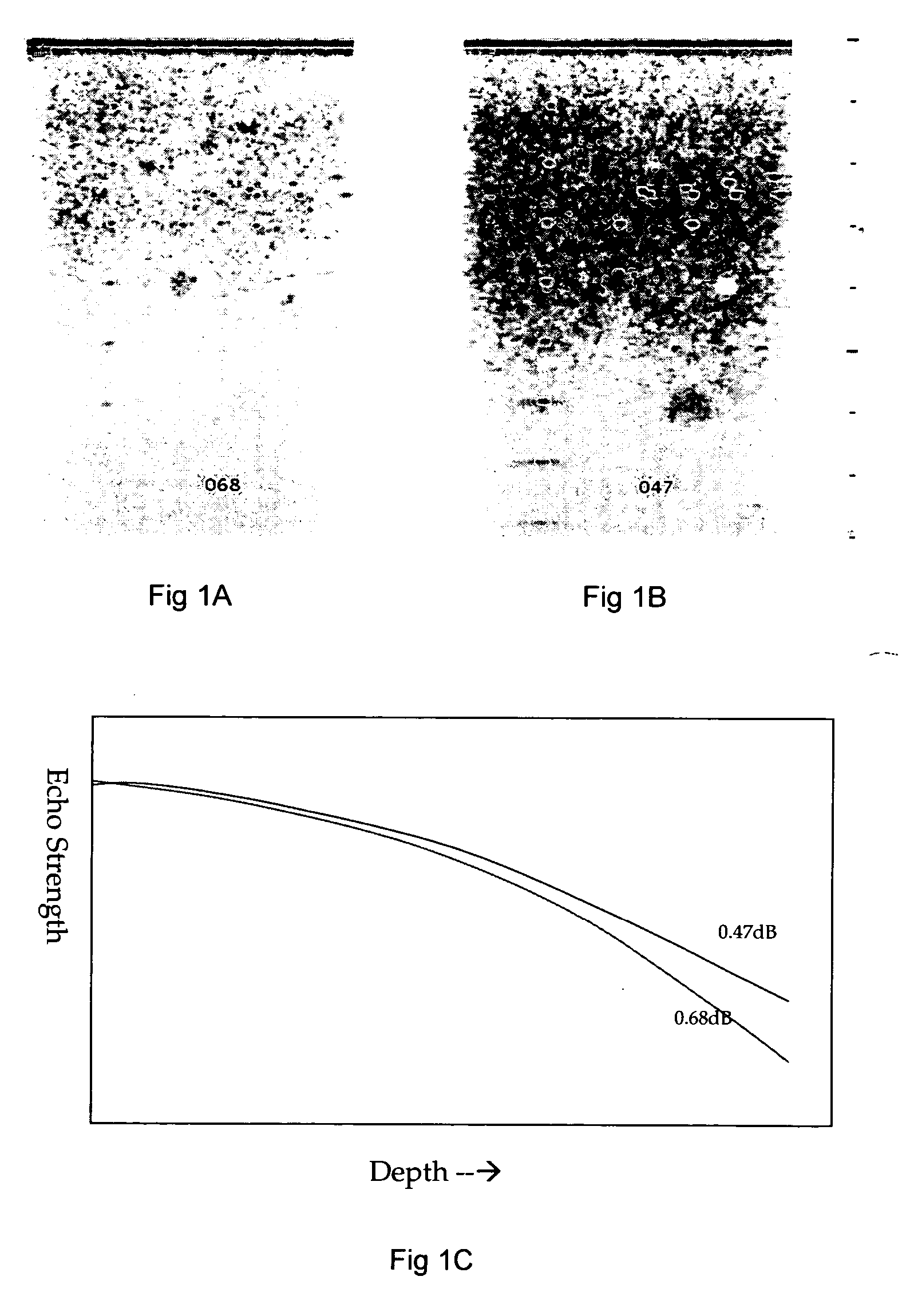
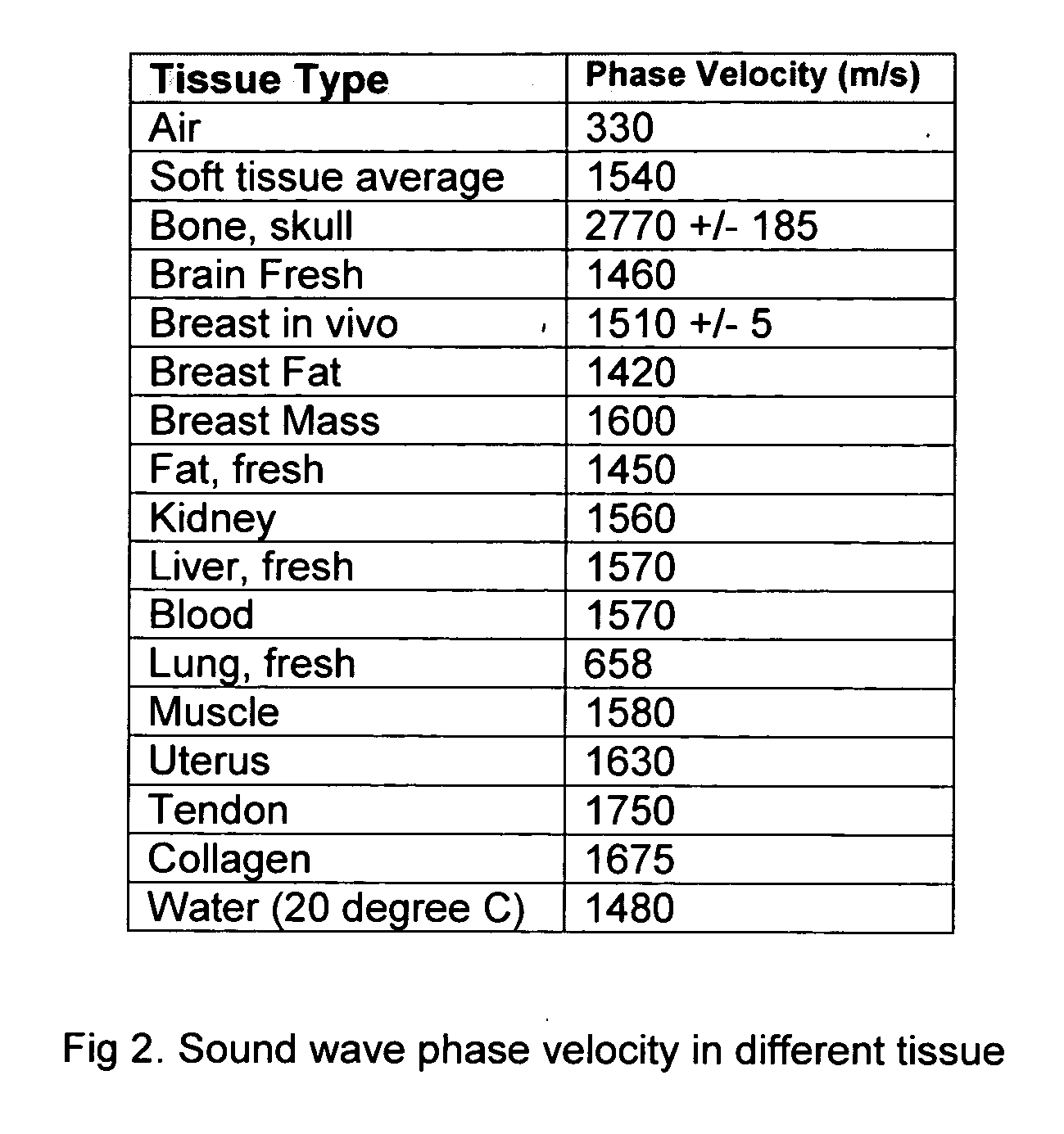
[0024] Referring to FIG. 2 for a table that lists media of different tissues for transmission of sound waves. Listed in the table on the right hand column in the table is also the corresponding phase velocity for each of these tissues. According to the listed data shown in FIG. 2, the sound travels in the media, and the phase velocity changes. Depending on the density of the media, the phase velocity varies. As the density of a media increases, the phase velocity of the sound waves transmitted in the media is also increased. Because of these changes, the phase velocity is employed as practical and useful measuring parameter to measure the variations of hardness in a soft tissue. However, the amplitude attenuation may or may not have direct relationship to the tissue hardness. It depends on the tissue homogeneity and absorption.

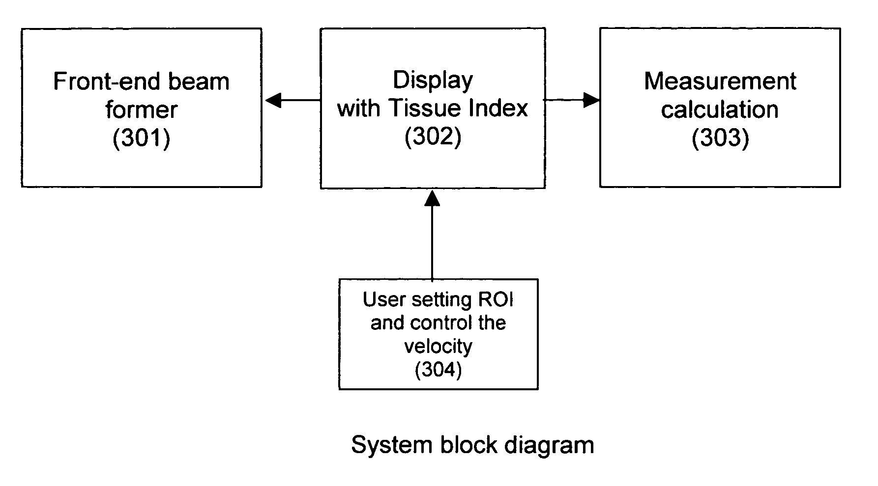
[0025]FIG. 3 is a functional block diagram for showing an ultrasound image scanning system implemented with user control functions to adjust the phase veloci...


PUM


Abstract

An ultrasonic image scanning system for scanning an organic object includes a beam former that provides a phase velocity adjustment function for producing an ultrasonic image with a programmable phase velocity. The ultrasonic image scanning system further includes a beam profile analysis function for calculating an optimal phase velocity with a user controller to adjust the phase velocity until a scan image of best image quality is achieved. Alternately, the system may provide an automatic phase velocity-scanning controller for automatically scanning through a range of phase velocities and selecting a best phase velocity generating a scanning image of a best quality. The system further includes a region of interest (ROI) controller for a user to select a region for scanning with a specific focal area for optimizing the phase velocity. The system may further provide a maximum gradient analyzer for selecting an image of a best quality in optimizing the phase velocity. A digital controller may also provide a real time programmable control by applying different control algorithms with combination of phase velocity and attenuation adjustment. A hardness computational processor is implemented to determine a tissue hardness using the phase velocity and in combination with the attenuation parameter.

Description

FIELD OF THE INVENTION [0001] This invention generally relates to system and method for carrying out a medical imaging process. More particularly, this invention relates to an ultrasonic imaging apparatus and method for providing the tissue hardness information through the image focus discrimination when those images are acquired by applying various phase velocities in the beam forming calculation. BACKGROUND OF THE INVENTION [0002] Even though the hardness of a biological tissue is an important parameter for detection of abnormal tissue growth to enable an early detection of diseases occurring to a biological organ and greatly improve the curing rates, there are still technical difficulties and limitations faced by those applying ultrasonic imaging technologies for accurately detect biological tissues of different hardness. Specifically, conventional technologies have attempted to obtain a hardness profile of an ultrasonic scanned image by assuming that the phase velocity, i.e. spe...

Claims


Application Information

Patent Timeline
Patent Type & Authority Applications(United States)
IPC IPC(8): A61B8/00
CPCA61B8/08A61B8/14G01S7/52049A61B8/587G01S7/52071A61B8/585G01S7/5205
Inventor LIN, SHENGTZWU, TONYHUE, PHUNG
Owner SONOWISE
Why Eureka
Try Eureka