System and Method For Robust Optic Disk Detection In Retinal Images Using Vessel Structure And Radon Transform

Inactive Publication Date: 2007-05-17
SIEMENS CORP
View PDF2 Cites 30 Cited by
  • Summary
  • Abstract
  • Description
  • Claims
  • Application Information

AI Technical Summary

Benefits of technology

[0008] In an exemplary embodiment of the present invention, a metod for optic disk detection in retinal images, comprises: extracting a vessel tree from a retinal image; locating an optic disk in the retinal image using the vessel tree; enhancing a contrast of the optic disk, removing

Problems solved by technology

However, the assumption that an optic disk is the brightest area in a retinal image does not always hold.
Even with healthy eyes, uneven illumination may cause some regions to be brighter, thus invalidating the assumption.
81-121, 2004, is computationally inte

Method used


Image

  • System and Method For Robust Optic Disk Detection In Retinal Images Using Vessel Structure And Radon Transform
  • System and Method For Robust Optic Disk Detection In Retinal Images Using Vessel Structure And Radon Transform
  • System and Method For Robust Optic Disk Detection In Retinal Images Using Vessel Structure And Radon Transform

Examples


Embodiment Construction

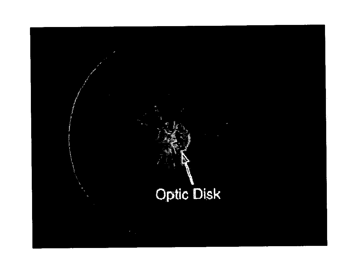
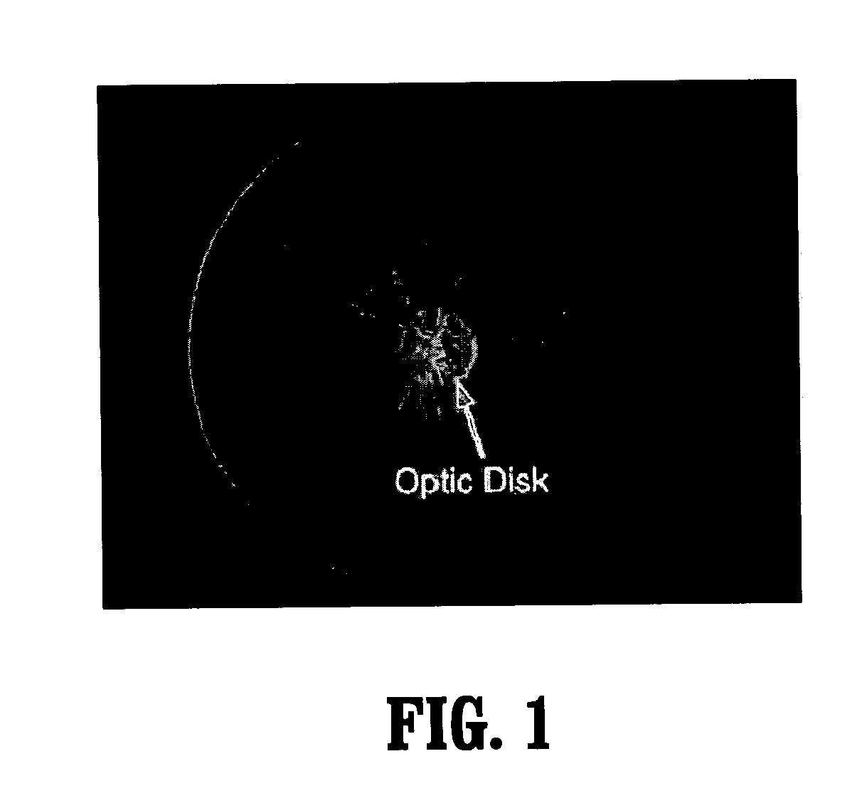
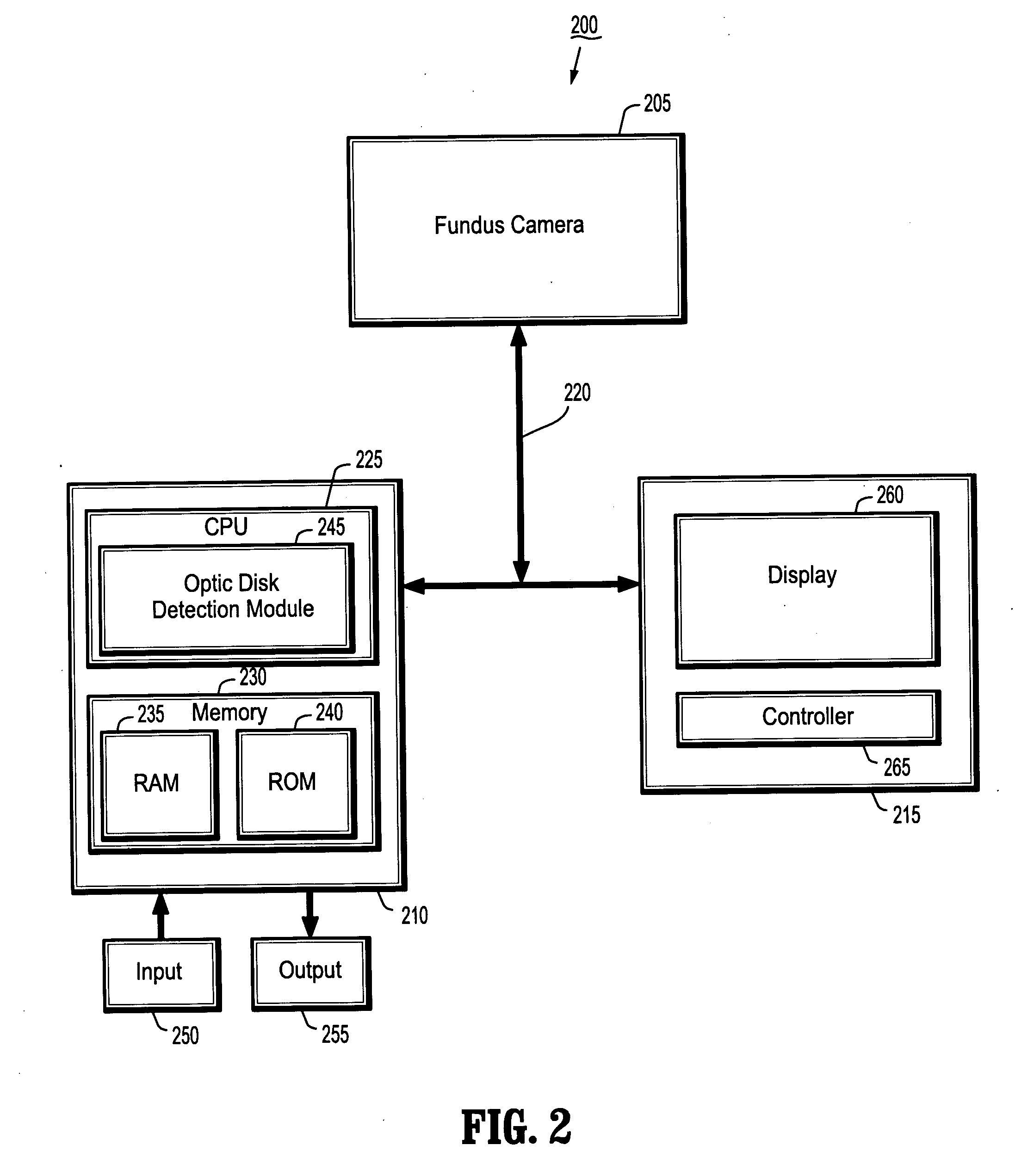
[0035]FIG. 2 illustrates a system 200 for robust optic disk detection in retinal images using vessel structure and radon transform according to an exemplary embodiment of the present invention. As shown in FIG. 2, the system 200 includes a fundus camera 205, a PC 210 and an operator's console 215 connected over a wired or wireless network 120.

[0036] The fundus camera 205 is a device used by optometrists, ophthalmologists, and other trained medical professional to photograph a patient's retina. The fundus camera 205 is generally used for monitoring progression of a disease, diagnosis of a disease (combined with retinal angiography), or in screening programs, where the photos can be analyzed later.

[0037] The PC 210, which may be a portable or laptop computer, includes a CPU 225 and a memory 230 connected to an input device 250 and an output device 255. The CPU 225 includes an optic disk detection module 245 that includes one or more methods for robust optic disk detection in retinal...


PUM


Abstract

A method for optic disk detection in retinal images, includes: extracting a vessel tree from a retinal image; locating an optic disk in the retinal image using the vessel tree; enhancing a contrast of the optic disk; removing vessels from the contrast enhanced optic disk; detecting a boundary of the optic disk with vessels removed by generating an edge map; projecting the edge map at a plurality of angles by using a radon transform; and estimating a radius and center of the optic disk using the projections.

Description

CROSS-REFERENCE TO RELATED APPLICATION [0001] This application claims the benefit of U.S. Provisional Application No. 60 / 726,047, filed Oct. 12, 2005, a copy of which is herein incorporated by reference.BACKGROUND OF THE INVENTION [0002] 1. Technical Field [0003] The present invention relates to optic disk detection in retinal images, and more particularly, to a system and method for robust optic disk detection in retinal images using vessel structure and radon transform. [0004] 2. Discussion of the Related Art [0005] Structural analysis of retinal images is critical to screening and diagnosis of eye diseases such as glaucoma, diabetic retinopathy and age-related macular degeneration. Estimating the location and size of an optic disk is needed in clinical applications such as measuring the progression of glaucoma as described in L. Bonomi and N. Orzalesi, Glaucoma: Concepts in Evolution: Morphometric and Functional Parameters in the Diagnosis and Management of Glaucoma. Kugler Ghend...

Claims


Application Information

Patent Timeline
IPC IPC(8): A61B3/10
CPCA61B3/12G06T7/0042G06T7/602G06T7/606G06T2207/30041G06T2207/30101G06T7/62G06T7/66G06T7/73
Inventor YAN, MICHELLE XIAO-HONGHUANG, KESCHULL, HANS
Owner SIEMENS CORP
Why Eureka
Try Eureka