Method for three dimensional multi-phase quantitative tissue evaluation

a multi-phase, tissue evaluation technology, applied in the field of imaging systems, can solve the problems of small volume imaging, limited observation of hyper-enhancement, perfusion and viability, and inability to assess the extent of cardiac injury to the entire organ

Inactive Publication Date: 2007-06-07
GENERAL ELECTRIC CO
View PDF9 Cites 39 Cited by
  • Summary
  • Abstract
  • Description
  • Claims
  • Application Information

AI Technical Summary

Problems solved by technology

However, in injured myocardium, the sarcolemmal membrane of myocytes become permeable allowing contrast agent to accumulate, which results in the observed hyper-enhancement.
Within CT-based research, perfusion and viability imaging is limited to small volumes of imaging.
Small volume imaging is not able to assess the extent of cardiac injury to the entire organ and, therefore, may be inadequate for routine clinical use.
Visualization and analysis of this complex array of data set can be quite time consuming for the physician.
Current methods of both acquisition and visualization do not provide adequate information in a seamless manner to the physician to enable a productive analysis of the images.
Some of the disadvantages associated with this type of analysis are related to the visualization of the injured / infracted region relatively compared to the rest of the imaging volume, including segmentation, volume analysis and visualization of the infarct throughout the cardiac phases.

Method used


Image

  • Method for three dimensional multi-phase quantitative tissue evaluation
  • Method for three dimensional multi-phase quantitative tissue evaluation
  • Method for three dimensional multi-phase quantitative tissue evaluation

Examples


Embodiment Construction

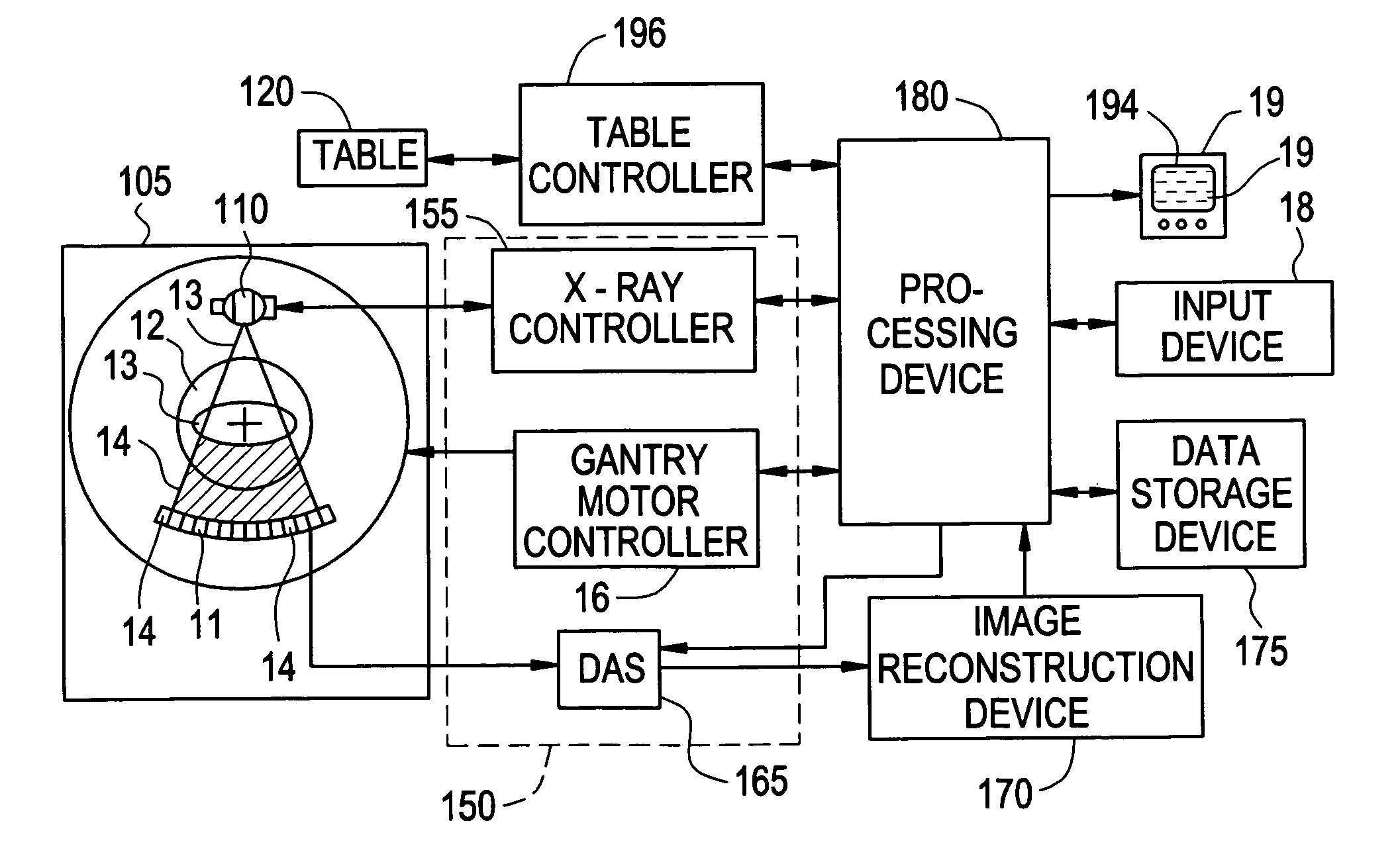
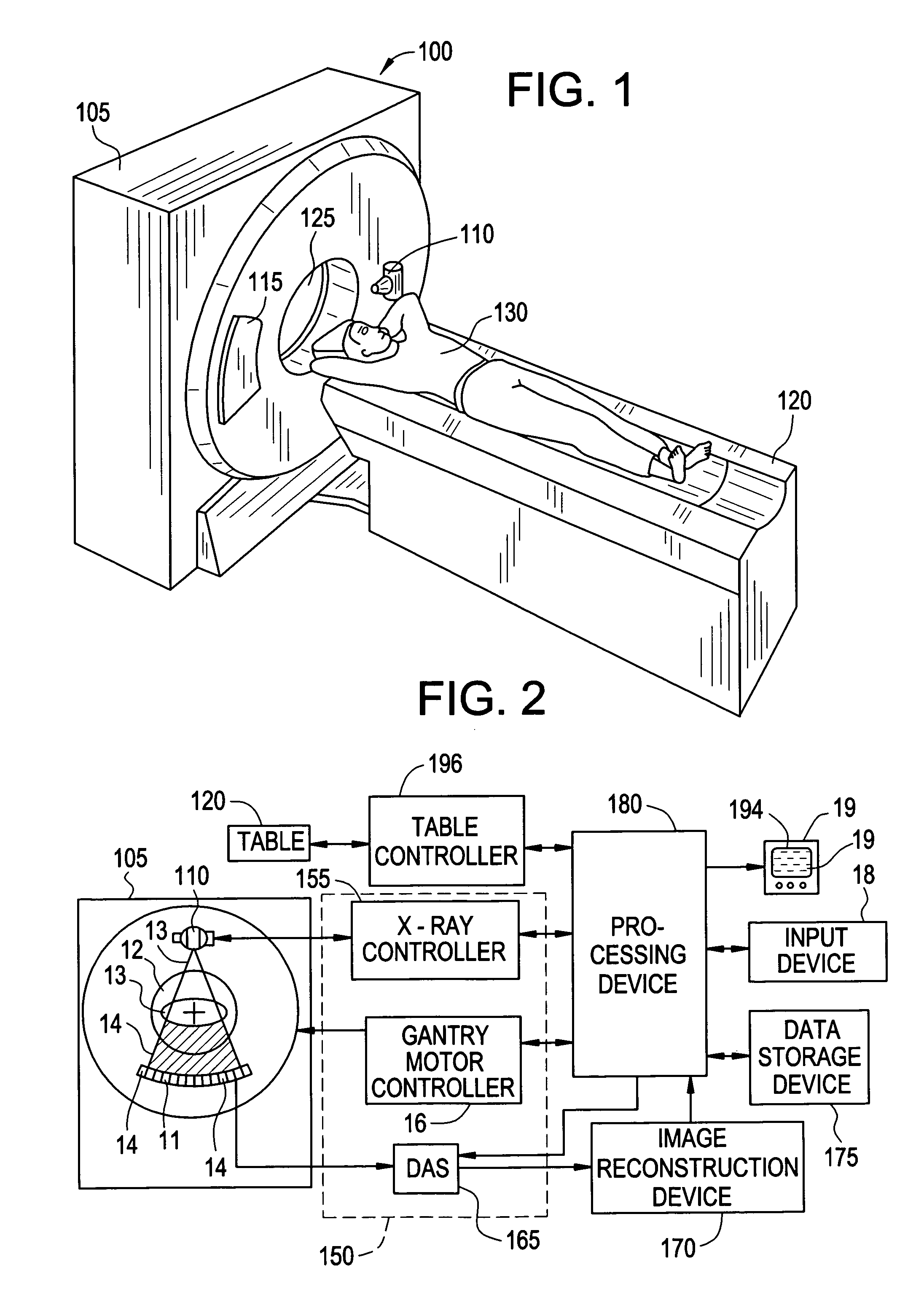
[0031] Disclosed herein in the exemplary embodiments are a system and methodologies that enable a streamlined workflow for 3D, multi-phasic quantitative perfusion and viability imaging on whole organ systems, with reference to a computed tomography (CT) imaging system. While an exemplary system and methodology of positioning an anatomical object relative to a CT imaging system is disclosed, it will be appreciated that such disclosure is illustrative only, and it should be understood that the method and system of the disclosed invention may readily be applied to other imaging systems, such as magnetic resonance (MR) or other scanning systems. Additionally, while the anatomical object disclosed is a heart and related myocardial tissue, it will also be appreciated that such disclosure is illustrative only, and the method and system of the disclosed invention may readily be applied to other anatomical objects, including, but not limited to a liver, brain, vasculature or kidney.

[0032] I...


PUM


Abstract

A method of evaluating tissue of an organ includes accessing image data and processing the image data to quantify at least one feature of interest in the tissue. The image data is derived from a computed tomography acquisition system and includes multiple phases of acquired whole organ data. Each phase is acquired within five gantry rotations of the acquisition system using an acquisition protocol.

Description

BACKGROUND OF THE INVENTION [0001] This invention relates generally to imaging systems and methods of using thereof, specifically to a method of imaging organ systems with a perfusion and / or a viability protocol. [0002] The diagnosis of myocardial tissue viability after a heart attack focuses on whether there will be functional improvement of dysfunctional myocardium after revascularization therapy. Both single photon emission computed tomography (SPECT) and contrast enhanced magnetic resonance imaging (MRI) have been able to measure the viability of tissue. SPECT is a relatively inexpensive method for functionally measuring the viability within the myocardium. In comparison, MRI is a better standard of measurement because of the spatial resolution that is available for definition of infarcts, and in particular, non-transmural infarcts. [0003] MRI uses both anatomical and functional methods to determine the viability of the myocardium. MRI also provides functional information about ...

Claims


Application Information

Patent Timeline
Patent Type & Authority Applications(United States)
IPC IPC(8): G06K9/00A61B5/05
CPCA61B5/7285A61B5/7289A61B6/032A61B6/4085A61B6/466A61B6/503A61B6/504A61B6/5217A61B6/5288A61B6/541A61B8/0891A61B6/5247G16H50/30
Inventor HOPPEL, BERNICE E.AVINASH, GOPAL B.PIACSEK, KELLY LYNN
Owner GENERAL ELECTRIC CO
Why Eureka
Try Eureka