Microfluidic systems for size based removal of red blood cells and platelets from blood

a microfluidic system and platelet technology, applied in the field of medical diagnostics and microfluidics, can solve the problems of large and expensive equipment, large sample volumes, and inability to accurately detect the presence of red blood cells and platelets in the blood,

Inactive Publication Date: 2007-07-12
THE GENERAL HOSPITAL CORP
View PDF99 Cites 193 Cited by
  • Summary
  • Abstract
  • Description
  • Claims
  • Application Information

AI Technical Summary

Benefits of technology

[0007] The invention features devices and methods for enriching a sample in one or more desired particles. An exemplary use of these devices and methods is for the enrichment of cells, e.g., white blood cells in a blood sample. In general, the methods of the invention employ a device that contains at least one sieve through which particles of a given size, shape, or deformability can pass. Devices of the invention have at least two outlets, and the sieve is placed such that a continuous flow of fluid can pass through the device without passing through the sieve. The devices also include a force generator for directing selected particles through the sieve. Such force generators employ, for example, diffusion, electrophoresis, dielectrophoresis, centrifugal force, or pressure-driven flow.

Problems solved by technology

Different flow cytometry and cell sorting methods are available, but these techniques typically employ large and expensive pieces of equipment, which require large volumes of sample and skilled operators.
Few attempts have been made, however, to miniaturize flow cytometers and cell sorters [5,6] that have yielded promising results which compare to the larger macroscale devices.

Method used


Image

  • Microfluidic systems for size based removal of red blood cells and platelets from blood
  • Microfluidic systems for size based removal of red blood cells and platelets from blood
  • Microfluidic systems for size based removal of red blood cells and platelets from blood

Examples


example 1

Diffusive Filter

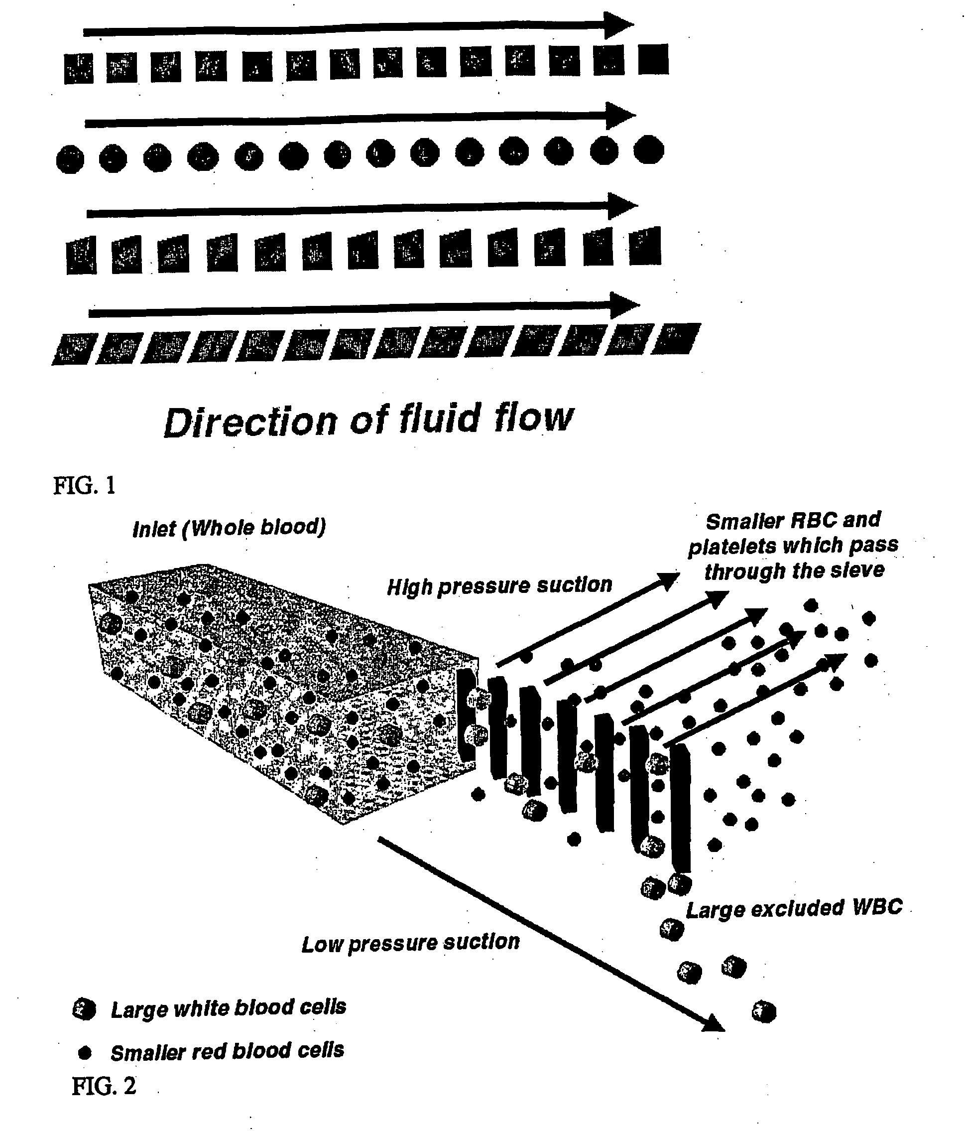
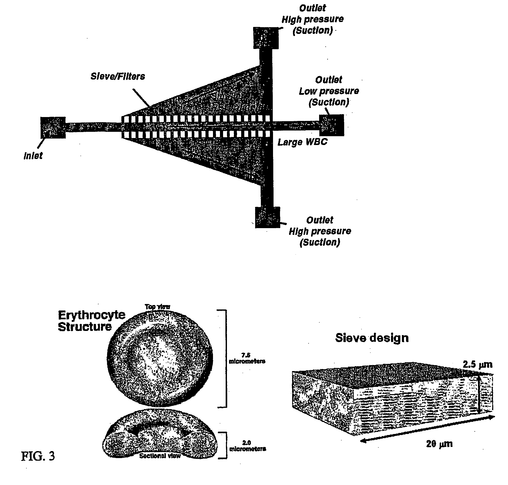
[0054] A device for size based separation of smaller RBCs and platelets from the larger WBCs was fabricated using simple soft lithography techniques (FIG. 13). A chrome photomask having the features and geometry of the device was fabricated and used to pattern a silicon wafer with a negative replica of the device in SU-8 photoresist This master was then used to fabricate PDMS channel and sieve structures using standard replica molding techniques. The PDMS device was bonded to a glass slide after treatment with O2 plasma. FIG. 13 shows a low magnification image of the channel structure with the diffuser geometry and sieves. The diffuser geometry is used to widen the laminar flow streamlines to ensure that the majority of the particles or cells flowing through the device will interact with the sieves. The smaller RBC and platelets pass through the sieves, and the larger WBCs are confined to the central channel. A higher magnification picture of the sieves is shown in ...

example 2

Electrophoresis

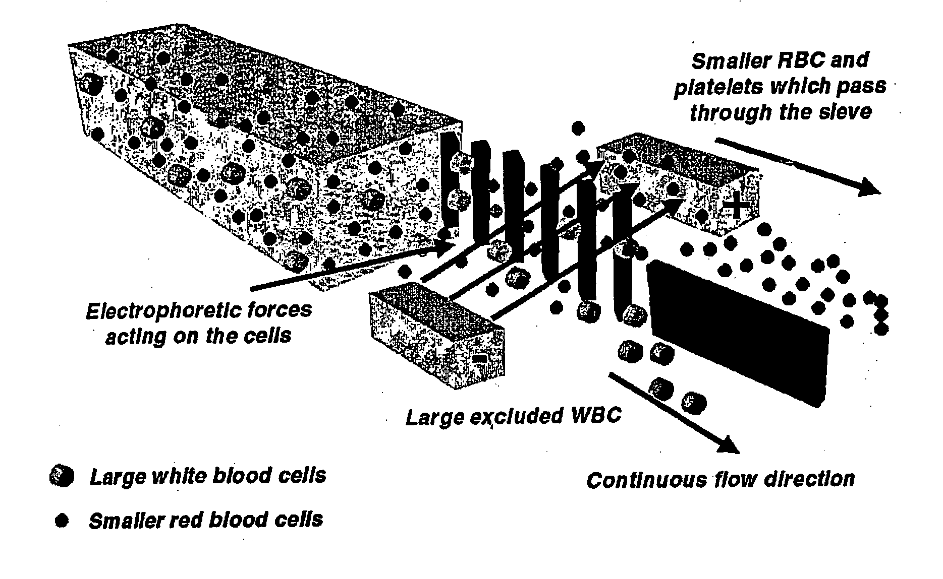
[0055] Electrophoresis can also be used to move cells across their laminar flow streamlines and ensure that all the cells or particles interact or come in contact with the sieves. The device was fabricated as in Example 1, but the PDMS is bonded to a glass slide having gold electrodes that were patterned photolithographically (FIG. 15). Electrophoresis is used to attract negatively charged cells towards the positively charged electrode. The smaller RBC and platelets pass through the sieves, while the larger WBCs are excluded. The WBCs are isolated and extracted through a separate port


PUM

PropertyMeasurementUnit
sizeaaaaaaaaaa
sizeaaaaaaaaaa
sizeaaaaaaaaaa

Abstract

The invention features devices and methods for enriching a sample in one or more desired particles. An exemplary use of these devices and methods is for the enrichment of cells, e.g., white blood cells in a blood sample. In general, the methods of the invention employ a device that contains at least one sieve through which particles of a given size, shape, or deformability can pass. Devices of the invention have at least two outlets, and the sieve is placed such that a continuous flow of fluid can pass through the device without passing through the sieve. The devices also include a force generator for directing selected particles through the sieve. Such force generators employ, for example, diffusion, electrophoresis, dielectrophoresis, centrifugal force, or pressure-driven flow.

Description

STATEMENT REGARDING FEDERAL SPONSORED RESEARCH [0001] This invention was made with Government support under Grant No. GM 62119 awarded by the NIH. The Government has certain rights in this invention.BACKGROUND OF THE INVENTION [0002] The invention relates to the fields of medical diagnostics and microfluidics. [0003] The study of disease of the blood, bone marrow, and related organs and tissues benefits from the molecular analysis of specific cells. The human body contains about five liters of blood that includes three types of cells that are found in different concentrations, red blood cells (RBCs), white blood cells (WBCs) and platelets. These cells can give insight into a variety of diseases. Disease identification may involve finding and isolating rare events, such as structural and morphological changes in specific WBCs. The first step towards this is isolation of particular cells, e.g., WBCs, from the blood sample. [0004] There are six different types of WBCs in blood, and the...

Claims


Application Information

Patent Timeline
Patent Type & Authority Applications(United States)
IPC IPC(8): B01L11/00B01L99/00B01L3/00G01N1/40G01N15/02G01N33/49
CPCA61M1/3633G01N2015/0294B01L3/502753B01L3/502761B01L2200/0647B01L2300/0681B01L2300/0816B01L2300/0861B01L2400/0409B01L2400/0421B01L2400/0424B01L2400/0487B03C5/005B03C5/024G01N1/4005G01N15/0272G01N33/491G01N2001/4016G01N2015/0288B01L3/50273
Inventor SETHU, PALANIAPPANTONER, MEHMET
Owner THE GENERAL HOSPITAL CORP
Why Eureka
Try Eureka