Method and Apparatus for Detecting the Presence of Dermal Melanin in Epithelial Tissue

a technology of epithelial tissue and dermal melanin, which is applied in the field of methods and apparatus for detecting the presence of dermal melanin in epithelial tissue, can solve the problems of large number of individuals with skin lesions, inaccurate analysis, and no longer possible calibration

Inactive Publication Date: 2007-07-12
MEDX HEALTH
View PDF9 Cites 28 Cited by
  • Summary
  • Abstract
  • Description
  • Claims
  • Application Information

AI Technical Summary

Problems solved by technology

This limits the use of such an approach to analyzing small areas of skin as once larger areas of tissue are imaged, over which the surface geometry of the imaged tissue varies, calibration is no longer possible and analysis becomes inaccurate.
However, due to the required calibration procedures, it is typically only possible to produce a map of dermal melanin over a small area of skin, currently 15 mm diameter.
Although effective, prior art techniques thus currently require detailed individual analysis of every suspect lesion on a given patient and thus rely on the clinician being able to quickly identify all potentially dangerous lesions.
While this is straight-forward for the majority of patients, some individuals present with large numbers of skin lesions.
Although very effective for characterising normal skin, the described technique in WO 04 / 010862 is however limited to obtaining measurements of melanin and blood concentrations.
The technique is not suitable for obtaining measurements of collagen as changes in collagen have an equal effect at every wavelength and therefore no effect on a ratio of two spectral measures.
Further disclosed techniques are unable to determine whether melanin is present only within the epidermis of the skin or whether melanin has penetrated into the dermis.

Method used


Image

  • Method and Apparatus for Detecting the Presence of Dermal Melanin in Epithelial Tissue
  • Method and Apparatus for Detecting the Presence of Dermal Melanin in Epithelial Tissue
  • Method and Apparatus for Detecting the Presence of Dermal Melanin in Epithelial Tissue

Examples


Embodiment Construction

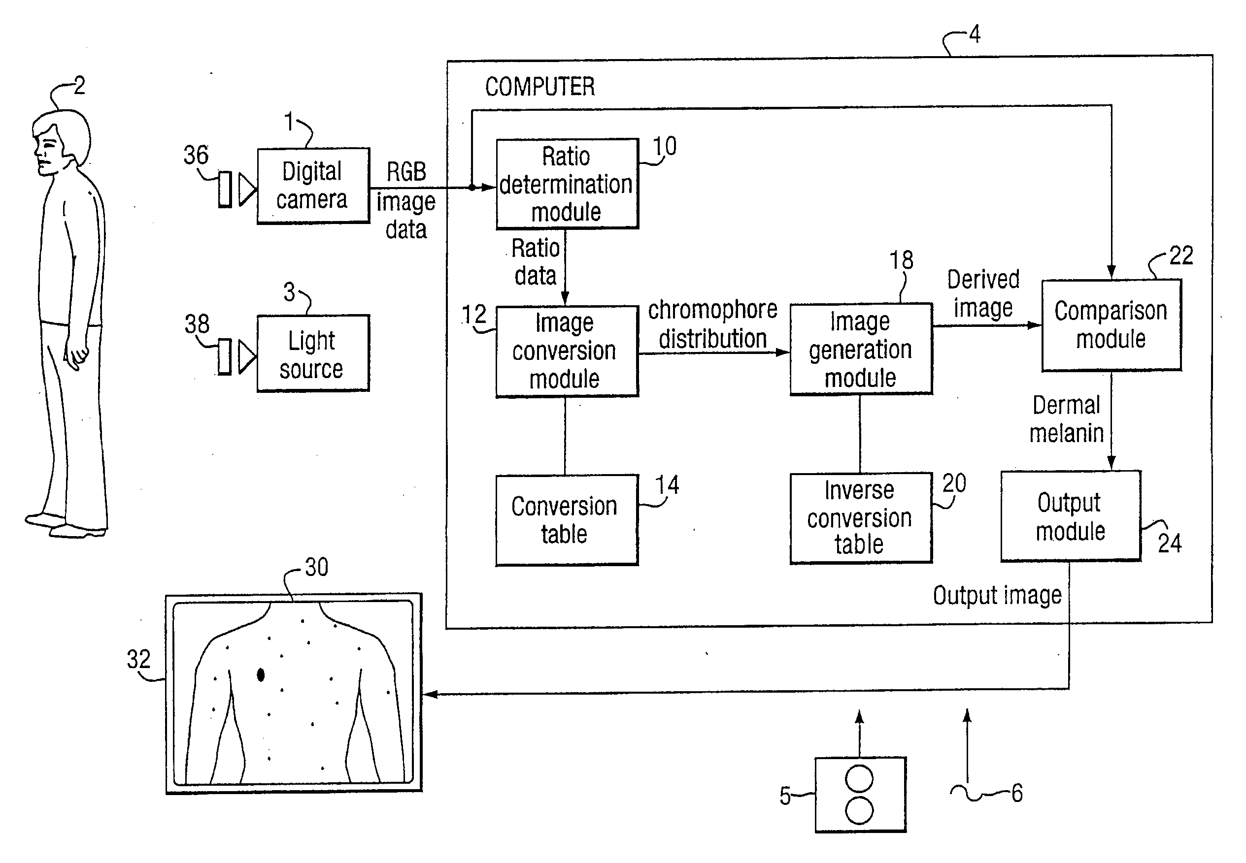
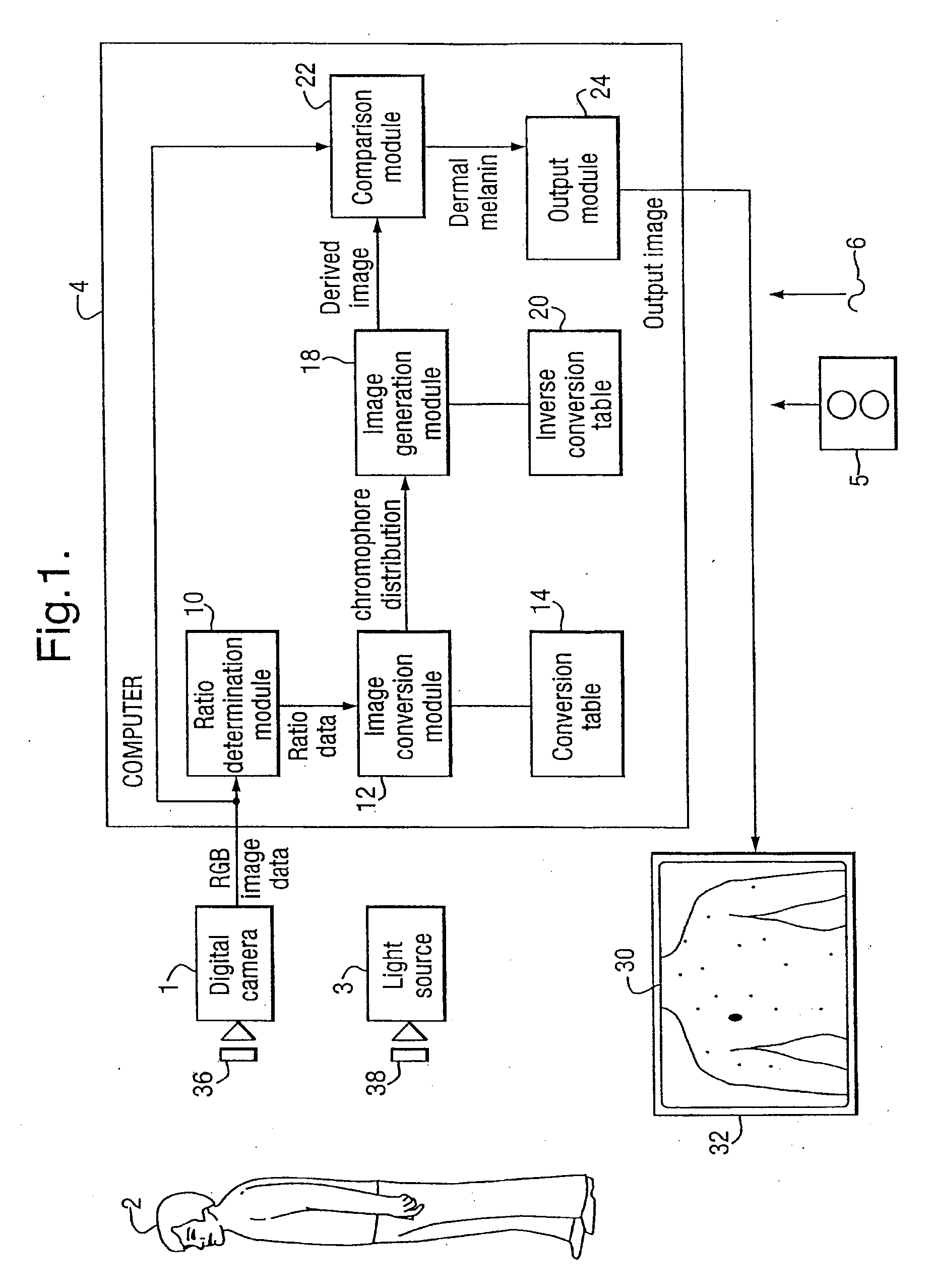
[0030]FIG. 1 is a schematic block diagram of an embodiment of the present invention. In accordance with this embodiment, a digital camera 1 comprising a conventional digital camera is provided which is arranged to obtain an image of an individual 2 illuminated by a light source 3. The images obtained by the digital camera 1 are then transmitted to a computer 4 which is configured by software either provided on a disk 5 or by receiving an electrical signal 6 by via a communications network to be configured into a number of functional modules 10-24 which cause the computer 4 to process the image data received from the digital camera 1 to generate an output image 30 which is shown on a display 32.

[0031] In the present embodiment the functional modules 10-24 comprise: a ratio determination module 10 for converting RGB image data into ratio data, an image conversion module 12 and a conversion table 14 for processing ratio data to generate data indicative of concentrations of blood and m...


PUM


Abstract

An image of an individual (2) is obtained using a digital camera (1). The image data is then processed by ratio determination module (10) and an image conversion module (12) to analyze the image and determine data representative of the distribution of blood and melanin in the skin of the imaged individual. This chromophore distribution data is then processed by an image generation module (18) which generates an image representative of the expected appearance of epithelial tissue having the determined distribution of chromophores where the epithelial tissue is under fixed illumination and has a flat spatial geometry and where all the identified melanin is present solely in the epidermis. The presence of dermal melanin can then be identified by comparing the original image data and the image derived from processing the calculated chromophore distributions and identifying where the ratio of these images differs by more than a threshold, the threshold being set at a level above the amount of variation expected due to lighting and geometry differences.

Description

CROSS-REFERENCE TO RELATED APPLICATION [0001] This application claims priority of U.K. Application No. 0526387.6, filed Dec. 23, 2005, which is hereby incorporated by reference in its entirety. FIELD OF THE INVENTION [0002] The present application concerns methods and apparatus for analysing epithelial tissue such as skin. In particular the present application concerns methods and apparatus for detecting the presence of dermal melanin in epithelial tissue. BACKGROUND OF THE INVENTION [0003] Over the last four decades there has been a world-wide increase in the incidence of melanoma. Although adequate sun protection has been identified as the first step toward preventing the occurrence of melanoma, early diagnosis and excision is the key to the survival of the many individuals who will still develop the disease. [0004] A range of techniques have been developed to assess pigmented lesions. With the most common of these, dermatoscopy, a hand held microscope is used to visualise morphol...

Claims


Application Information

Patent Timeline
Patent Type & Authority Applications(United States)
IPC IPC(8): A61B6/00
CPCA61B5/443A61B5/0059G06T7/0012
Inventor PREECE, STEPHEN JOHNCOTTON, SYMON D'OYLY
Owner MEDX HEALTH
Why Eureka
Try Eureka