Pelvic floor function diagnostic and therapeutic station and uses thereof

Active Publication Date: 2007-08-09
RGT UNIV OF CALIFORNIA
View PDF3 Cites 31 Cited by
  • Summary
  • Abstract
  • Description
  • Claims
  • Application Information

AI Technical Summary

Benefits of technology

[0013] In yet another embodiment there are three surface electrodes on the catheter holder. The surface electrodes can measure the electromyographic (EMG)

Problems solved by technology

Pelvic floor muscle weakness can thus cause prolapse of pelvic organs and incontinence of urine, feces and poor vaginal closure.
However none of these devices measure constrictor and elevator functions of pelvic floor muscles precisely.
Furthermore, these devices cannot measure the length tension curve of the constrictor and elevator function of the pelvic floor muscle.
There are two major problems with vaginal pressure measurements.
First, air filled balloons and water filled balloons, used by most of the investigators, do not measure absolute pressures.
None of the techniques used in the published literature made any attempt to distinguish pressures in different portions of the vaginal canal.
Techniques used by other investigators also did not make any attempt to distinguishing pressures from different segments of vaginal canal.
This device however does not measure the constrictor and elevator function of the pelvic floor muscles.
However none of th

Method used


Image

  • Pelvic floor function diagnostic and therapeutic station and uses thereof
  • Pelvic floor function diagnostic and therapeutic station and uses thereof
  • Pelvic floor function diagnostic and therapeutic station and uses thereof

Examples


Example

EXAMPLE 1

Side-hole Manometry and Sleeve Sensor Technique to Measure Vaginal Pressure

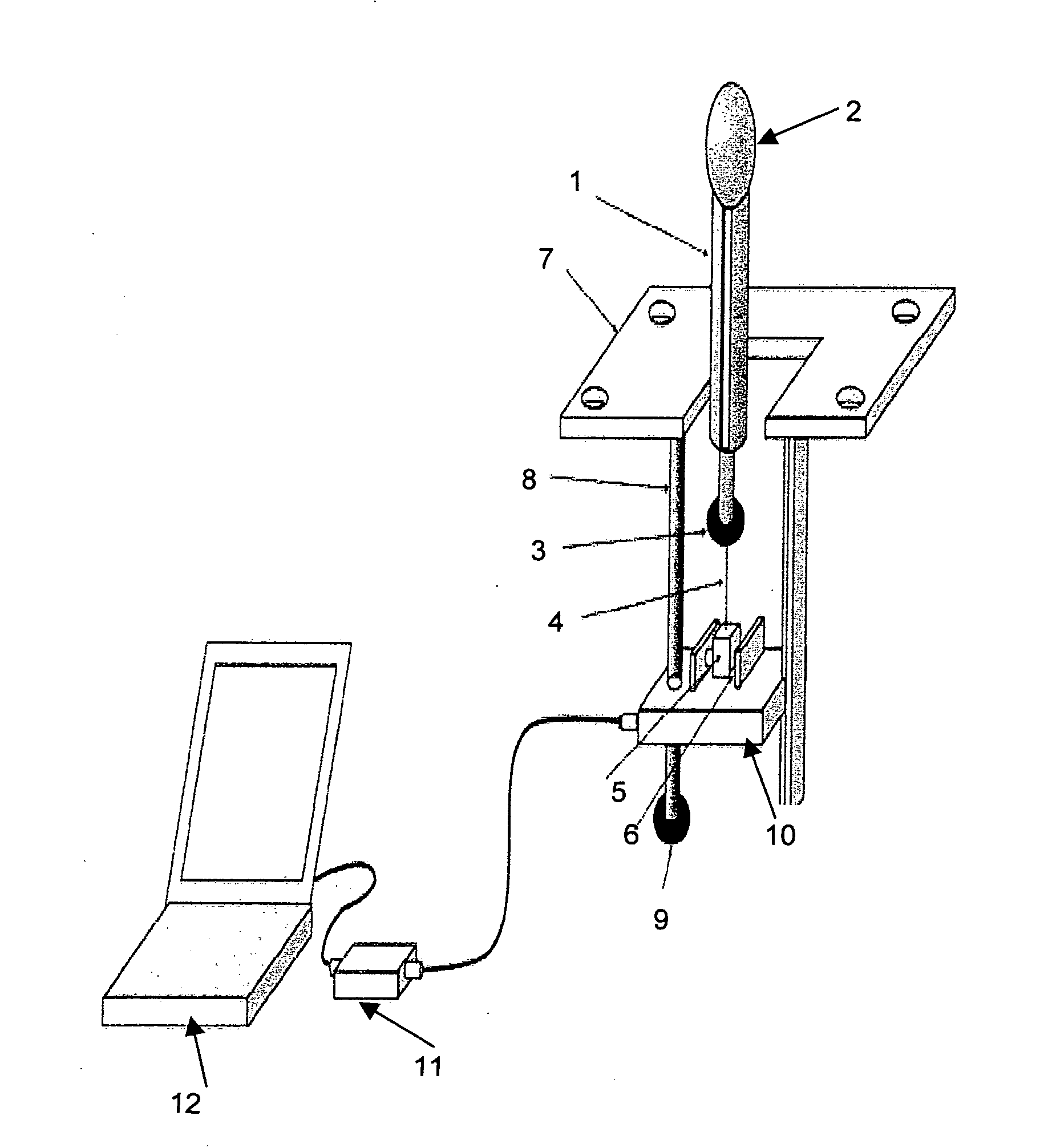
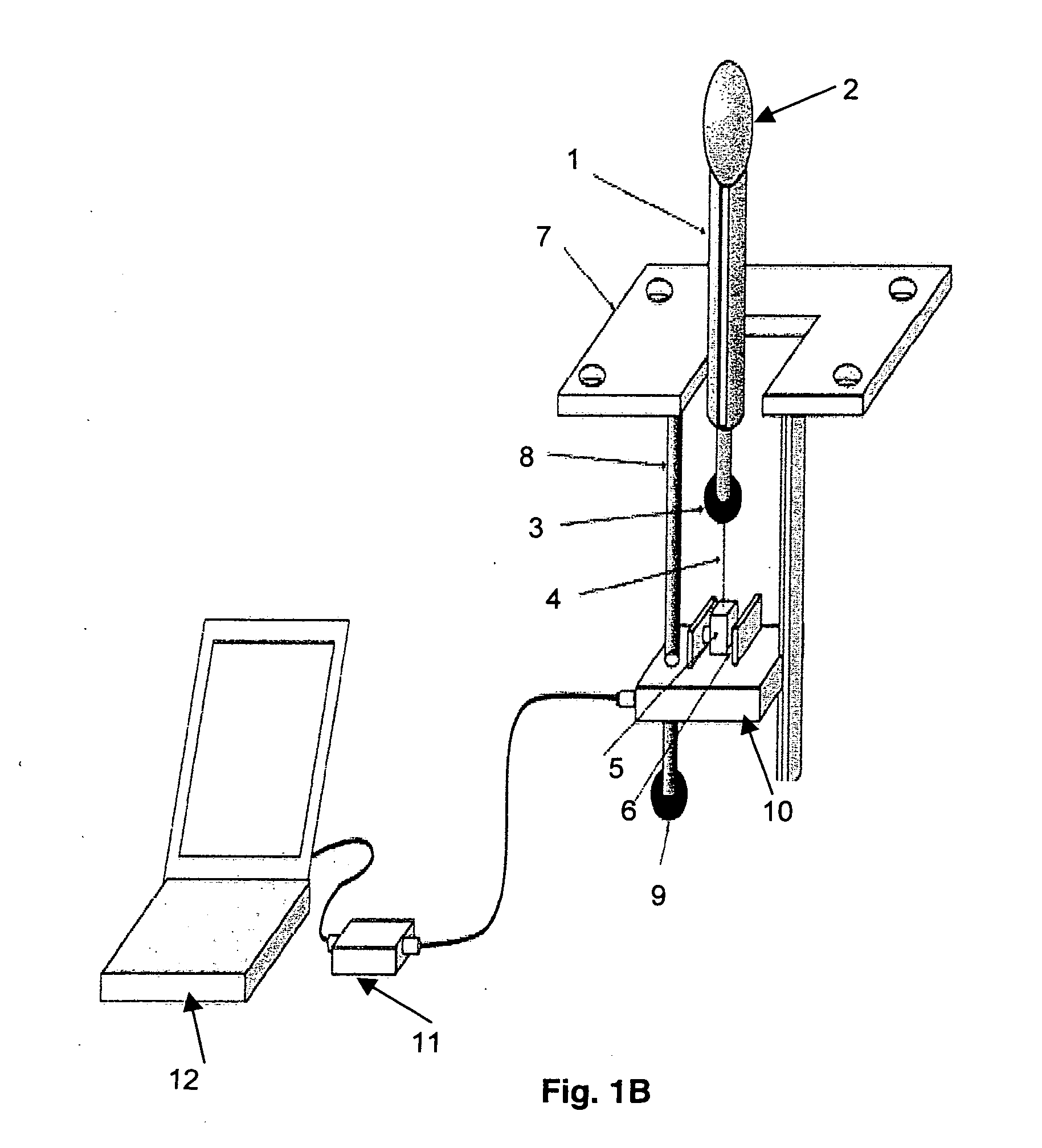
[0050] Elevator function of pelvic diaphragm causes cranial movement of vaginal high pressure zone (HPZ) during contraction but the manometry catheter may not necessarily move with it which can cause motion artifact in the side hole pressure recordings. The second problem with side-hole pressure recording technique is that the vaginal high pressure zone is distributed in a bell shaped manner, i.e., the maximum or peak pressure is located in the center of high pressure zone. It is extremely difficult to keep a side hole at the location of peak pressure, especially when it is moving in the cranio-caudal direction. Both of the limitations of side hole pressure recording technique can be overcome by a sleeve sensor, (3). Sleeve sensor is usually 6 cm in length and it records peak pressure along its length, irrespective of the high pressure zone location on the sleeve. The problem with sleeve sensor ho...

Example

EXAMPLE 2

Vaginal Probe Size and Vaginal Pressure Measurement

[0052]FIG. 5 illustrates an example of a catheter holder to hold the sleeve sensor used in the instant device. A standard size sleeve sensor can be placed inside the holder and the two (holder+sleeve sensor) are placed inside the vagina. Pressure sensor surface of sleeve sensor faces anterior direction (towards the pubic bone) if one wants to record the anterior vaginal pressure. Holders of different sizes are designed such that irrespective of the size of the holder, a standard 4 mm sleeve catheter fits into the holder. By using different size holders, one can increase the length of the puborectalis muscle and at the same time record pressure at rest and during pelvic floor muscle squeeze.

[0053] The following references are cited herein: [0054] 1. Constantinou, et al. Pressure components in the Female Urethra: Female Incontinence. New York, N.Y.: Allan R Liss Inc. 113-120 (1981). [0055] 2. Sapsford, et al. Arch. Phys. ...


PUM


Abstract

The instant invention provides a pelvic floor function diagnostic and therapeutic station, which includes a vaginal/ano-rectal plug and station. The important components of the station are a catheter holder with a pressure measuring (sleeve sensor or other alterative) to measure the constrictor function of the pelvic floor muscle, a vertical force transducer system to measure the elevator function of the pelvic floor muscle and a modular support table or a chair to hold the subject position. The diagnostic and therapeutic station thus allows for simultaneous measurement of the constrictor and elevator functions of the pelvic diaphragm. The station can also be used to treat impaired pelvic floor function via biofeedback therapy.

Description

BACKGROUND OF THE INVENTION [0001] 1. Field of the Invention [0002] The present invention relates generally to the field of diagnosis and therapy of pelvic floor muscle disorders. More specifically, the present invention relates to pelvic floor function diagnostic and therapeutic station that can be used for diagnosis and treatment of pelvic floor muscle disorders. [0003] 2. Description of the Related Art [0004] The pelvic floor muscles (PFM) form the floor of the pelvic basin and performs two major functions, an elevator function for pelvic organs and constrictor function for three orifices, urethra, vagina and anal canal. The elevator function of the pelvic floor supports the pelvic viscera and is probably important for the prevention of rectal, vaginal and bladder prolapse. Constrictor function, on the other hand is involved in the closure mechanism of urethra, vagina and anal canal. In individuals with normal pelvic muscle function, the pelvic floor muscle contracts simultaneous...

Claims


Application Information

Patent Timeline
IPC IPC(8): A61B5/103
CPCA61B5/04882A63B2230/60A61B5/486A61B5/6887A61B5/702A61N1/36007A63B71/0622A63B21/023A63B21/1488A63B23/20A63B2021/1609A63B2071/0683A63B2213/004A63B2220/51A63B2220/56A61B5/227A63B21/1609A63B21/4045A61B5/391
Inventor MITTAL, RAVINDER K.LIU, JIANMINNAGER, CHARLES W.
Owner RGT UNIV OF CALIFORNIA
Why Eureka
Try Eureka