X-ray device and image-processing method

a technology of x-ray and image processing, applied in the direction of instruments, patient positioning for diagnostics, applications, etc., can solve the problems of large quantity of information to be managed, certain calcium deposits or certain opacities are not spotted, and new tomosynthesis mammography devices have limitations, etc., to facilitate the detection of radiological signs, improve the quality of life, and facilitate the identification

Active Publication Date: 2007-08-16
GENERAL ELECTRIC CO
View PDF27 Cites 19 Cited by
  • Summary
  • Abstract
  • Description
  • Claims
  • Application Information

AI Technical Summary

Benefits of technology

[0014] The goal of the invention is precisely to overcome the drawbacks of the techniques explained here above. To this end, the invention proposes techniques for the acquisition of the projection images associated with image-processing methods used to facilitate the detection of radiological signs in the projection image, by means of series of 3D data. With these image-processing methods, the radiological signs are easier to identify in the projection image, thus enabling more efficient examination.

Problems solved by technology

However, it can happen that certain calcium deposits or certain opacities are not spotted.
In particular, since mammography images are the result of projections, they represent superimposed structures that disturb the visibility of the structures of the breast, sometimes leading to a falsely positive interpretation when a superimposition of these structures resembles opacity or to a falsely negative interpretation when the structures obscure the visibility of a lesion.
However, this new tomosynthesis mammography device has limitations.
Consequently, the quantity of information to be managed is very great.
Similarly, access to a piece of information having clinical interest takes much more time since this information is sought sequentially in the image volume.
Furthermore, this type of device has a methodology of use completely different from the methodology of currently used mammography devices.
These new methodologies with which the practitioners are unfamiliar have not yet been adopted.
This is chiefly due to the fact that these new methodologies have not yet been in existence for very long and that the clinical efficiency of these new devices has not yet been fully demonstrated.
However, the new tomosynthesis mammography devices cannot have a very high frequency of use since the time of access to the information of clinical interest is very great.
Furthermore, this type of device does not guarantee screening success because such success depends essentially on the time spent in locating information of clinical interest.
Another problem, which is more specific to mammography but could occur in other fields, is related to the need to be able to analyze radiological signs, which are clinically interesting between 100 μm and 500 μm.
This problem of spatial resolution is critical for tomosynthesis mammography devices.
The devices then cannot be used to obtain adequate image quality for very fine analysis of the radiology signs.

Method used


Image

  • X-ray device and image-processing method
  • X-ray device and image-processing method
  • X-ray device and image-processing method

Examples


Embodiment Construction

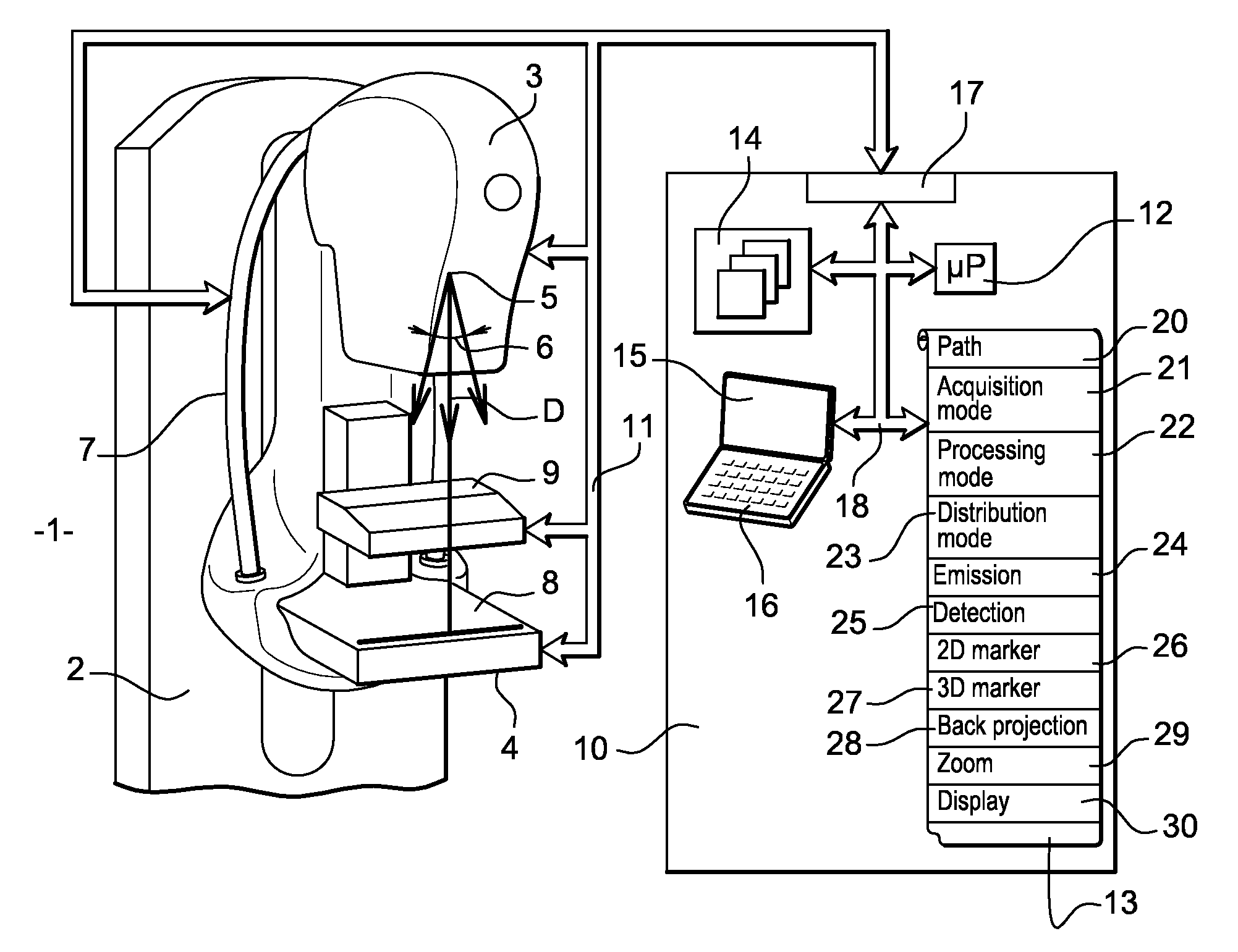
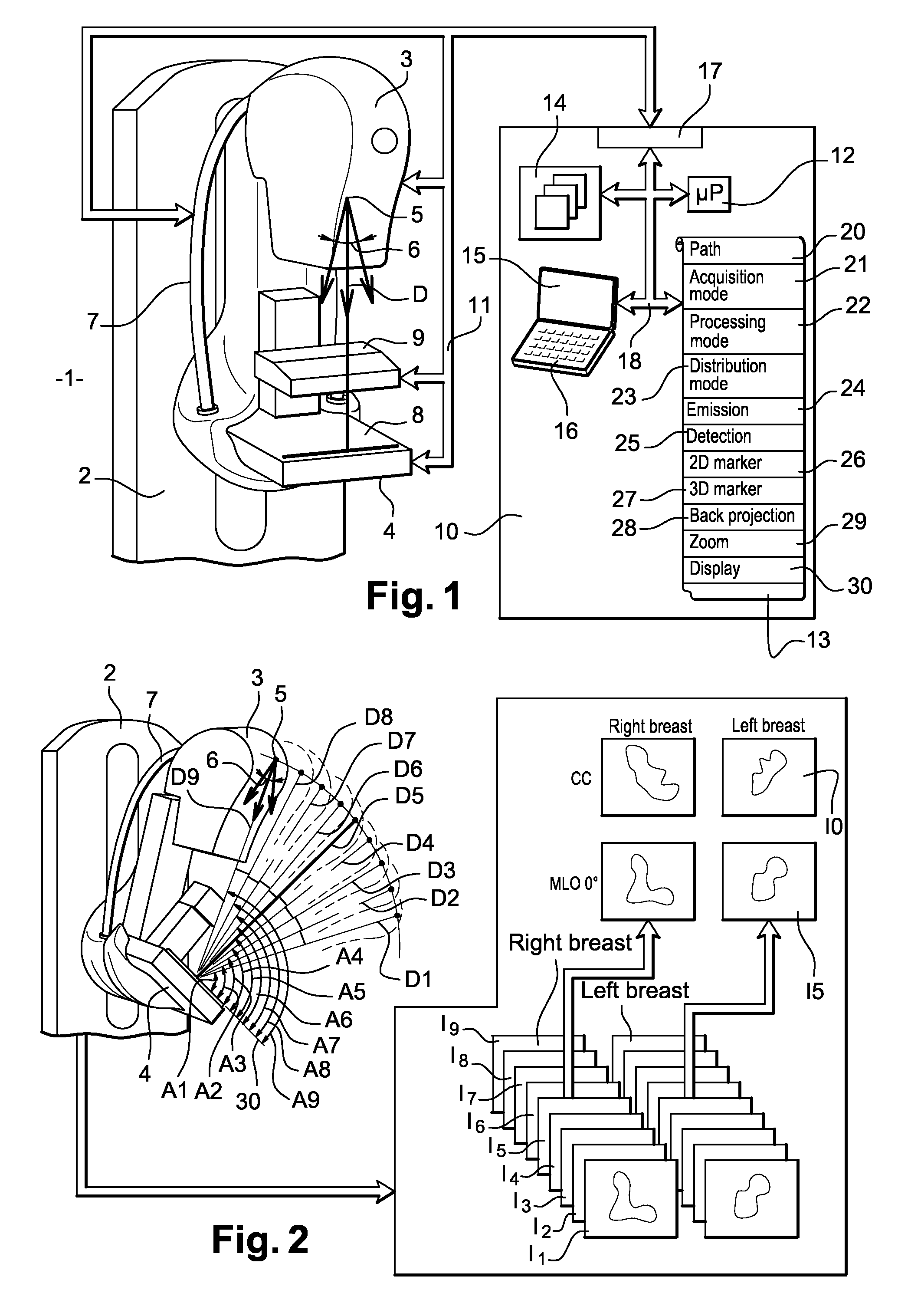
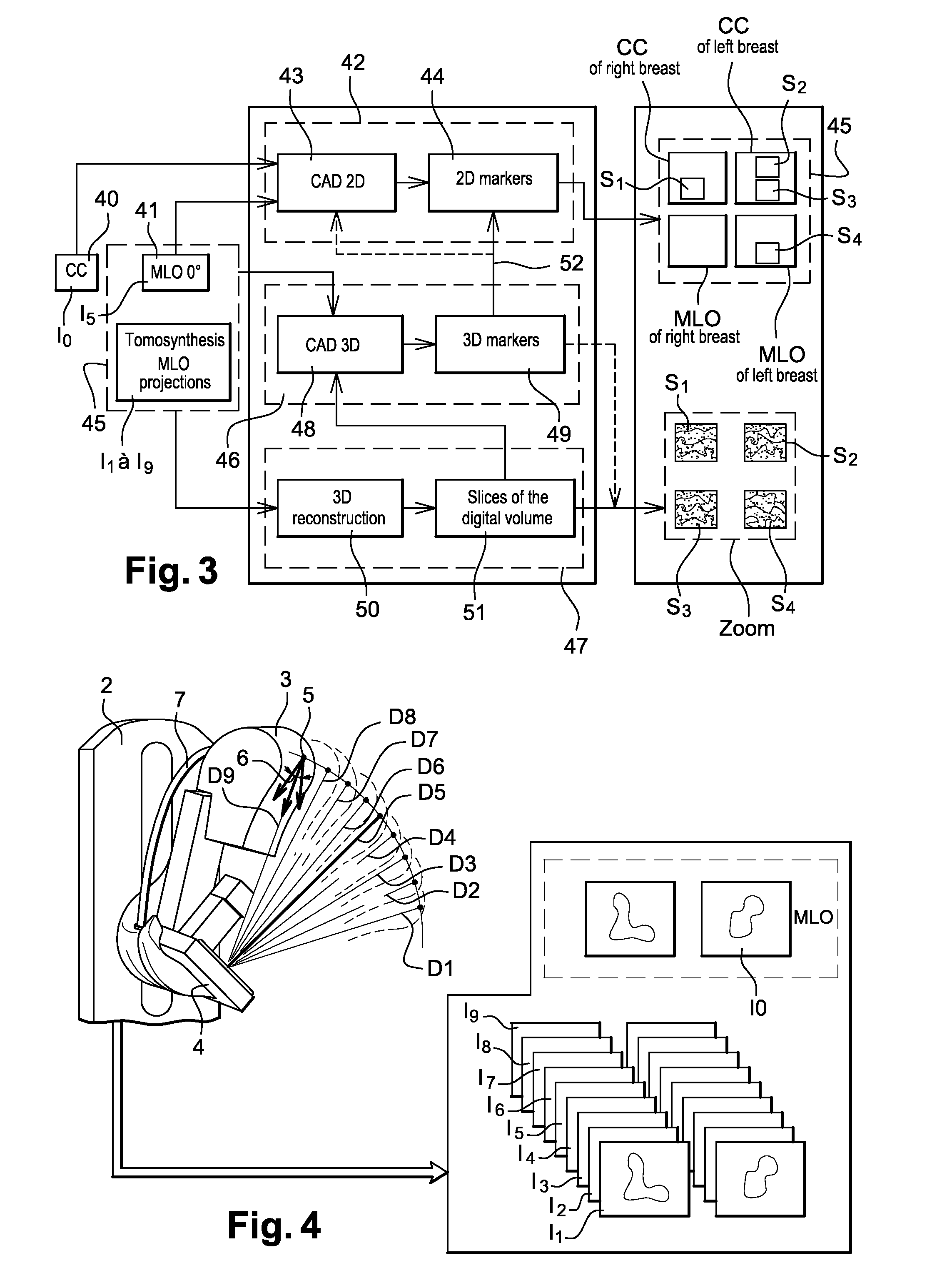
[0042]FIG. 1 shows an X-ray device, especially a mammography machine, according to the invention. This X-ray device 1 has a vertical column 2. On this vertical column, there is a hinged arm 7 bearing an X-ray-emitting tube 3 and a detector 4 capable of detecting the X-rays emitted by the tube 3. This arm 7 may be oriented vertically, horizontally or obliquely. The tube 3 is provided with a focus 5 which is the X-ray emitting focus. This focus 5 emits an X-ray beam 6 along the direction of emission D.

[0043] The arm 7 is hinged on the vertical column 2 in such a way that it enables the tube 3 to be shifted along a path in the shape of an arc of a circle while leaving the detector 4 immobile. Other arrangements are possible by which the tube can be shifted in a plane or in a sphere portion. The tube 3 can then occupy different positions distributed in swiveling between two extreme positions. These two positions are for example symmetrical to each other relative to the perpendicular to...


PUM


Abstract

An x-ray device comprises means for the production of at least one standard projection image of the object in which presumed suspect zones corresponding to radiological signs are represented by markers. The device comprises means for the production of a digital volume of markers in which 3D markers are created in order to represent presumed suspect zones of the object. It also comprises means of re-projection of the 3D markers in the standard projection image in order to confirm the presence of the markers or eliminate or add the markers of the projection image is necessary.

Description

CROSS REFERENCE TO RELATED APPLICATION [0001] This application claims priority to French Application No. 65 50565 filed on Feb. 16, 2006. FIELD OF THE INVENTION [0002] An object of the present invention is an X-ray device which, in one particular exemplary embodiment, is a mammography device. It can be applied to special advantage but not exclusively in medical imaging and in non-destructive X-ray controls. [0003] It is an aim of the invention to acquire at least one radiography image projection at the same time as a series of projections for tomosynthesis processing. [0004] It is another aim of the invention to improve the ergonomy of use of an X-ray device of this kind to make it both simpler and faster to use. [0005] It is also an aim of the present invention to set up a 3D display of any 2D zone of the projection image having clinical interest. DESCRIPTION OF THE PRIOR ART [0006] Today, mammography is widely used for the early detection of lesions associated with breast cancer. ...

Claims


Application Information

Patent Timeline
Patent Type & Authority Applications(United States)
IPC IPC(8): A61B6/04A61B6/00A61B6/02
CPCG06T2211/436G06T11/008
Inventor MULLER, SERGE LOUIS WILFRIDPETERS, GEROBERNARD, SYLVAINIORDACHE, RAZVAN GABRIEL
Owner GENERAL ELECTRIC CO
Why Eureka
Try Eureka