Reagents and methods for cancer prognosis and pathological staging
a cancer prognosis and pathological technology, applied in the field of reagents and methods for assessing colorectal cancer progression in an individual, can solve the problems of difficult development of robust diagnostic candidate biomarkers for targeted therapies, affecting normal cells as well as cancer cells, and affecting normal cell developmen
- Summary
- Abstract
- Description
- Claims
- Application Information
AI Technical Summary
Benefits of technology
Problems solved by technology
Method used
Image
Examples
example 1
Immunohistochemical Staining of Downstream Molecules in EGF Pathway in Colorectal Tumor Progression
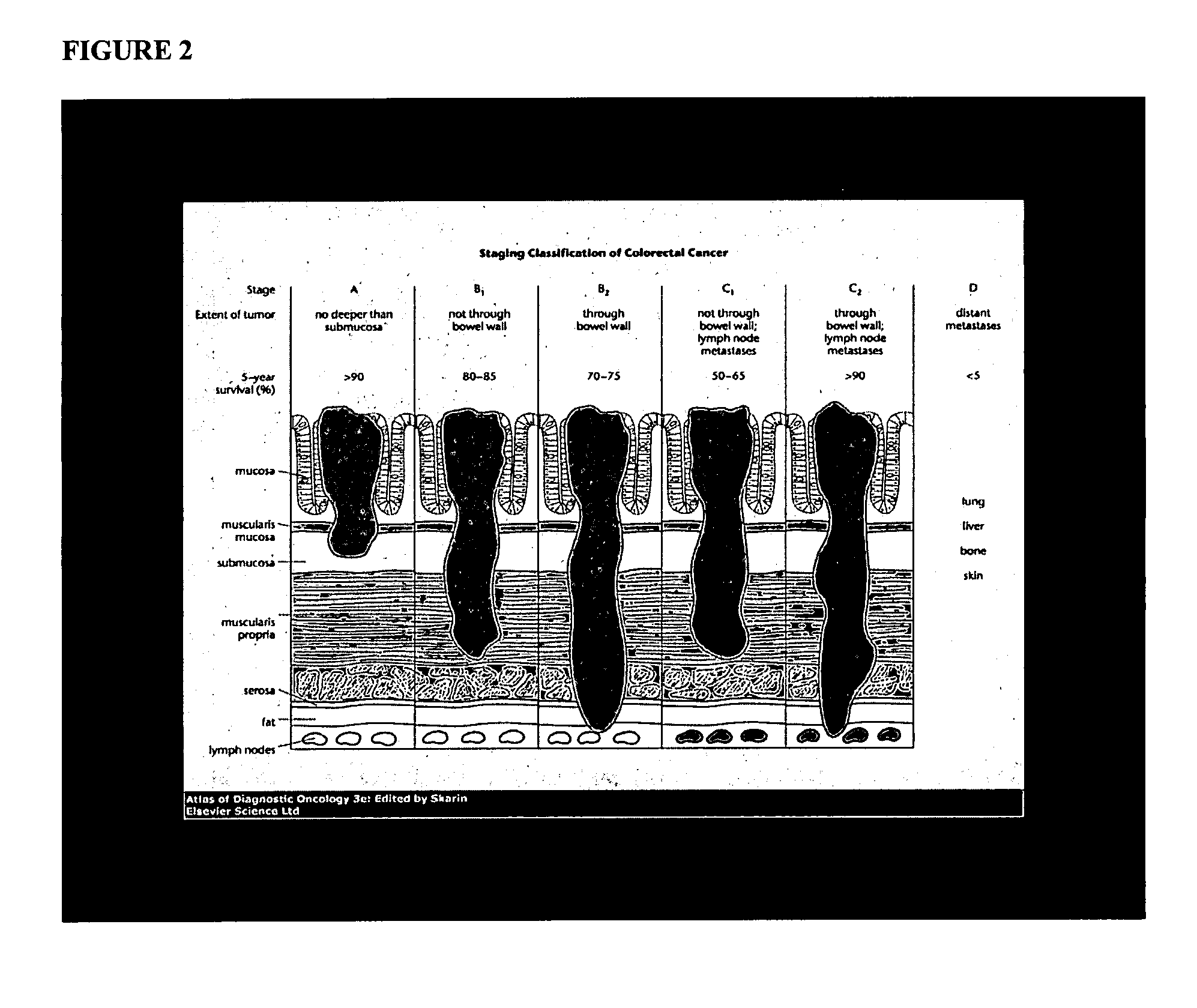
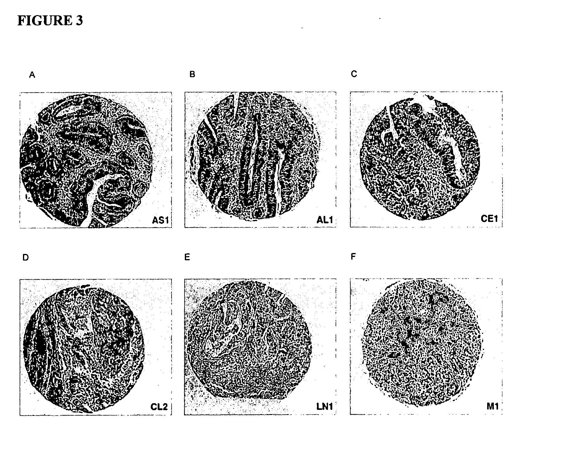
[0093] In order to determine whether biomarker profiles could be identified for colorectal cancer that correlate with pathology staging of carcinomas, the expression levels of biomarkers linked to the expression of EGFR in colorectal cancer cases was examined using a commercially available tissue array and tissue samples from individual cases. The biomarkers were assessed using immunohistochemistry (“IHC”).
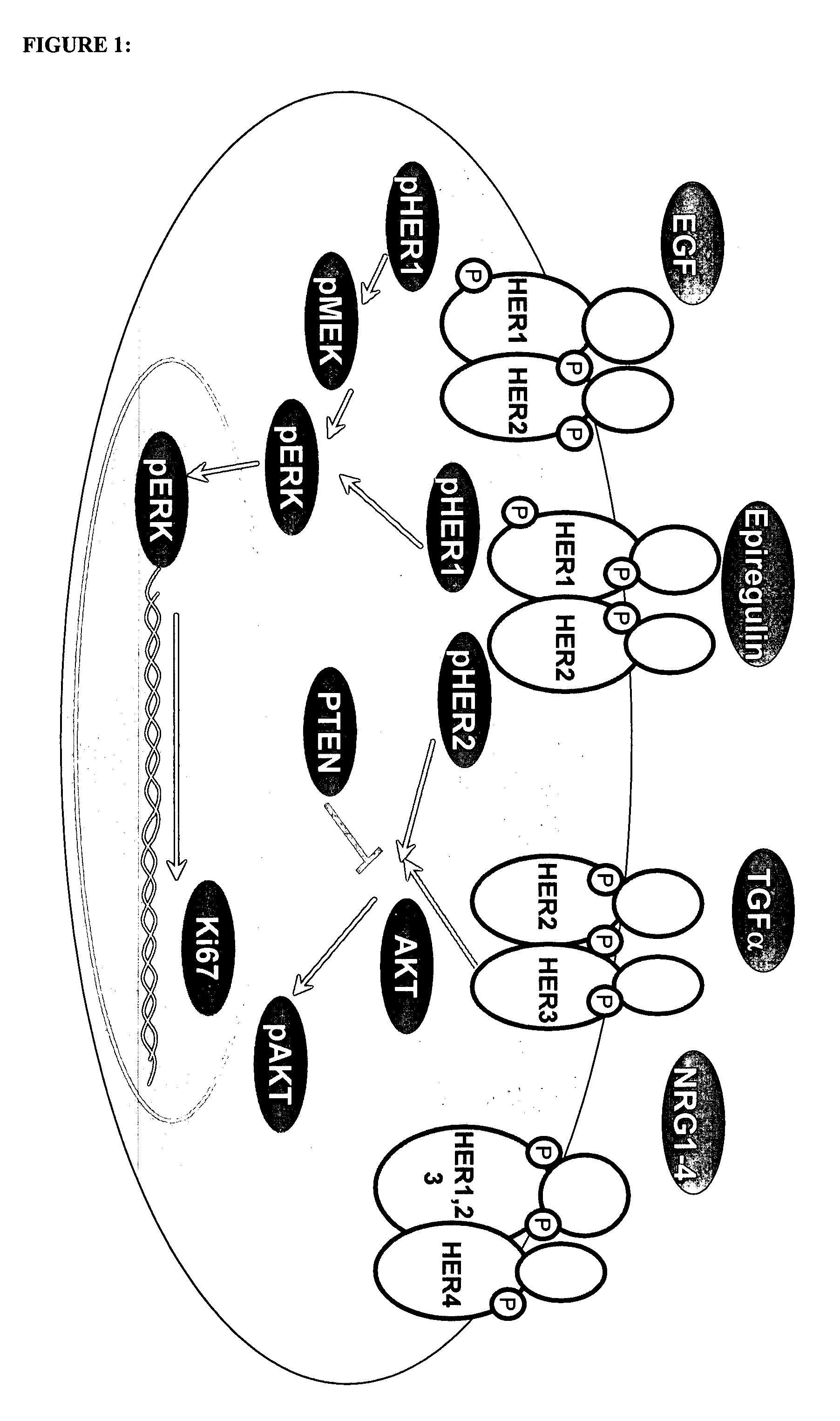
[0094] The Ventana Medical Systems' murine antibody clone, 3C6, was used to detect HER1 / EGFR expression by immunohistochemistry. The 3C6 clone reacts with the extra-cellular domain of the receptor. Other biomarkers investigated were pHER1, PTEN, pAKT, pMEK, Ki67, and pERK. pTYR was also assessed as a surrogate of activation. All reagents were used as described below and in TABLE 1, and according to specified package inserts.
[0095] The performance of probes and antibodies to detect...
example 2
Correlation Between EGFR Gene Copy Number and Colorectal Cancer Tumor Staging Using In Situ Hybridization
[0106] Fluorescent in situ hybridization (FISH) for EGFR was preformed on single slide sections from individual cases and a multi-tissue array in order to assess gene status in colorectal cancer progression.
[0107] Using dual color FISH, the number of EGFR gene copies per cell was evaluated in formalin-fixed, paraffin-embedded (FFPE) single-slide sections and in a multi-tissue array (FIG. 12). The details of the single slide sections and multi-tissue array are outlined in Example 1.
[0108] In-situ hybridization detection of the EGFR gene was conducted with either probes from, Ventana (Spectrum Orange labeled), Invitrogen (SPOTLight™-EGFR, DIG labeled), or Vysis (Spectrum Orange EGFR and Spectrum Green CEP 7 labeled probes). The EGFR probes from Ventana and Zymed were detected with fully automated protocols on the Ventana Discovery®XT. Detection of the Vysis probes was semi-autom...
example 3
Heterogeneity in Expression Levels of EGFR Pathway Molecules Within a Tumor
[0110] The expression levels of EGFR pathway molecules were assessed in colorectal cancer case study to evaluate possible heterogeneity in the expression levels of these molecules in a single tumor.
[0111] A 41 year old man presenting with rectal bleeding and abdominal cramping was diagnosied with a 3 cm rectal mass. Distal colon resection after radiotherapy revealed a well differentiated rectal adeno-carcinoma of stage T3. Single slide sections from various regions of the tumor were prepared as detailed in Example 1. IHC was performed to determine expression levels for EGFR, HER-2, pAKT, Ki67, Survivin and VEGF as described in Example 1. Survivan was detected with an antibody from Novus (NB500-201) by incubating for 2 hrs at room temperature. VEGF was detected using an antibody from Santa Cruz (SC-7269) by including for 1 hr at room temperature.
[0112]FIGS. 14-19 show that expression levels of tumor bio-mar...
PUM
| Property | Measurement | Unit |
|---|---|---|
| thick | aaaaa | aaaaa |
| acid | aaaaa | aaaaa |
| affinity | aaaaa | aaaaa |
Abstract
Description
Claims
Application Information
 - R&D
- Intellectual Property
- Life Sciences
- Materials
- Tech Scout
- Unparalleled Data Quality
- Higher Quality Content
- 60% Fewer Hallucinations
Browse by: Latest US Patents, China's latest patents, Technical Efficacy Thesaurus, Application Domain, Technology Topic, Popular Technical Reports.
© 2025 PatSnap. All rights reserved.Legal|Privacy policy|Modern Slavery Act Transparency Statement|Sitemap|About US| Contact US: help@patsnap.com


