
Fibrin sealant delivery device including pressure monitoring, and method and kits thereof

a delivery device and fibrin sealant technology, applied in the field of fibrin sealant, can solve the problems that the commercially available fibrin sealant devices lack desirable safety features that can benefit physicians and patients, and achieve the effects of reducing the amount of material, facilitating extended pain relief, and heightened safety

Inactive Publication Date: 2007-09-13
PAUZA KEVIN
View PDF23 Cites 68 Cited by
  • Summary
  • Abstract
  • Description
  • Claims
  • Application Information

AI Technical Summary

Benefits of technology

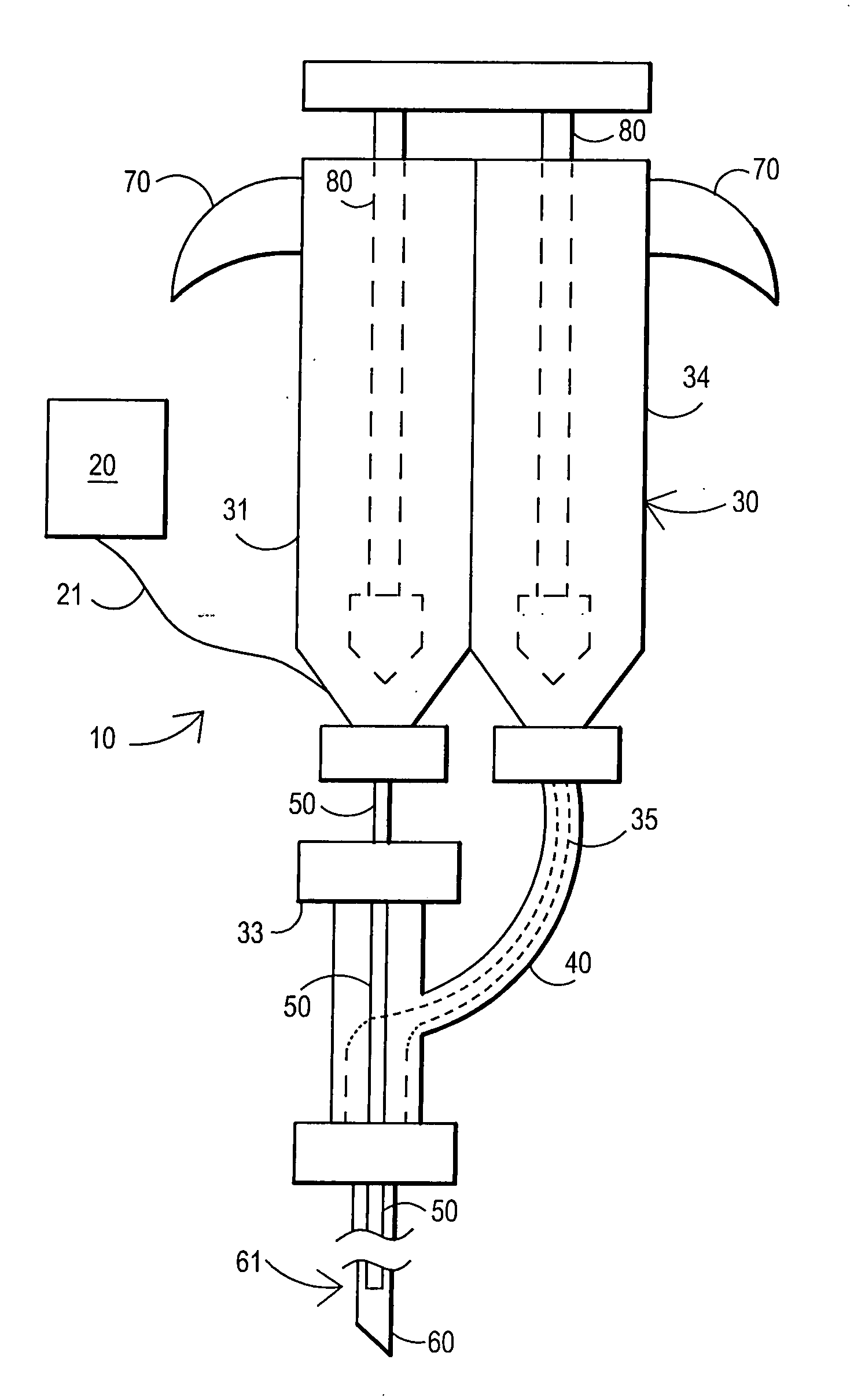
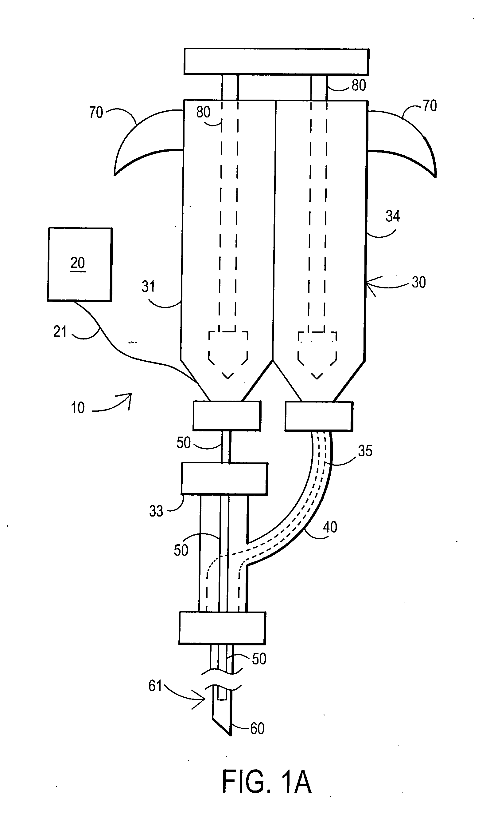
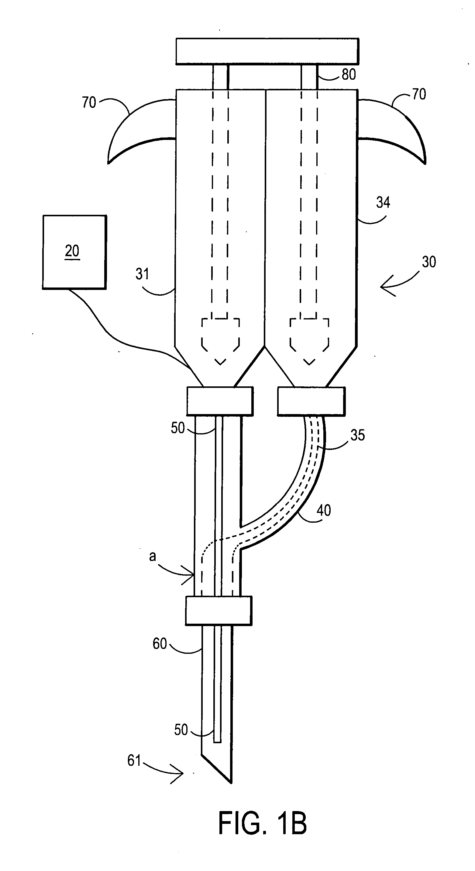
[0015] The method can be practiced so that the disc is injected with the biologic sealant at multiple positions of the disc. In one embodiment, the injecting occurs by inserting an introducer needle having a tip into the intra-discal space to a position adjacent to the at least one defect, inserting a second needle or a polymeric catheter through the introducer needle (optionally up to but not beyond the tip of the introducer needle), and injecting the biologic sealant through the second needle or polymeric catheter while monitoring the pressure of the sealant being injected. Alternatively, one component is injected through the introducer needle, and a second component of the biologic sealant is injected through the second needle or polymeric catheter. The invention thus includes a method of delivering a biologic sealant to a spinal area while monitoring the pressure of the fluid being delivered, wherein the pressure is monitored using an electronic pressure monitor. This invention provides improved safety for the patient as the surgeon can precisely monitor pressure of the fluid and thus, indirectly, the pressure in the disc thereby allowing the surgeon to avoid overpressurization of the disc that could lead to rupture or other damage.
[0022] The defect repaired during the practice of this invention can be a tear of the annulus fibrosus, a fissure in the annulus fibrosus, the fibrous capsule of a spinal joint and the like. This treatment serves to reduce the amount of material from the nucleus pulposus that leaks through the defect(s) in the annulus fibrosus, and or the potential in-growth of granular tissue and coincidental innervation which may be a source of pain not normally present in a healthy joint. Alternately, this treatment may insulate innervated granular tissue from the effects of nucleus pulposus. The presence of this innervated granular tissue sometimes found within the annulus at the site of an anular defect or tear, is believed to be a common physiologic healing response. Advantageously, injection of the fibrin sealant can also serve to restore normal disc (or joint) height and physiologic hydrostatic pressure, key components to disc health. It should be understood that normal physiologic hydrostatic pressure can vary from person to person, and that the treatment may produce near-normal hydrostatic pressure. As used herein, normal physiologic pressure encompasses this range of pressures. In one embodiment, neither the nucleus pulposus nor the annulus fibrosus has been heated in the body to stiffen the disc either prior to or concurrent with the injection, such as discussed in for example U.S. Pat. No. 6,095,149. In one embodiment, in the practice of this invention the nucleus pulposus has not been removed by surgery, such as in the case of a total or partial discectomy or by nucleoplasty for a herniated disc.
[0023] Advantageously, the method and kit of this invention facilitate extended pain relief for patients with discogenic pain, wherein for example nucleus pulposus leaks out of the disc through defects (e.g. tears or fissures) in the annulus fibrosus. The pressure monitor provides a heightened level of safety whereby the physician can measure pressure in real time so as to avoid over-pressurizing a disc being treated. Likewise, the physician can observe the pressure reading in conjunction with injection of the fibrin sealant to determine whether the disc is being sealed and whether sufficient fibrin sealant has been injected. In this way the physician can use the delivery device as a diagnostic tool to assess whether the disc is treatable.
[0024] Additionally, the method and kit of this invention facilitate extended pain relief for patients with other spinal joint pain, wherein for example of the potential in-growth of granular tissue and coincidental innervation which may be a source of pain not normally present in a healthy joint. Alternately, this treatment may insulate innervated granular tissue from the effects of nucleus pulposus.

Problems solved by technology

However, the inventors have recognized that a problem exists in that existing commercially available fibrin sealant devices lack desirable safety features that can benefit physicians and patients.

Method used


Image

  • Fibrin sealant delivery device including pressure monitoring, and method and kits thereof
  • Fibrin sealant delivery device including pressure monitoring, and method and kits thereof
  • Fibrin sealant delivery device including pressure monitoring, and method and kits thereof

Examples


example 1

Fluoroscopic Guided Intra-Discal Injection

[0093] After sterile preparation, an introducer needle is advanced in oblique projection to a superior articular process. A curved spinal needle is advanced through the introducer needle into the disc. Both anterior-posterior and lateral fluoroscopic projections are used to confirm proper needle placement. If the needle placement needs to be adjusted, placement is again confirmed fluoroscopically. A contrast agent is injected to confirm needle placement. In patients with chemical radiculitis, the contrast agent can be observed to be leaking through the annular fissures and / or intra-discal pathology, thus permitting their identification. Once the needle is properly positioned in the intra-discal space, the fibrin sealant (or its components) is injected using the syringe system of this invention having a pressure monitor. Pressure is monitored to ensure that the disc is not over-pressurized. The fibrin sealant is observed to force the contras...


PUM


Abstract

Apparatus for delivering biologic sealant device that includes a pressure monitor coupled to the delivery device to measure pressure within the device. A method of treating a disc using the device as well as a kit including the device is described.

Description

[0001] This application claims priority to U.S. provisional application No. 60 / 623,600, filed Oct. 29, 2004 and is a continuation-in-part of U.S. application Ser. No. 11 / 205,760, filed Aug. 17, 2005, of U.S. application Ser. No. 11 / 205,784, filed Aug. 17, 2005, and of U.S. application Ser. No. 11 / 205,775, filed Aug. 17, 2005, and to U.S. provisional application No. 60 / 764,019, filed Feb. 1, 2006, and to U.S. provisional application No. 60 / 854,413, filed Oct. 24, 2006, all of which are incorporated herein by reference.BACKGROUND OF THE INVENTION [0002] The present invention relates generally to the use of fibrin sealant whereby the sealant is delivered such as by injection to the spinal area, and more particularly through use of a delivery device that includes a pressure monitor. [0003] Fibrin sealants, and glues, are well known and are used extensively in various clinical settings. Such sealants are indicated as adjuncts to hemostasis in surgeries when control of bleeding by convent...

Claims


Application Information

Patent Timeline
Patent Type & Authority Applications(United States)
IPC IPC(8): A61M5/19
CPCA61B17/00491A61M2005/3201A61B2019/464A61M5/19A61M5/2425A61M5/31575A61M5/3158A61M5/31581A61M5/31585A61M5/31593A61M5/31596A61M5/486A61M25/0032A61M2005/3114A61M2005/3152A61B2017/00495A61B2090/064
Inventor RICHARDS, MARKBURKINSHAW, BRIAN D.PAUZA, KEVINROGAN, JAMES B.
Owner PAUZA KEVIN
Learn More