
Method for deploying a medical device

Inactive Publication Date: 2007-09-27
ETHICON ENDO SURGERY INC
View PDF9 Cites 40 Cited by
  • Summary
  • Abstract
  • Description
  • Claims
  • Application Information

AI Technical Summary

Benefits of technology

[0017] In one embodiment, the method includes the steps of inserting a flexible grasping instrument (such as a flexible forceps) into a proximal opening of a working channel of the endoscope; extending the flexible grasping instrument through a distal opening of the working channel of the endoscope; grasping the distal portion of the flexible element

Problems solved by technology

As a result, the accessory devices used by a physician can be limited in size by the diameter of the accessory channel of the scope used.
Additionally, the physician may be limited to a single accessory device when using the standard endoscope having one working channel.
One disadvantages of such large diameter / multiple working channel endoscopes can be that such devices can be relatively expensive.
Further, such large diameter / multiple working channel endoscopes can have an outer diameter that makes the endoscope relatively stiff, or otherwise difficult to intubate.
One disadvantage of known systems is the potential for the distal end of a device used externally of an endoscope to move, which may cause the accessory to lack precision or the ability to be maintained within a desired field of view of the imaging capability of the endoscope.
Such a non-circular endoscope may be disadvantageous from the point of view of cost, complexity, or ease in cleaning / sterilization.

Method used


Image

  • Method for deploying a medical device
  • Method for deploying a medical device
  • Method for deploying a medical device

Examples


Embodiment Construction

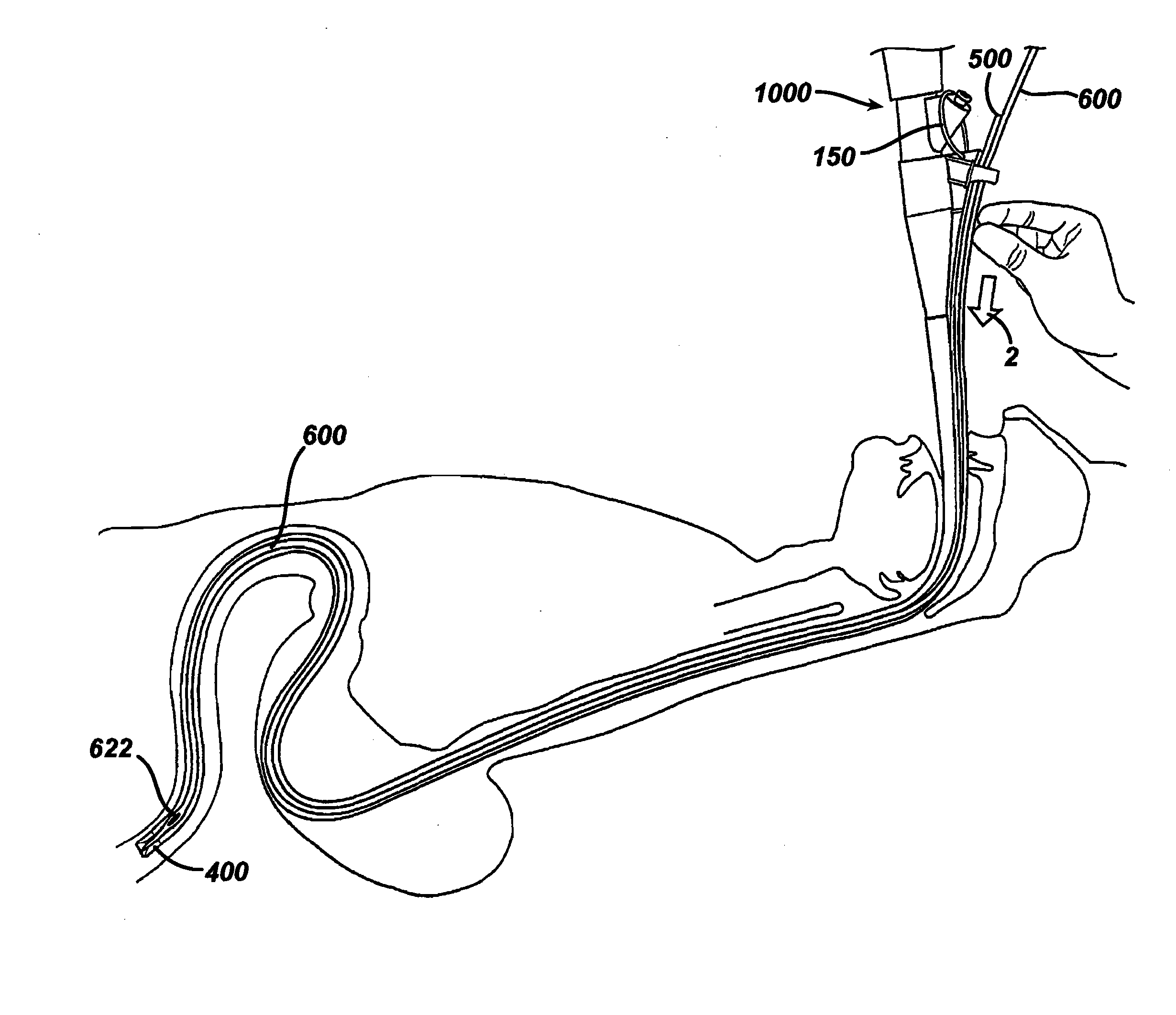
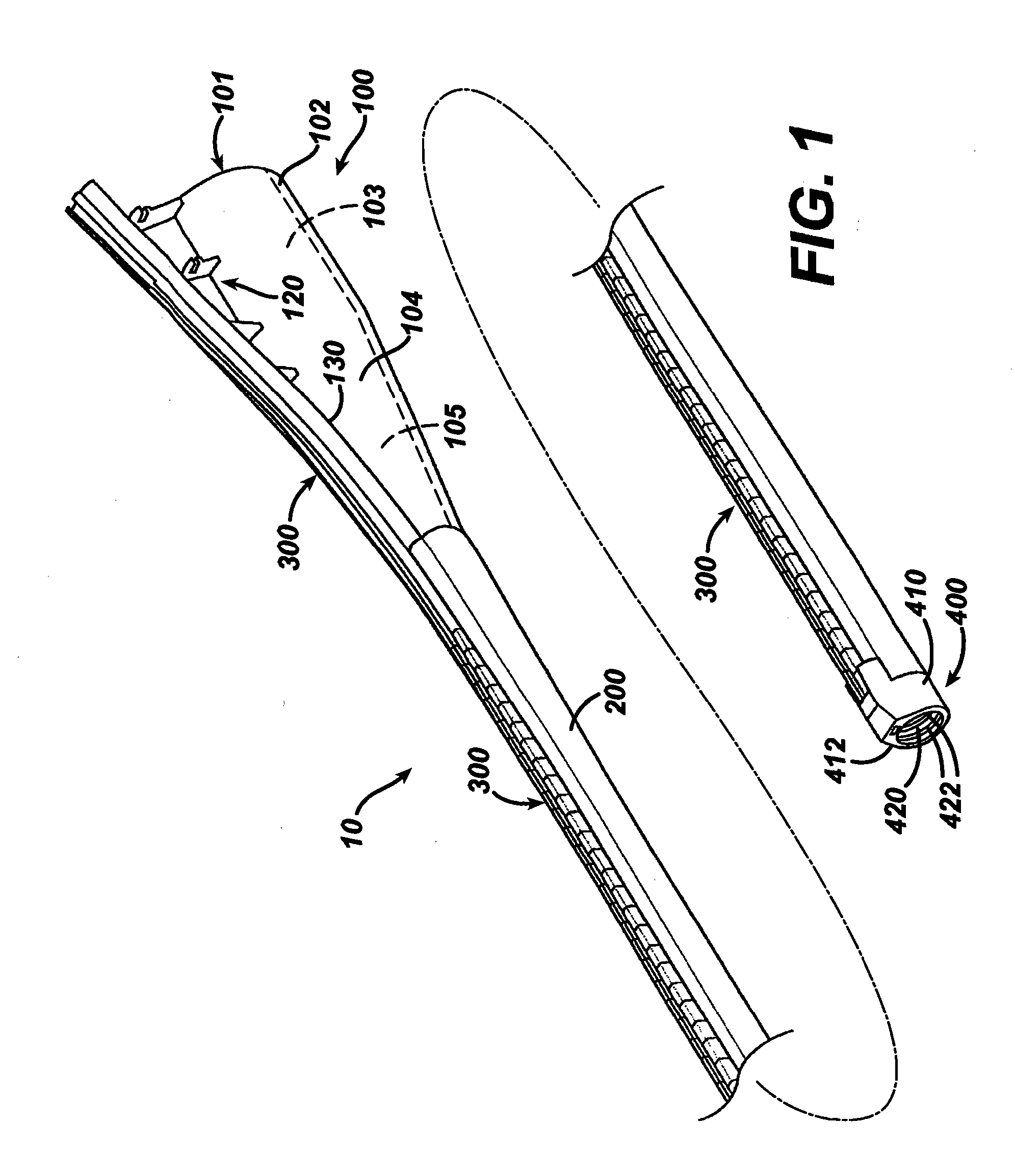
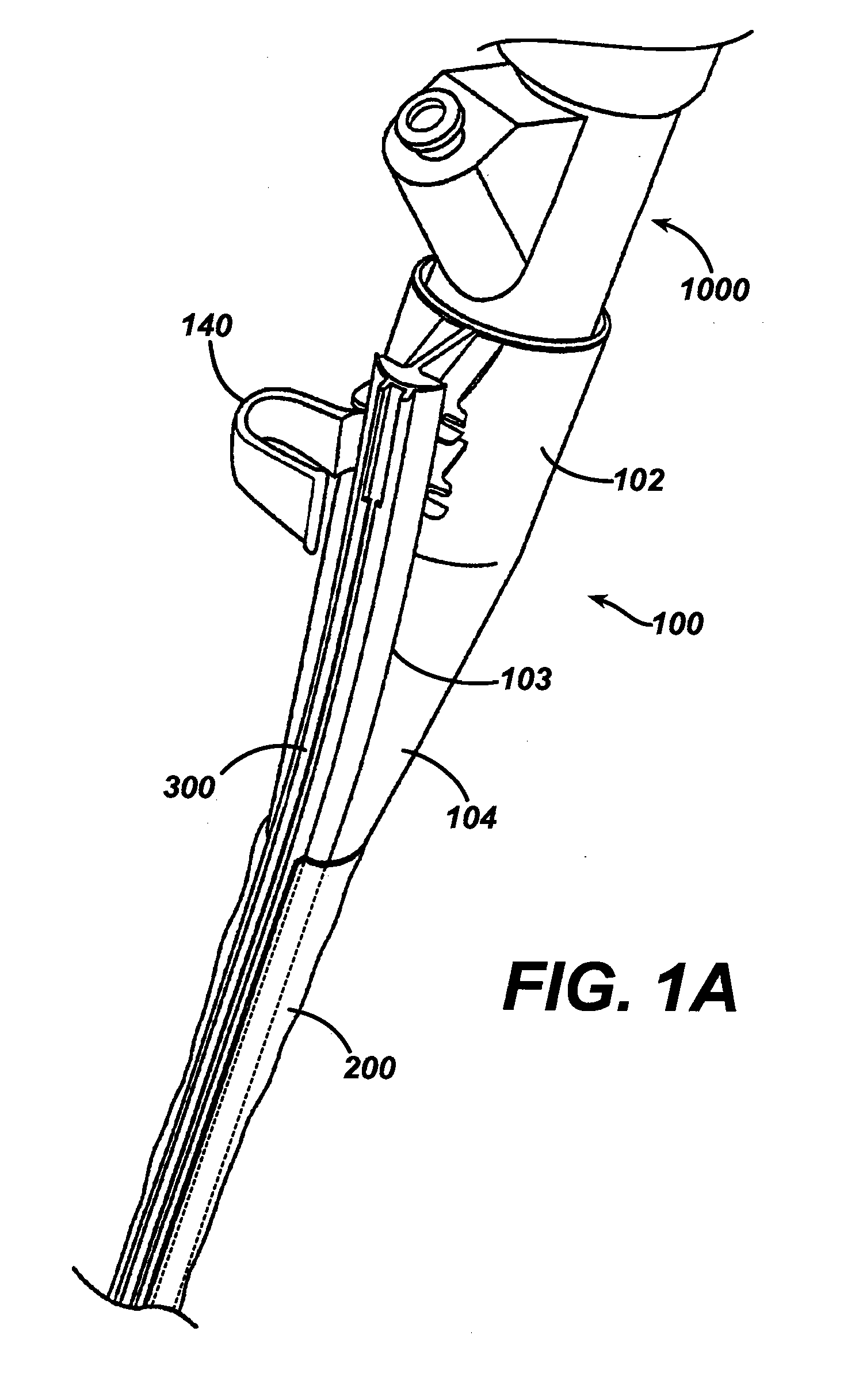
[0081]FIGS. 1-49 are included in U.S. Ser. No. 11 / 128,733 filed May 13, 2005, to which this application claims priority. FIGS. 1 and 2 illustrate a medical apparatus 10 according to one embodiment of the present invention. In one embodiment, apparatus 10 can include a handle 100, a flexible catheter or sheath 200 extending from handle 100, a flexible track 300 disposed on the sheath 200, and an endcap 400 disposed at the distal end of sheath 200. Handle 100 and flexible sheath200 can each be sized to receive an endoscope therethrough.

[0082] Apparatus 10 can also include a carrier 500 which is adapted to slidably engage track 300, as shown in FIG. 2. Endcap 400 can be sized and shaped to engage the distal end of an endoscope, such as an endoscope 1000 as shown in FIG. 2. Endoscope 1000 can be any commercially available endoscope, such as a gastroscope or colonoscope having an articulating distal section, and including a viewing element 1100 and a working channel 1200. Any suitable e...


PUM


Abstract

A medical apparatus and method useful for positioning a tubular member such as a feeding tube, is disclosed. In one application, the method can include providing a flexible element releasably coupled to the feeding tube, positioning the endoscope within the patient's body, positioning the feeding tube at a desired location in the patient's body; and retracting the flexible element and the endoscope from the patient's body while maintaining the feeding tube in the desired location.

Description

CROSS REFERENCE TO RELATED APPLICATIONS [0001] This application incorporates by reference and is a continuation in part of, and claims priority to U.S. Ser. No. 11 / 128,733 filed May 13, 2005, which is a continuation in part of U.S. Ser. Nos. 10 / 440,957 (published as US 2004 / 0230095); 10 / 440,660 (published as US 2004 / 0230096) and U.S. Ser. No. 10 / 440,956 (published US 2004 / 0230097); each filed May 16, 2003.FIELD OF THE INVENTION [0002] The present invention is related generally to medical devices and more particularly to devices and methods useful in endoscopic procedures. BACKGROUND OF THE INVENTION [0003] Minimally invasive procedures are desirable because such procedures can reduce pain and provide relatively quick recovery times as compared with conventional open medical procedures. Many minimally invasive procedures are performed with an endoscope (including without limitation laparoscopes). Such procedures permit a physician to position, manipulate, and view medical instruments...

Claims


Application Information

Patent Timeline
IPC IPC(8): A61B1/273
CPCA61B1/00073A61B1/00135A61B1/00154A61B1/018A61B1/2736A61B17/3415A61J15/0019A61J15/0007A61J15/0026A61J15/0069A61J15/0073A61J15/0061A61J15/0023A61J15/003
Inventor STEFANCHIK, DAVID
Owner ETHICON ENDO SURGERY INC
Features
  • Generate Ideas
  • Intellectual Property
  • Life Sciences
  • Materials
  • Tech Scout
  • Unparalleled Data Quality
  • Higher Quality Content
  • 60% Fewer Hallucinations
Learn More