Shape-sensing expandable member

a technology of expandable members and shape sensors, which is applied in the field of minimally invasive medical devices, can solve the problems of inability to characterize the space within the body, inaccurate limited use of information that is conveyed back to the practitioner, so as to minimize the undesired side effects of perennial lacerations, maintain and increase inflation, and maintain the effect of inflation control

Active Publication Date: 2009-01-01
NIDUS MEDICAL
View PDF4 Cites 90 Cited by
  • Summary
  • Abstract
  • Description
  • Claims
  • Application Information

AI Technical Summary

Benefits of technology

[0009]After receiving the sensory information from the inflated expandable device and after processing that information, an implant customized for the target region, or one otherwise appropriate for the target region, can be selected and delivered to the target region. By example, a stent, occlusion device, or other prosthetic can be selected to fit the target region and delivered to the region and deployed. Also by example, where a biotherapeutic material (e.g. a gel or therapeutic emulsion) is to be delivered to a target region, the expandable apparatus may provide sufficient information as to the volume of the space so as to know how much of the semi-solid agent is needed to fill the region. Thus, the apparatus is a useful adjunct for any minimally invasive surgical therapy or other minimally invasive procedure, especially where the procedure is better facilitated by knowing the size of the space into which the implant is directed.
[0010]The apparatus comprises an expandable member that can be inflated within the body using, for example, an inflation lumen for infusing fluid or gas into the expandable member. Thus, the member can be an expandable member such as a balloon adapted to expand by inflation of liquid or gas. The expandable member is delivered in a deflated state to the target region. The expandable member may be compliant so that the shape it takes on upon inflation can conform to the space of the target region. Optimally, the inflated expandable member may substantially fill a volume of the space at the target region, in order that the dimensional information relayed about the space is as accurate as possible.
[0015]Once the apparatus is placed in the target region and expanded, the transduction mechanisms allow for the accurate determination of the distance between any two sensors. With multiple transducers sensing distances between multiple other transducers, a picture of the volume and space of the region can be developed. For the purpose of relaying sufficient information to a microprocessor to generate an accurate picture of the space and volume of the target region, the expandable member will have a plurality of transducers positioned within the member or outside the member. In some embodiments, the apparatus will most optimally have several internal transducers, and at least one external transducer. Each transducer relates to at least one other transducer within the expandable member or to the one or more external transducers, so that information regarding such parameters as the distance between two transducers can be relayed to a microprocessor. Optimal sampling of information may be achieved where every transducer communicates with every other transducer to develop sufficient sensory information so that accurate characterization of the three-dimensional space within the target region is achieved.
[0020]The apparatus may typically have mechanisms for controlling various aspects of the apparatus. Mechanisms for controlling the apparatus elements may typically reside outside the body during any such minimally invasive procedure. One type of controlling mechanism is a handle external to the body. A control mechanism can be available to the system for controlling various aspects of the apparatus such as steering the expandable member to the target region, inflating the member with air or fluid such as saline and also deflating the member and withdrawing the member. In addition, a control mechanism can control or readjust transducer position in some or all of the transducers. It is possible that the apparatus will be positioned in the space of the target region, and expanded, and a first set of readings taken from the transducers. Thereafter, some slight repositioning of the apparatus can occur, in order to take another set of measurements that can be averaged with the first set. The handle, or other control means, can be used to facilitate this movement of the apparatus, and request for a second (or subsequent readings) from the transducers.
[0021]In some embodiments, it may be desirable to maintain control on the degree of inflation of the member, e.g. if the cavity or space is expanding, for example during labor in childbirth. In this instance, the apparatus may include mechanisms for maintaining an expanding pressure on the expandable member, particularly while the space itself is expanding. Accordingly, the apparatus could be configured to maintain a certain expanding pressure on the expandable member. With the maintenance of pressure on the space by continuously (albeit slowly) inflating the expandable member, the apparatus can maintain its position within a space that is expanding, even while it continues to relay information about the volume of the space to the microprocessor. An apparatus with this continuously expanding capability could be placed at the cervical opening during child birth to monitor the cervical dilation preceding vaginal delivery.
[0022]Another usefulness of the ability of the apparatus to maintain and increase the inflation on the expandable member would be to exert pressure in a space and cause desired physiological response thereby. For example, the inflating apparatus could be used not only to measure an expansion of the cervical or vaginal space, but also to gently expand the cervix to minimize the undesired side effects of perennial lacerations which can occur when the cervix expands quickly.

Problems solved by technology

However, due to the nature of minimally invasive procedures a physician typically relies on additional technology to gather the needed information about the target location while conducting the procedure.
A major limitation of many of these visualization technologies is their inability to characterize the space within the body.
Many of these systems image two-dimensional planes or create two-dimensional projections of a three-dimension object, and therefore the information that is conveyed back to the practitioner can be of limited use or is inaccurate with respect to the three-dimensional character of the space.

Method used


Image

  • Shape-sensing expandable member
  • Shape-sensing expandable member
  • Shape-sensing expandable member

Examples


Embodiment Construction

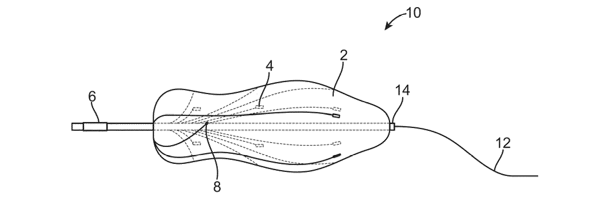
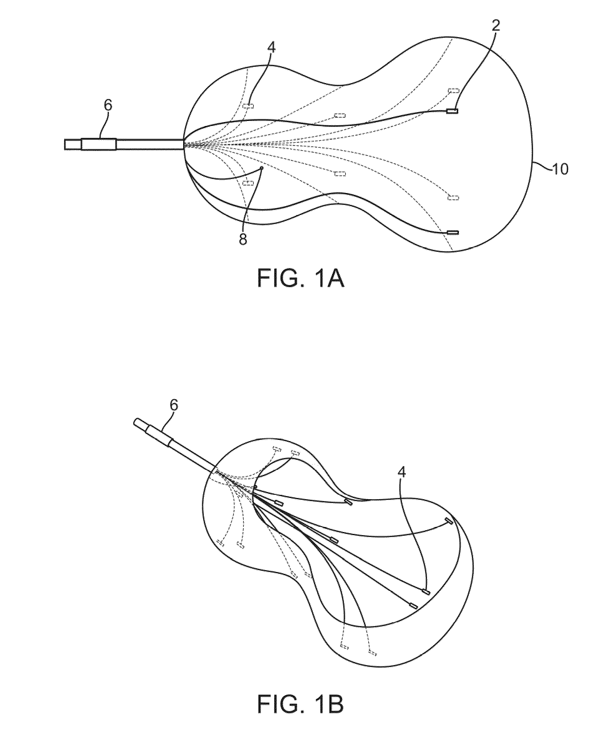
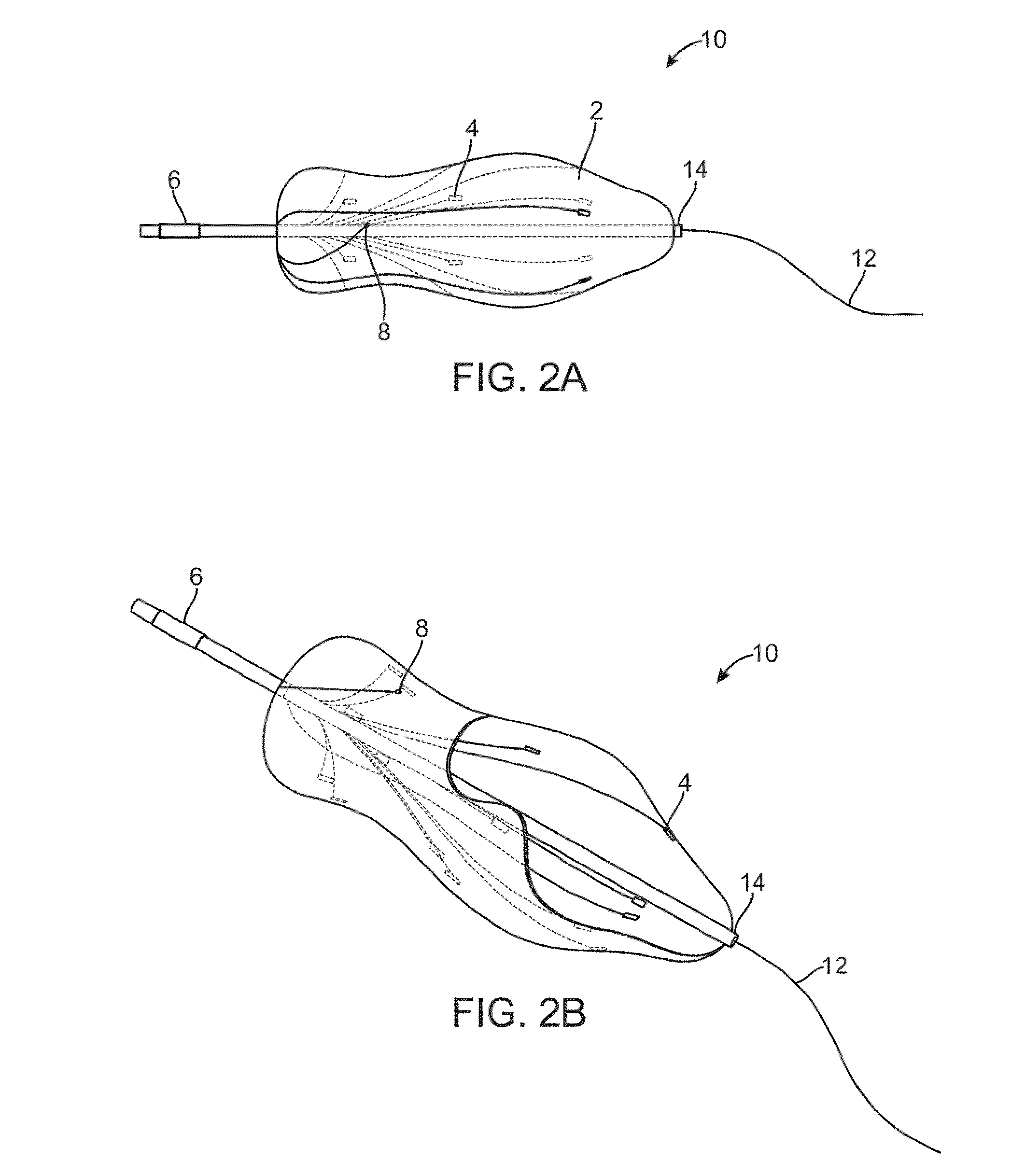
[0043]FIGS. 1A and 1B show side and perspective views, respectively, of an expandable member 10 in its inflated state. Expandable member 10 may be made out of highly distensible elastomeric compliant material such as latex, an elastomeric block copolymer, or a thermoplastic elastomeric material, and therefore adapted to inflation with fluid or gas. The expandable member 10 can also be made out of other materials that are non-compliant, such as PET (polyethylene terpthalate), polyurethane or silicone. In this case, where the material is not expandable the member may have excess material when deflated and that material is fillable with the fluid or gas infusion medium. Expandable member 10 is generally designed to be conformable to the shape of a hollow body cavity, and will in any event occupy a three-dimensional space upon expansion. In order that the expandable member optimally sense the volume and other three-dimensional characteristics of the space within a target region of the b...


PUM


Abstract

An expandable member for characterizing a three-dimensional space within a mammalian body is described. The expandable member is delivered to a target region in a deflated state where it is expanded by inflation of the member. Sensory transducers that contact the member relay sensory information generated when the member is in an expanded state to a microprocessor located outside the body. Using the sensory information, a data-driven picture that characterizes the three-dimensional space within the body is created with a microprocessor. The apparatus is useful in preparation for minimally invasive surgical therapy.

Description

CROSS-REFERENCE TO RELATED APPLICATIONS[0001]This application claims priority to U.S. Prov. Pat. App. 60 / 914,014 filed Apr. 25, 2007, which is incorporated herein by reference in its entirety.FIELD OF THE INVENTION[0002]The present invention relates generally to a minimally invasive medical apparatus for determining characteristics of a space within a mammalian body and methods of using this apparatus.BACKGROUND OF THE INVENTION[0003]Minimally-invasive surgical procedures and interventions reduce recovery time and post-operative pain by reducing the size of the incision and intra-operative blood loss. However, due to the nature of minimally invasive procedures a physician typically relies on additional technology to gather the needed information about the target location while conducting the procedure. Current technology to support visualization during minimally invasive procedures includes imaging systems in the visual light spectrum, ultrasound imaging, infrared imaging, and x-ray...

Claims


Application Information

Patent Timeline
Patent Type & Authority Applications(United States)
IPC IPC(8): A61B6/12
CPCA61B5/1076A61B5/1077A61B5/435A61B2562/168A61B2562/046A61B2562/164A61B2562/0209A61B2562/0215
Inventor SAADAT, VAHIDMALCHANO, ZACHARY J.
Owner NIDUS MEDICAL
Why Eureka
Try Eureka