Medical image diagnostic apparatus, picture archiving communication system server, image reference apparatus, and medical image diagnostic system

a diagnostic apparatus and image technology, applied in the field of picture archiving communication system server, image reference equipment, medical image diagnostic equipment, etc., can solve the problems of insufficient information described above to determine the test method, the inability to acquire information required for the present test, and the inability of test technicians to figure out information on the photographing position, the reconstruction range, etc., to achieve efficient utilization of shared information and high precision

Active Publication Date: 2007-10-11
TOSHIBA MEDICAL SYST CORP
View PDF6 Cites 50 Cited by
  • Summary
  • Abstract
  • Description
  • Claims
  • Application Information

AI Technical Summary

Benefits of technology

[0014] The present invention is designed in consideration of the above situation, and it is an object of the present invention to provide a medical image diagnostic apparatus, a picture archiving communication system server, an image reference a...

Problems solved by technology

However, information described above may not be sufficient to determine a test method, a range/direction to be photographed, and a photographing condition.
However, a known medical image diagnostic system has the following problems.
Accordingly, there occurs a problem that information required for the present test cannot be acquired, there is an item set by imagination based on a past image or the like, or a photographing condition where the same sett...

Method used


Image

  • Medical image diagnostic apparatus, picture archiving communication system server, image reference apparatus, and medical image diagnostic system
  • Medical image diagnostic apparatus, picture archiving communication system server, image reference apparatus, and medical image diagnostic system
  • Medical image diagnostic apparatus, picture archiving communication system server, image reference apparatus, and medical image diagnostic system

Examples


first embodiment

[0054] (Medical Image Diagnostic System)

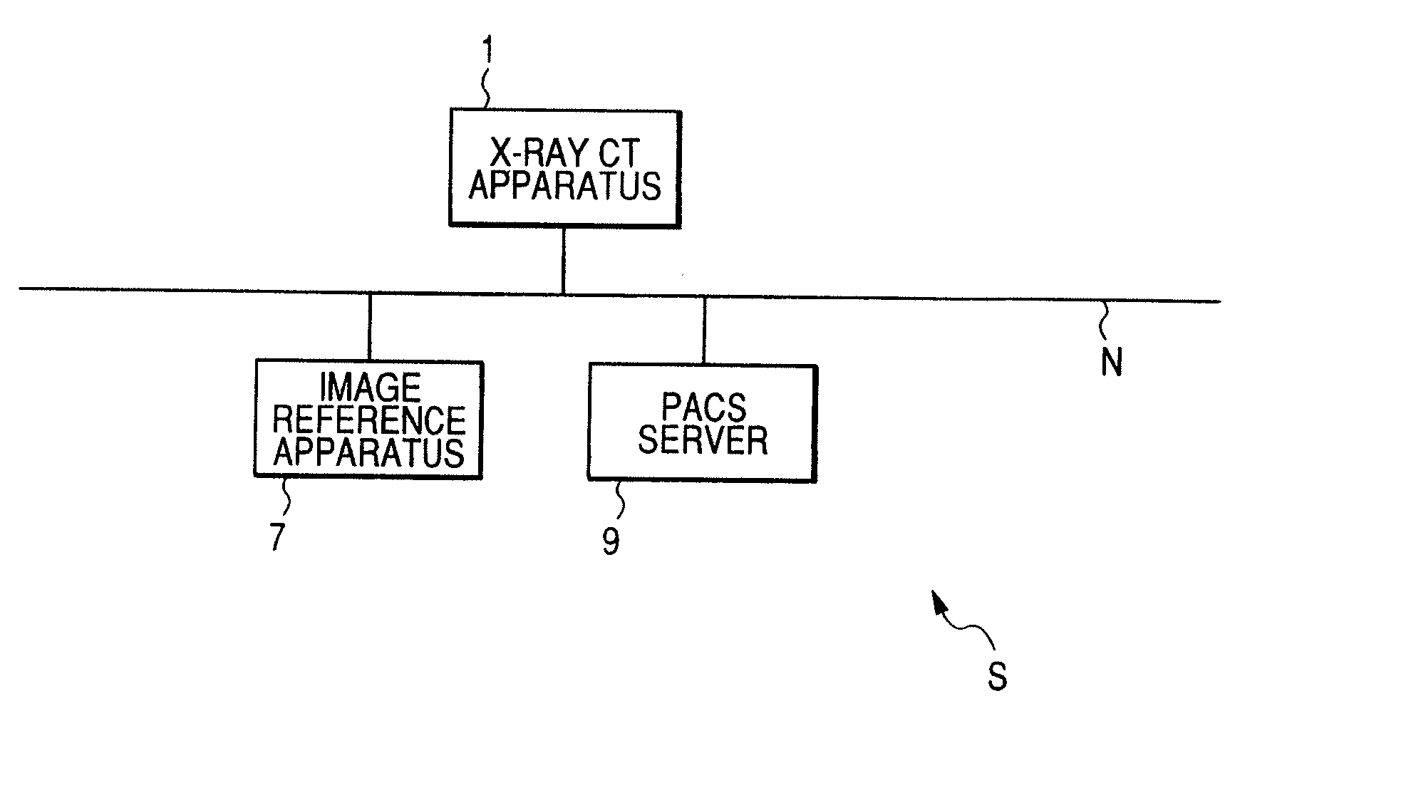
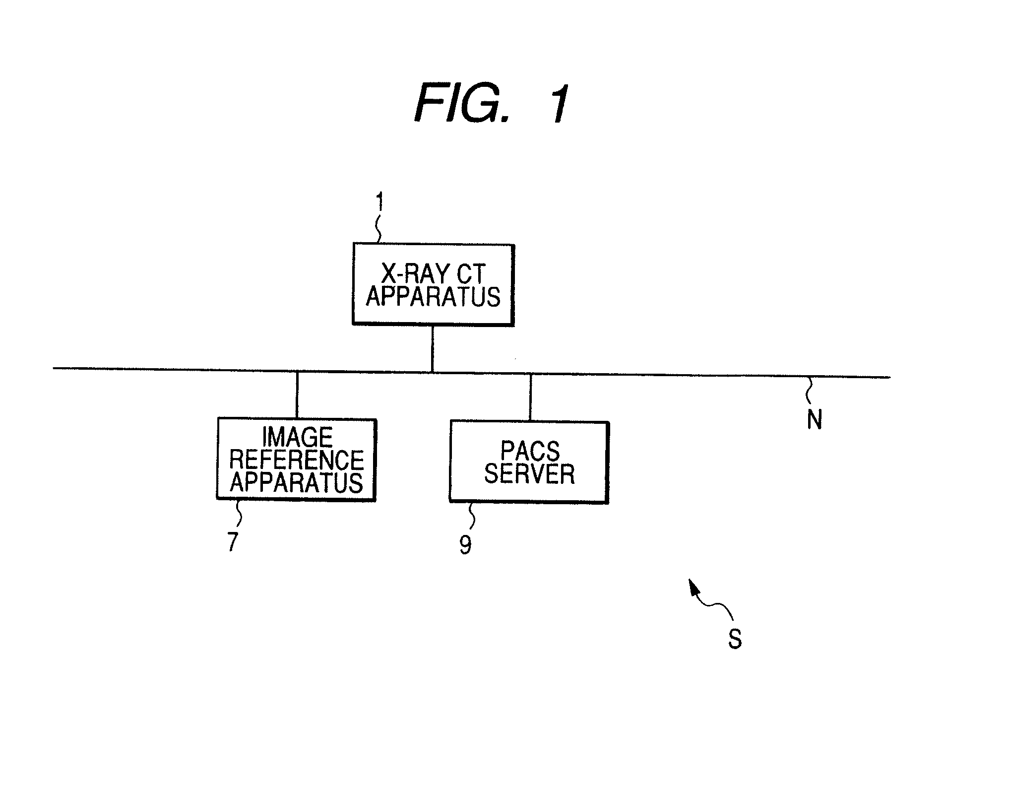
[0055]FIG. 1 is a view illustrating the configuration of a medical image diagnostic system S according to the present embodiment. As shown in the drawing, the medical image diagnostic system S is build by using a hospital information system (HIS) and includes a medical image diagnostic apparatus (X-ray computerized tomography apparatus 1; hereinafter, referred to as an ‘X-ray CT apparatus 1’), an image reference apparatus 7, and a PACS server 9, which are all connected to a network N.

[0056] In addition, in FIG. 1, an example is shown in which the X-ray CT apparatus 1 is used as a medical image diagnostic apparatus. However, the technical scope of the present invention is not limited thereto. For example, the present invention may be applied to other apparatuses that determine the photographing position by the use of a bed, such as a magnetic resonance imaging apparatus (MRI apparatus), an X-ray diagnostic apparatus, or a nuclear medicine dia...

third example

[0133] Next, a process of creating a shared object in the image reference apparatus 7 will be described. The process of creating a shared object refers to creating a shared object by using the setting of image processing parameters used for creation of a key image in, for example, an image reading step, the position of the key image within a photographing range, and the like.

[0134]FIG. 9 is a timing chart illustrating a flow of a process performed by the image reference apparatus 7 in creating a shared object. As shown in the drawing, first, the control unit 71 transmits to the PACS server 9 a request for acquisition of an image data group belonging to the series to be read, in order to perform an image reading process (step S13). The control unit 91 of the PACS server 9 receives the acquisition request and reads out a corresponding image data group from the image storage unit 95a (step S14). The read image data group is transmitted to the image reference apparatus 7 by the transmi...

second embodiment

[0145] Next, a second embodiment of the present invention will be described. A medical image diagnostic system S according to the present embodiment has a support function (hereinafter, referred to as a ‘support function using a shared object’) of simplifying access to past information (for example, a key image at the time of a previous test) used in condition setting and image reading (and report creation accompanied thereby) of various medical-related apparatuses, which are used for an image diagnosis, using the shared object created by the method described in the first embodiment, for example.

[0146]FIG. 10 is a view illustrating the configuration of the medical image diagnostic system S according to the present embodiment. When the configuration shown in the drawing is compared with that in FIG. 1, the configuration shown in the drawing is different from that in FIG. 1 in that an X-ray CT apparatus 3 having a support function using a shared object is further provided and the ima...


PUM


Abstract

A shared object is newly created in a unified format with respect to past medical information that is effective in a photographing step or a report creating step. Since the shared object can include a position determination image, unique object information, body coordinates information, a photographing condition, a image creating condition, and key image information, it is possible to automatically set the same photographing condition, photographing range, a tomographic position to be photographed, and image creating condition as those in a past test by using the information described above.

Description

CROSS REFERENCE TO RELATED APPLICATIONS [0001] This is a Continuation Application of PCT Application No. PCT / JP2006 / 323618, filed Nov. 27, 2006, which was published under PCT Article 21(2) in Japanese. [0002] This application is based upon and claims the benefit of priority from prior Japanese Patent Application No. 2005-341087, filed Nov. 25, 2005, the entire contents of which are incorporated herein by reference.BACKGROUND OF THE INVENTION [0003] 1. Field of the Invention [0004] The present invention relates to a medical image diagnostic apparatus that creates an image from data obtained by scanning a tested body placed on a bed, a picture archiving communication system server (PACS server) connected to a medical image diagnostic apparatus, an image reference apparatus for observing an image acquired by a medical image diagnostic apparatus, and a medical image diagnostic system having the medical image diagnostic apparatus, the picture archiving communication system server, and th...

Claims


Application Information

Patent Timeline
IPC IPC(8): A61B5/05G16H30/20G16H30/40G16H50/20
CPCA61B6/032A61B6/488G06F19/321G01R33/543G01R33/546A61B6/545G16H50/20G16H30/20G16H30/40G16H40/63
Inventor KAMINAGA, SHIGEOOZAKI, MASAHIROMASUZAWA, TAKASHIIWASA, AKIRAKANAZAWA, HITOSHI
Owner TOSHIBA MEDICAL SYST CORP
Why Eureka
Try Eureka