Immunoassay product and process

a technology of immunoassay and product, applied in the field of immunoassay product and process, can solve the problems of high noise and low detection level, waste of reagents, long process, etc., and achieve the effect of reducing the starting volume of such liquids

Active Publication Date: 2007-10-18
MILLIPORE CORP
View PDF21 Cites 27 Cited by
  • Summary
  • Abstract
  • Description
  • Claims
  • Application Information

AI Technical Summary

Benefits of technology

[0009] A rapid, efficient and convenient method to detect one or more biological entities on a blotting membrane is provided. The detection can relate to the position, nature or amount of the biological substance on one or more membranes. The invention method involves a pressure assisted regiment, selected from positive pressure or a vacuum assisted regiment for the supply and removal of reagents and permits washing of the contaminants from substances embedded in the membrane to be detected using very low volumes of liquid. This method enables completion of the blocking, washing and antibody binding steps in about 30-45 minutes without comprising blot quality.
[0012] In another aspect, the invention is directed to an apparatus useful in conducting the method of the invention. The device is comprises of several layers including a porous support layer below the one or more layers of blotting membrane, a flow distributor above the blotting membrane(s) and a well on the flow distributor to contain the liquid to the desired area and to allow for lower starting volumes of such liquid. Preferably, the flow distributor is a non-binding or low binding porous membrane such as a 0.22 micron membrane.

Problems solved by technology

The “spots” are then detected by, at a minimum, by blocking the membrane with a protein or detergent solution to reduce non-specific binding (which otherwise leads to a high level of noise and low level of detection).
It is a lengthy process that most researchers dislike and which consumes (wastes) a large volume of reagents.
The system requires large volumes of liquid in order to operate, is cumbersome to employ and is still quite time consuming.
However, they discount this effort as it is only available in small volume applications and still is uncontrollable.

Method used


Image

  • Immunoassay product and process
  • Immunoassay product and process
  • Immunoassay product and process

Examples


example

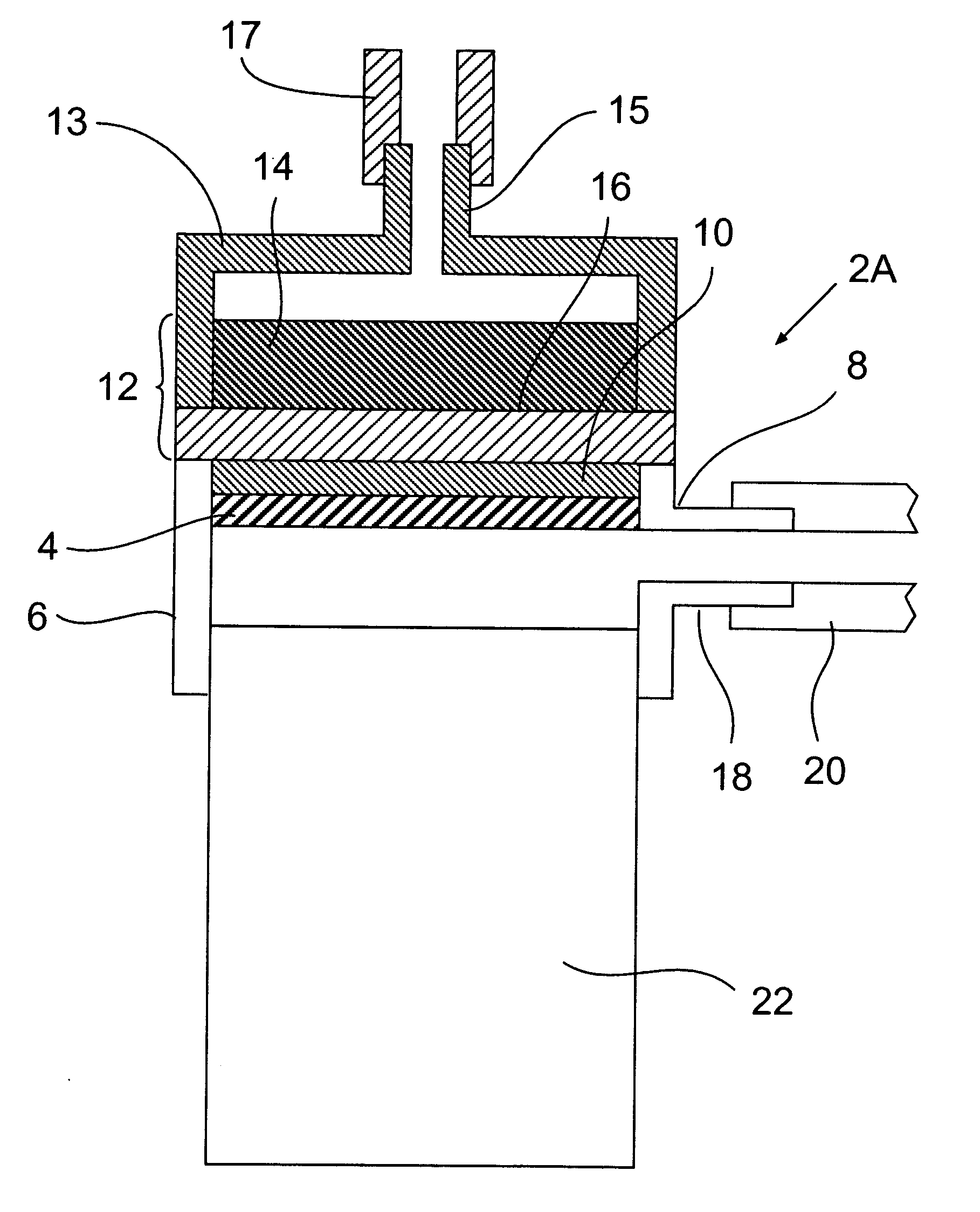
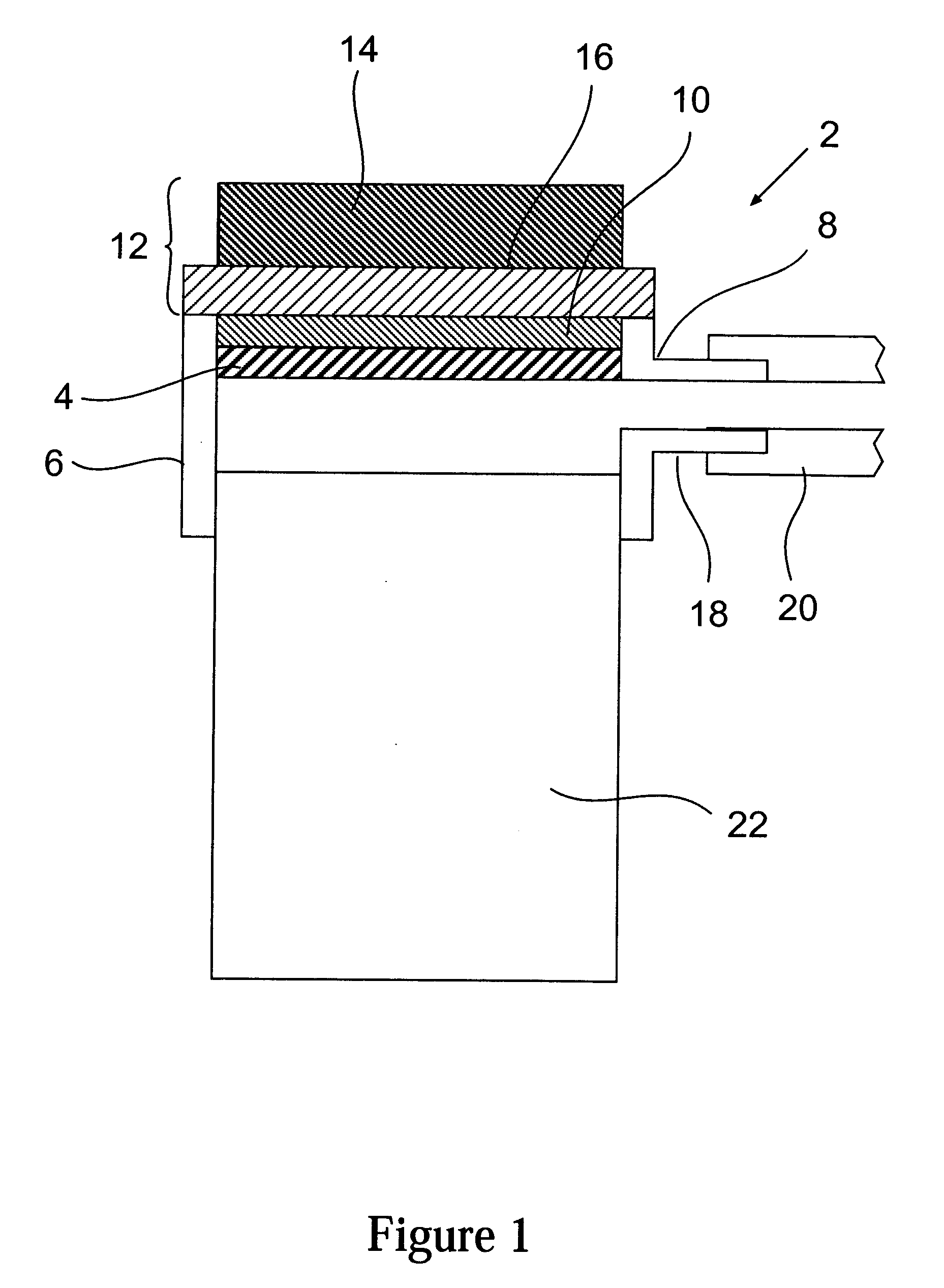
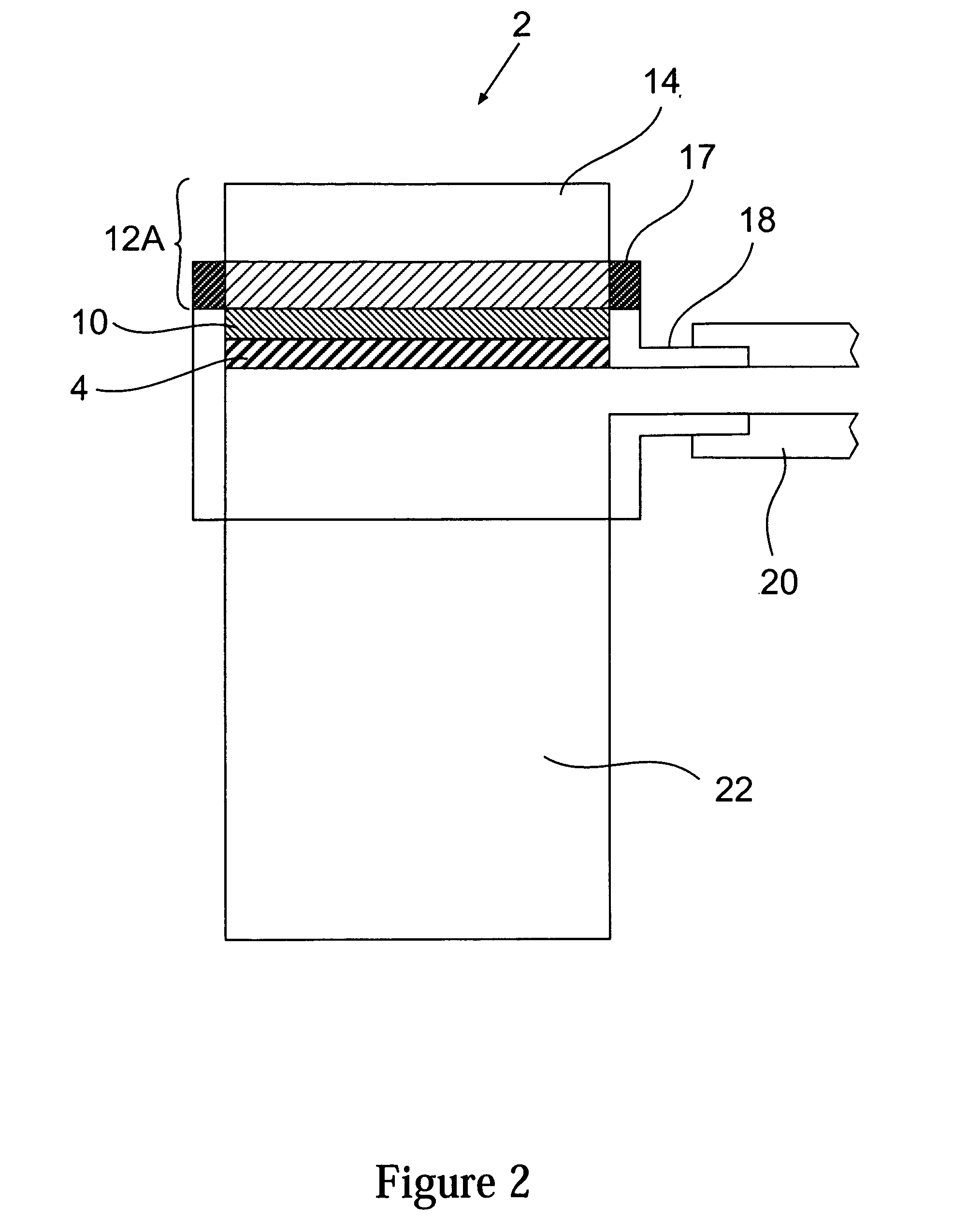
[0075] The device of FIG. 1 was made using the base of a STERICUP® device (available from Millipore Corporation of Billerica, Mass.) as the vacuum manifold, a piece of Porex® porous plastic as the porous support, a Durapore® hydrophilic membrane (GVPP), 0.22 micron pore size as the flow distributor and a polystryrene strip (typically 4 mm larger than the membrane size. For example, 76 mm (L)×86 mm (W)×25 mm (H) for 72 mm×82 mm) well formed by bending the strip at 90° to form four corners and sealing the ends of the strip to each other using (3211 light cure adhesive, available from Loctite). The well was glued to the membrane surface using the Loctite 3211 light cure adhesive.

[0076] The base was connected to a valved vacuum line via its vacuum port.

[0077] A prewet (prewet in 100% methanol, then in water) blotting membrane (IMMOBILION™ Western blotting membrane available from Millipore Corporation of Billerica, Mass.) containing a sample of bovine liver lysate was placed on the bas...


PUM

PropertyMeasurementUnit
sizesaaaaaaaaaa
sizesaaaaaaaaaa
sizesaaaaaaaaaa

Abstract

A rapid, efficient and convenient method to detect one or more biological entities on a blotting membrane is provided. The detection can relate to the position, nature or amount of the biological substance on one or more membranes. The invention method involves a pressure assisted regiment (such as vacuum or positive gas pressure) for the supply and removal of reagents and permits washing of the contaminants from substances embedded in the membrane to be detected using very low volumes of liquid. This method enables completion of the blocking, washing and antibody binding steps in about 30 minutes without comprising blot quality. In another aspect, the invention is directed to an apparatus useful in conducting the method of the invention. The device is comprised of several layers including a porous support layer below the blotting membrane(s), a flow distributor above the blotting membrane(s) and a well on the flow distributor to contain the liquid to the desired area and to allow for lower starting volumes of such liquid. Preferably, the flow distributor is a non-binding or low binding hydrophilic porous membrane such as a 0.22 micron membrane.

Description

CROSS-REFERENCE TO RELATED APPLICATIONS [0001] This application claims the benefit of U.S. Provisional Application No. 60 / 795,452, filed on Apr. 27, 2006 and U.S. Provisional Application No.: 60 / 732,994, filed on Nov. 3, 2005. [0002] The invention relates to a laboratory device and process of using the device to detect the position of or presence / absence of substances that are contained in a blotting membrane. More particularly, it concerns a technique for applying reagents and wash solutions to a blotting membrane to accomplish this detection quickly via the use of a vacuum or positive pressure.BACKGROUND OF THE INVENTION [0003] The use of gel electrophoresis is currently the ubiquitous technique for the separation of biological materials. (Nonbiological materials can also be separated using gels or other chromatographic supports as well, but the scope of effort with regard to biologicals is greater.) Typical applications include separation of nucleic acid fragments of various size...

Claims


Application Information

Patent Timeline
Patent Type & Authority Applications(United States)
IPC IPC(8): G01N33/53B01J19/00
CPCB01L3/50255B01L2400/0487G01N33/54366Y10T436/2575G01N33/561G01N33/5302G01N33/54393
Inventor MABUCHI, MASAHARUKIMURA, HIROKOEMERICK, MARCCLARK, PHILLIPGREENIZEN, KURT
Owner MILLIPORE CORP
Why Eureka
Try Eureka