Reflection Microscope for Examination of the Corneal Endothelium and Method of Operating Same

a technology of reflection microscope and endothelium, which is applied in the field of new noncontact endothelium reflection microscope apparatus, can solve the problems of complex apparatus, loss of transparency, and sensitive balance of the corneal transparency, and achieve the effects of reducing the use of electronic components, and increasing reliability

Active Publication Date: 2007-11-15
C S O SRL
View PDF4 Cites 33 Cited by
  • Summary
  • Abstract
  • Description
  • Claims
  • Application Information

AI Technical Summary

Benefits of technology

[0013] The apparatus according to the invention permits to have the endothelium test without use of sensors, photosensors or other devices placed onto a reflected optical path. A high

Problems solved by technology

The cornea transparency is subjected to a very delicate balance, and a number of diseases can produce a loss of the transparency.
The above described system, with the use of a focusing detec

Method used


Image

  • Reflection Microscope for Examination of the Corneal Endothelium and Method of Operating Same
  • Reflection Microscope for Examination of the Corneal Endothelium and Method of Operating Same
  • Reflection Microscope for Examination of the Corneal Endothelium and Method of Operating Same

Examples


Embodiment Construction

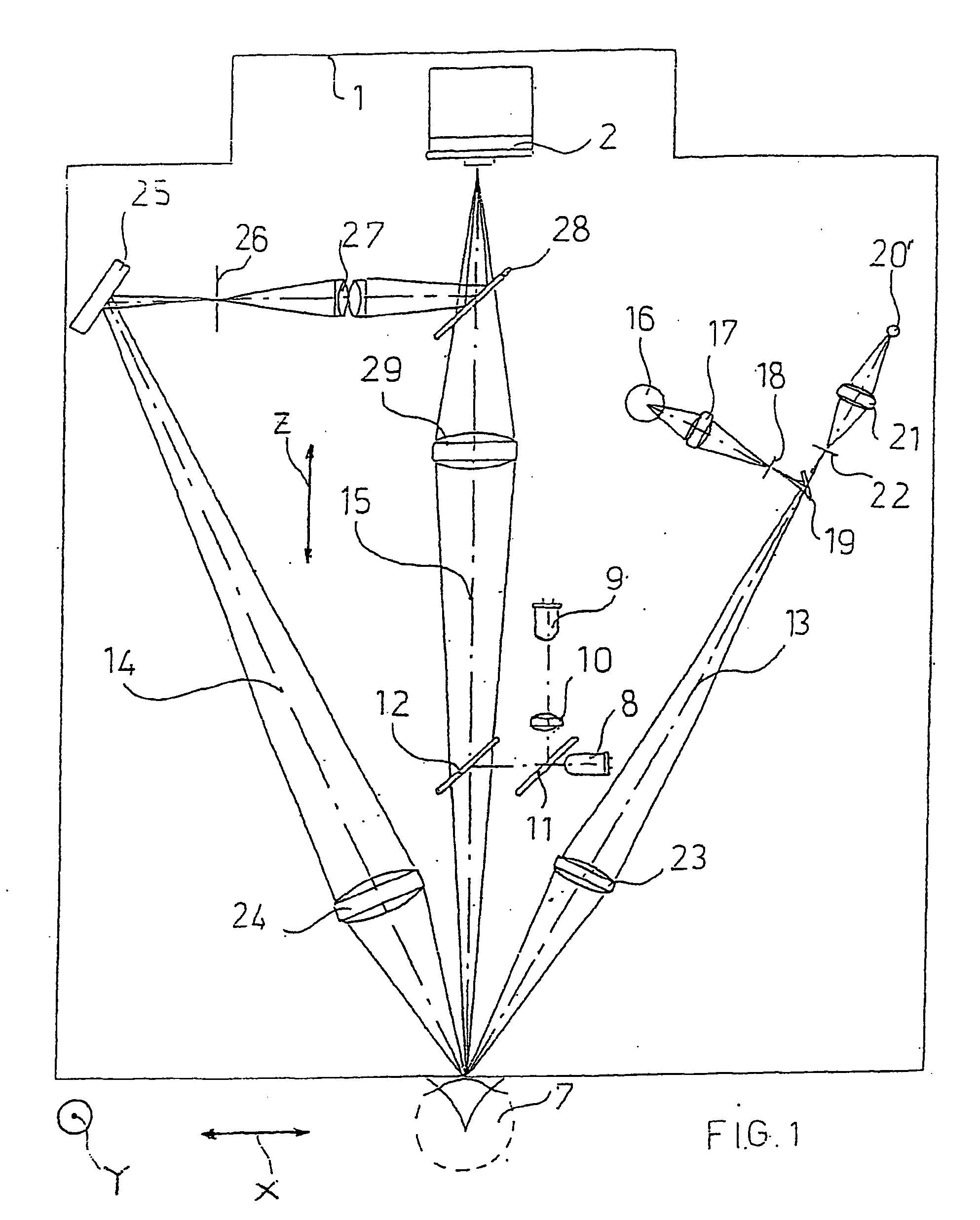
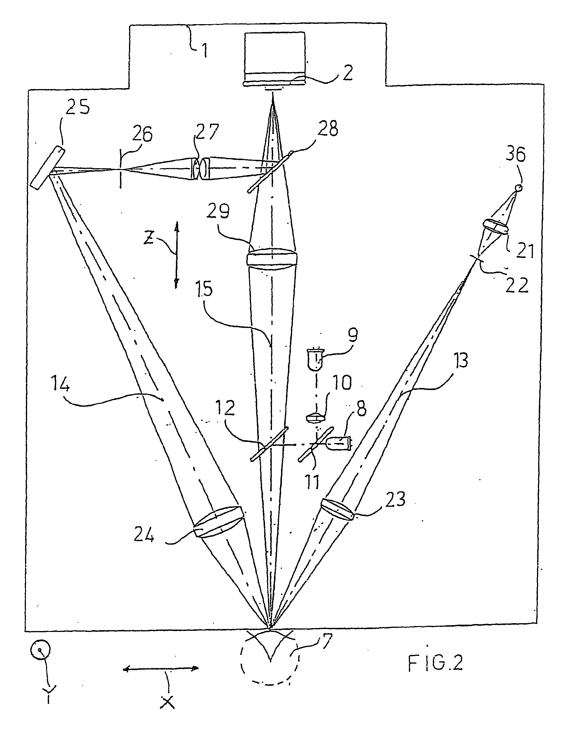
[0022] Referring to FIGS. 1 to 3, the apparatus according to the invention comprises a movable optical head or microscope 1 provided with a CCD high speed camera 2, i.e. a monochrome digital camera with shooting capacity of at least one hundred frames per second with FireWire high speed data output, i.e. with IEEE 1394 port or equivalent.

[0023] The high speed camera 2 is directly connected to a CPU unit 3. The unit 3 comprises a controller 4, e.g. a 65XX type controller produced by the company National Instruments (United States, Texas) or equivalent. The controller 4 controls a power driver board 5, so that the signal coming from the CPU unit 3 is suitable for driving electric DC motors 6 as described hereinafter.

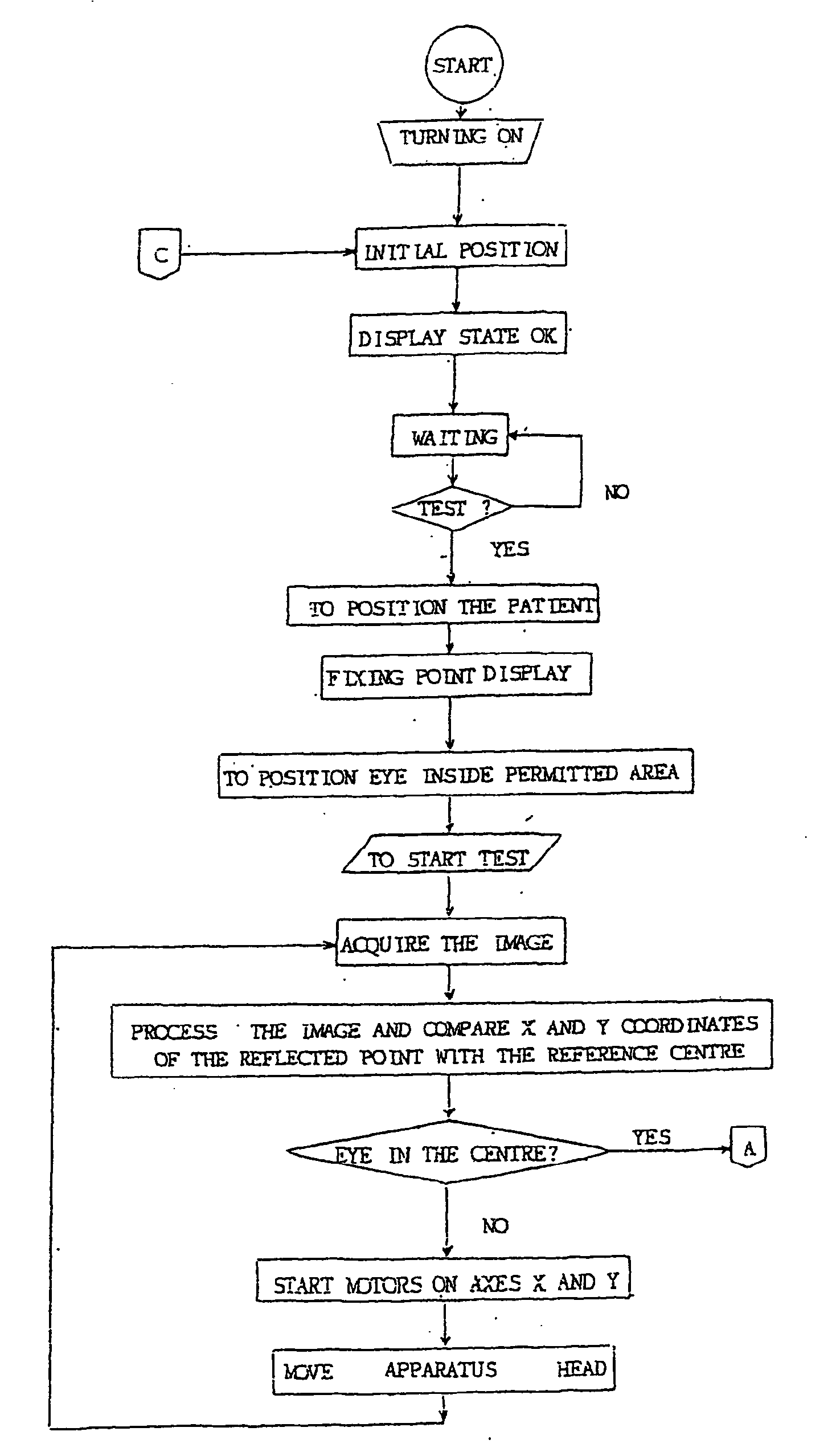
[0024] The function of the motors 6 is to set in position the microscope 1 with the camera 2, following to automatic control by the CPU unit 3 so that the eye center 7 to be examined is found. Such a finding is obtained via a reflection onto the cornea surface of the lig...


PUM


Abstract

An apparatus and a method for operating an endothelium reflection microscope. The apparatus comprises an optical head including: an illuminating system, an eye-front observation optical system along a central channel in which an alignment-use light spot is received and imaged by a camera comprising a digital optical sensor, and an enlarged-imaging optical system for enlarged observation or photographing of the subject part of the digital camera. The apparatus further comprises a drive for moving the optical head and a CPU controller for automatically controlling the drive, the illuminating system and the eyefront optical system. The method comprises an endothelium image acquisition procedure in which the grey level inside a check area of the camera sensor is constantly checked during the advancement along an advancement direction (Z-); when the grey level reaches a predetermined threshold value, a delay time (Δt) is triggered; and when the delay time (Δt) lapses, the acquisition of one or more images of the endothelium by the digital camera is enabled.

Description

FIELD OF THE INVENTION [0001] The present invention refers to a new non-contact endothelium reflection microscope apparatus that permits to automatically obtain the endothelium image and to display clinically useful parameters such as the number and density of the cells, shape, surface, minimum, maximum and medium area, standard deviation, variation coefficient, percentage of cells of various shapes, area distribution histogram, perimeter distribution histogram. BACKGROUND OF THE INVENTION [0002] The endothelium is the most internal layer of the tissues forming the cornea, and consists of a single layer of flat polygonal cells. The endothelium function is to adjust the water contents, permitting a suitable hydration of the cornea. The shape and amount of the cells influence the quality of the vision. The cornea transparency is subjected to a very delicate balance, and a number of diseases can produce a loss of the transparency. [0003] The endothelium cells are of hexagonal shape in ...

Claims


Application Information

Patent Timeline
IPC IPC(8): A61B3/15
CPCA61B3/14A61B3/135
Inventor MURA, SERGIO
Owner C S O SRL
Why Eureka
Try Eureka