Ovarian cancer cell and myeloma cell surface glycoproteins, antibodies thereto, and uses thereof

a technology of ovarian cancer and myeloma, which is applied in the field of ovarian cancer cell and myeloma cell surface glycoprotein, antibodies thereto, can solve the problems of low cell proliferation rate and multi-, ineffective therapy, and only slightly prolonged overall survival, so as to achieve the effect of monitoring the effectiveness of anti-cancer therapies

Inactive Publication Date: 2007-11-22
CAERUS THERAPEUTICS INC
View PDF1 Cites 4 Cited by
  • Summary
  • Abstract
  • Description
  • Claims
  • Application Information

AI Technical Summary

Benefits of technology

[0025] It is a further aspect of the invention to permit the detection of the cell surface glycoproteins described herein in a sample of bodily fluid, to aid in the diagnosis of multiple myeloma, ovarian cancer, or other cancer cells expressing a glycoprotein with the epitope recognized by the antibodies herein, by the detection of the glycoprotein antigen shed from cancer cells into the bodily fluid, such as blood. Furthermore, the stage of the disease may be monitored and the effectiveness of anti-cancer therapies can be monitored by determining the level or changes over time of the level of shed surface glycoprotein in a bodily fluid such as blood.

Problems solved by technology

The limitations of effective therapy for MM are primarily associated with a low cell proliferation rate and multi-drug resistance.
However, overall survival has only been slightly prolonged, and no evidence for a cure has been obtained.
Less than 0.1% of women are affected by hereditary ovarian cancer syndrome, but these women may face a 40% lifetime risk of developing ovarian cancer.
Although pelvic examinations can occasionally detect ovarian cancer, small, early-stage ovarian tumors are often not detected by palpation due to the deep anatomic location of the ovary.
Thus, ovarian cancers detected by pelvic examination are generally advanced and associated with poor survival.
The pelvic examination may also produce false positives when benign adnexal masses (e.g., functional cysts) are found.
The Pap smear may occasionally reveal malignant ovarian cells, but it is not considered to be a valid screening test for ovarian carcinoma.
Evidence is limited on whether tumor markers become elevated early enough in the natural history of occult ovarian cancer to provide adequate sensitivity for screening.
Tumor markers may have limited specificity.

Method used


Image

  • Ovarian cancer cell and myeloma cell surface glycoproteins, antibodies thereto, and uses thereof
  • Ovarian cancer cell and myeloma cell surface glycoproteins, antibodies thereto, and uses thereof
  • Ovarian cancer cell and myeloma cell surface glycoproteins, antibodies thereto, and uses thereof

Examples


example 1

Preparation and Screening of Hybridomas

[0091] 1. Sources of cells Human myeloma cell lines (U266, OPM, RPMI1860, KR12 and NCl H929), and chronic myelogenic leukemic cell line (K562) were purchased from the American Type Culture Collection (ATCC®). Fresh human ovarian cancer, breast cancer, and liver cancer specimens were used. Cell lines of prostate cancer, LnCap (ATCC®); neuroblastoma cell line, NCl H2106 (ATCC®); and a cervical cancer, Caski (ATCC®) were also evaluated, as well as an EBV-transformed B cell tumor, Namalwa (ATCC®).

[0092] 2. Immunization Mice were immunized with a pool of plasmacytoma cells, U266, RPMI1860 and OPM (5×10⁶ total in 50 μl containing Ribi adjuvant, 50%), in the left footpad, and with K562 cells (5×10⁶ total in 50 μl containing Ribi adjuvant, 50%) in the right footpad. The immunization was repeated after 14 days. The left popliteal lymph node was removed and the extracted cells were fused 3 days after the second immunization.

[0093] 3. Generation of B c...

example 2

Specificity Assessment of Monoclonal Antibodies

[0100] Cell surface staining using a panel of monoclonal antibodies Jul. 16, 1999 analyzed by flow cytometry is depicted in FIG. 4. A strong staining of plasmacytoma cells by MA69 (also called VAC69 herein) is demonstrated in panel F, while negative staining was demonstrated for the control cell lines, including human B cell tumor lines IM9 and HT (IM9 with isotype control, panel A; IM9 with MA69, panel B; HT with isotype control, panel C; HT with MA69, panel D) and myeloma cell line U266 with an isotype control monoclonal antibody (panel E). Furthermore, peripheral blood cells (PBMC) from normal individuals showed insignificant binding to the antibodies.

[0101] Hybridoma cell line IMM002.69.47.4 which produces monoclonal antibody MA69 was deposited on Aug. 3, 1999, with the American Type Culture Collection, 10801 University Boulevard, Manassas Va. 20110-2209, and has been assigned Accession No. PTA-450.

example 3

Binding of Monoclonal Antibody to Cell Surface Glycoprotein

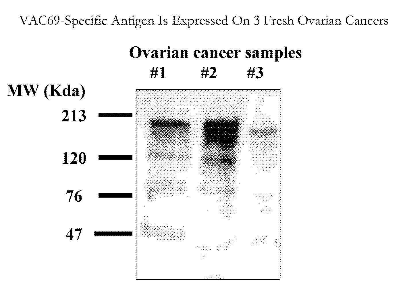
[0102] B cell hybridoma culture designated MA69 (also VAC69 herein) was shown to detect a distinct band on myeloma membranes from five human multiple myeloma cell lines (RPMI1860, U266, KR-12, OPM-1 and NCl H929) using Western blots. Four of the five myeloma cell lines showed binding to a cell surface glycoprotein with an approximate molecular weight of between about 78 and about 120 kDa. The PBMC serving as a negative control did not show binding to the antibody. In addition, no staining of two human B cell tumors (HT and IM9) was observed (FIG. 5).

[0103]FIG. 6 graphically presents the results of a repeat of the experiment described in FIG. 5 using a cellular ELISA method. In this experiment, the MA69 detected a distinct band in 5 out of 5 myeloma cell lines with varying intensities. The control membrane preparations consisting of normal PBMC and a human B cell tumor (HT) were not stained by the antibody.


PUM


Abstract

The present invention is directed to cell surface antigens found on myeloma cells and on ovarian cancer cells that are recognized by monoclonal antibodies, and antibody binding fragments thereof, as described. The monoclonal antibodies of the invention are capable of being used for therapeutic, screening, diagnostic and cell purification purposes. A representative and exemplified monoclonal antibody of the present invention recognizes and binds to an epitope common to a surface antigen that is expressed on multiple myeloma cells and to a surface antigen that is expressed on ovarian cancer cells. The function of this monoclonal antibody both in vivo and in vitro is demonstrated.

Description

CROSS-REFERENCE TO RELATED APPLICATIONS [0001] This application is a Divisional Application of U.S. patent application Ser. No. 10 / 049,608, filed Sep. 16, 2002, which is the national phase application under 35 U.S.C. §371 of the International Application Serial No. PCT / US2000 / 21574, filed Aug. 8, 2000, which is a continuation-in-part application of U.S. application Ser. No. 09 / 374,367, filed Aug. 13, 1999, all of which are incorporated herein by reference as if fully set forth herein.FIELD OF THE INVENTION [0002] The application is related to new surface glycoproteins of human myeloma cells and human ovarian tumor cells, monoclonal antibodies thereto, and methods of diagnosis and treatment of myeloma and ovarian cancer based thereon. BACKGROUND OF THE INVENTION [0003] Multiple myeloma (MM) embodies a plasma cell disorder characterized by neoplastic proliferation of a single clone of plasma cells engaged in the production of a monoclonal immunoglobulin, usually monoclonal IgG or IgA....

Claims


Application Information

Patent Timeline
Patent Type & Authority Applications(United States)
IPC IPC(8): A61K51/10C07K16/30A61K35/28G01N33/532A61K39/00A61K39/395A61K45/00A61K49/00A61K51/00A61P15/00A61P19/00A61P35/00A61P37/02C07K14/705C12N5/10C12N15/02C12P21/08G01N33/574
CPCA61K2039/505G01N2800/52C07K16/3069C07K16/3061A61P15/00A61P19/00A61P35/00A61P37/02
Inventor GELBER, COHAVA
Owner CAERUS THERAPEUTICS INC
Why Eureka
Try Eureka