Respiratory gated image fusion of computed tomography 3D images and live fluoroscopy images

a computed tomography and respiratory gate technology, applied in the field of advanced respiratory gated image fusion system, can solve the problems of limited space required for time-resolved ct data acquisition, respiratory motion can introduce significant challenges, and lack of real-time 3d support, so as to reduce the negative impact of respiratory related, efficient and safer intervention, and improve accuracy

Inactive Publication Date: 2007-11-22
SIEMENS HEALTHCARE GMBH
View PDF31 Cites 32 Cited by
  • Summary
  • Abstract
  • Description
  • Claims
  • Application Information

AI Technical Summary

Benefits of technology

[0006]The present invention provides new and improved methods and systems for providing real-time image guidance by uniquely combining the capabilities of two well-known imaging modalities, anatomical 3D data from computer tomography (CT) and real time data from live fluoroscopy images provided by an angiography/fluoroscopy system to ensure a more efficient and safer intervention in treating punctures, drainages and biopsies in the abdominal and thoracic areas of the human body.
[

Problems solved by technology

Respiratory motion can introduce significant challenges during non-vascular interventions like RF ablations, biopsies and radiotherapy treatment.
The challenges that are introduced during these types of interventions are related to the manner in which they are supported.
One drawback of using ultrasound or CT guidance is the lack of real time 3

Method used


Image

  • Respiratory gated image fusion of computed tomography 3D images and live fluoroscopy images
  • Respiratory gated image fusion of computed tomography 3D images and live fluoroscopy images
  • Respiratory gated image fusion of computed tomography 3D images and live fluoroscopy images

Examples


Embodiment Construction

[0012]As required, detailed embodiments of the present inventions are disclosed herein; however, it is to be understood that the disclosed embodiments are merely exemplary of the invention, which may be embodied in various forms. Therefore, specific structural and functional details disclosed herein are not to be interpreted as limiting, but merely as a basis for the claims and as a representative basis for teaching one skilled in the art to variously employ the present invention in virtually any appropriately detailed structure.

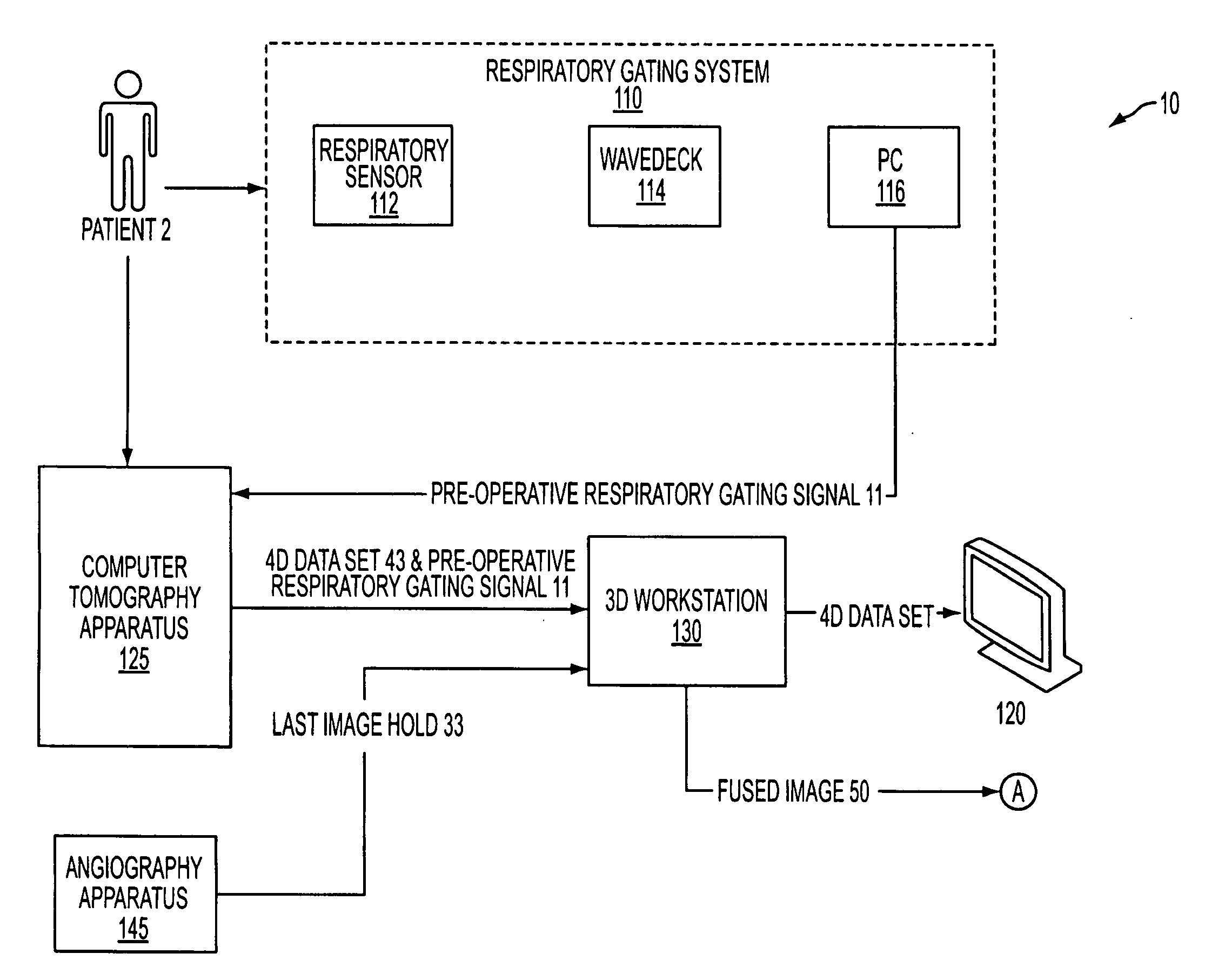
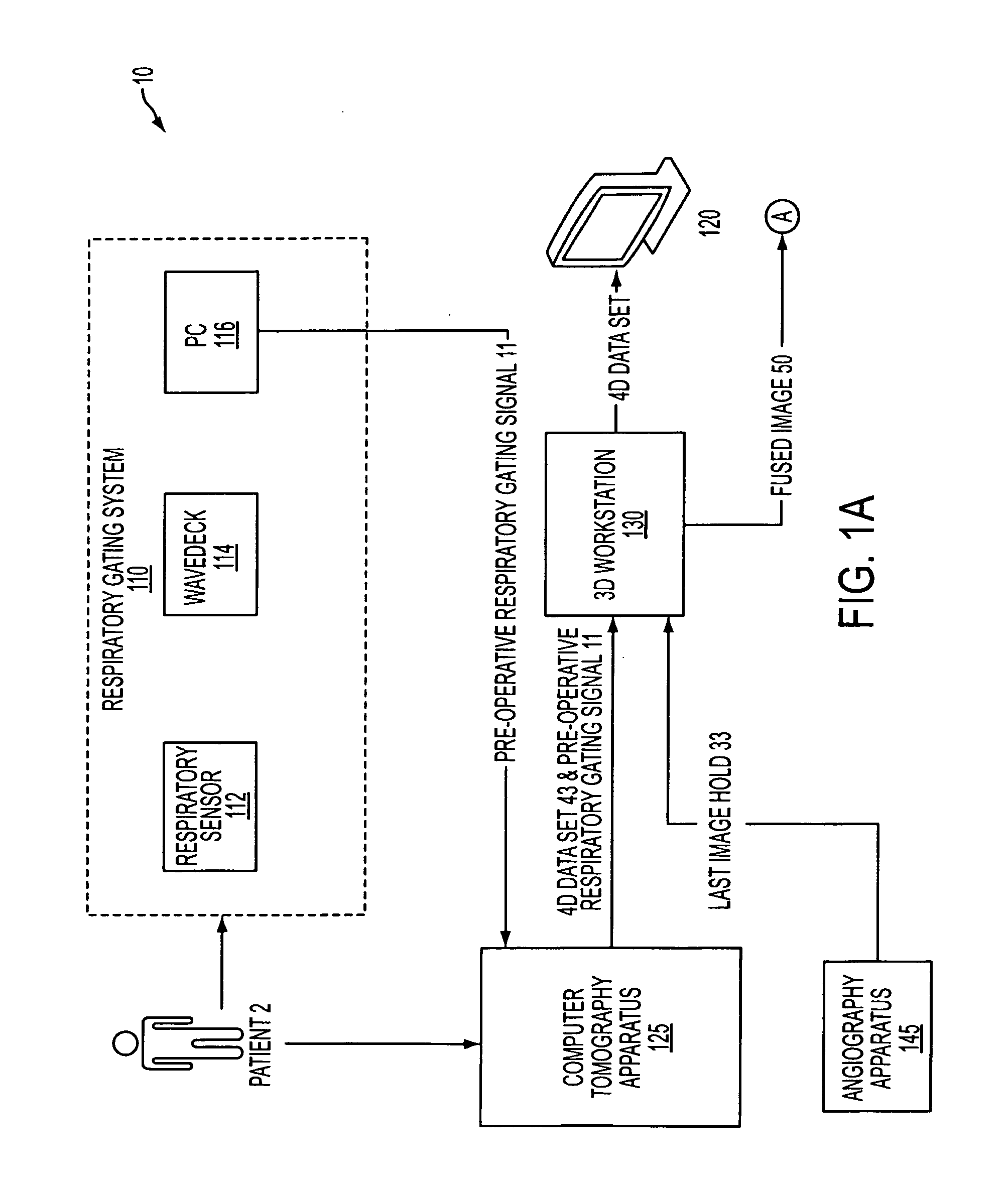
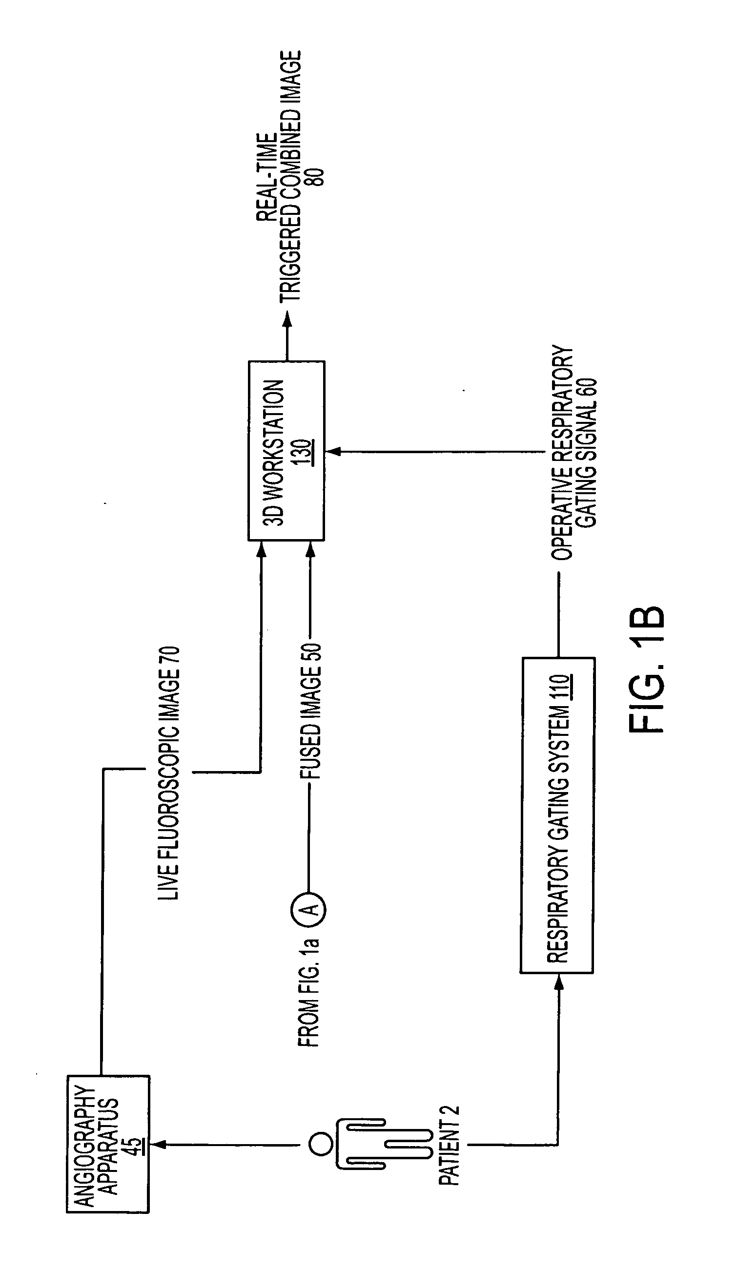
[0013]Referring now to FIGS. 1a &1b, according to an embodiment of the present invention, a system 10 for implementing the present invention can comprise, inter alia, a respiratory gating sub-system 110 comprising a respiratory sensor 112, a wave-deck device 114 and a personal computer (PC) 116. The system 10 can further comprise computer tomography (CT) apparatus 125, a 3D workstation 130, a display 120 and angiography apparatus 145.

[0014]System 10 may incl...


PUM


Abstract

A system and method is provided directed to improved real-time image guidance by uniquely combining the capabilities of two well-known imaging modalities, anatomical 3D data from computer tomography (CT) and real time data from live fluoroscopy images provided by an angiography/fluoroscopy system to ensure a more efficient and safer intervention in treating punctures, drainages and biopsies in the abdominal and thoracic areas of the human body.

Description

BACKGROUND[0001]1. Field of the Invention[0002]The present invention is generally directed to medical imaging. More particularly, the present invention is directed to an advanced respiratory-gated image fusion system and methods for use in the image-assisted treatment of punctures, drainages and biopsies in the abdominal and thoracic areas of the human body.[0003]2. Background of the Invention[0004]Respiratory motion can introduce significant challenges during non-vascular interventions like RF ablations, biopsies and radiotherapy treatment. The challenges that are introduced during these types of interventions are related to the manner in which they are supported. These types of interventions are presently supported by ultrasound guidance or CT guidance. One drawback of using ultrasound or CT guidance is the lack of real time 3D support. To reconcile this lack of real time 3D support, i.e., the time-varying discrepancy between the navigation positions and the pre-operative image da...

Claims


Application Information

Patent Timeline
IPC IPC(8): A61B6/00A61B5/05
CPCA61B5/0816A61B6/032A61B6/541A61B6/5235A61B6/504
Inventor LOTHERT, MARK
Owner SIEMENS HEALTHCARE GMBH
Why Eureka
Try Eureka