Device for Tissue Diagnosis and Spatial Tissue Mapping

a tissue diagnosis and tissue technology, applied in the field of tissue diagnosis and spatial tissue mapping, can solve the problems of inexact positioning of tissue removal instruments, inability to accurately detect cancer near the surface, and low measurement performance of previous attempts to use electrical measurements for tissue diagnosis

Inactive Publication Date: 2007-11-29
MILLER CRAIG JAMES
View PDF12 Cites 59 Cited by
  • Summary
  • Abstract
  • Description
  • Claims
  • Application Information

AI Technical Summary

Problems solved by technology

For the most part previous attempts to use electrical measurements for tissue diagnosis have suffered from low performance as measured by their combined sensitivity, which is the ability to correctly identify abnormal tissue, and specificity, which is the ability to correctly identify normal tissue.
Previous workers who have attempted to use electrical impedance of tissue for the detection of cancer near the surface have not been successful because they have primarily measured the bulk impedance of tissue averaged over a depth far exceeding the region of interest.
The positioning of the tissue removal instrument is difficult and inexact because it is hand-guided.
If one examination shows abnormal tissue and a repeat examination does not, it is not possible to know if the abnormal tissue has repaired itself or whether the repeat examination simply missed the initial abnormal tissue area.
In the case of cervical tissue changes, disease is slow to develop, usually taking more than ten years to progress from initial abnormalities to invasive cancer.
Because the location and extent of the abnormality are unknown using current cell collection methods, these natural tissue repair processes cannot be monitored except by expensive diagnostic procedures.
This leads to repeat examinations and expensive sophisticated diagnostic procedures.
No immediate results at the time of examination are available to the patient.
This produces patient anxiety and creates the opportunity for laboratory data mix-ups in patient records.
Patient follow-up can be difficult because patients must be contacted and scheduled for subsequent retesting or further diagnostic work.
The delay in laboratory processing is a large problem for migrant communities and underdeveloped areas where social and economic issues limit the opportunities for scheduling multiple medical visits.
Each location tested by such methods is uncorrelated to other location readings.
Surface cell collection often does not detect the initial deeper tissue changes.

Method used


Image

  • Device for Tissue Diagnosis and Spatial Tissue Mapping
  • Device for Tissue Diagnosis and Spatial Tissue Mapping
  • Device for Tissue Diagnosis and Spatial Tissue Mapping

Examples


embodiment

Preferred Embodiment

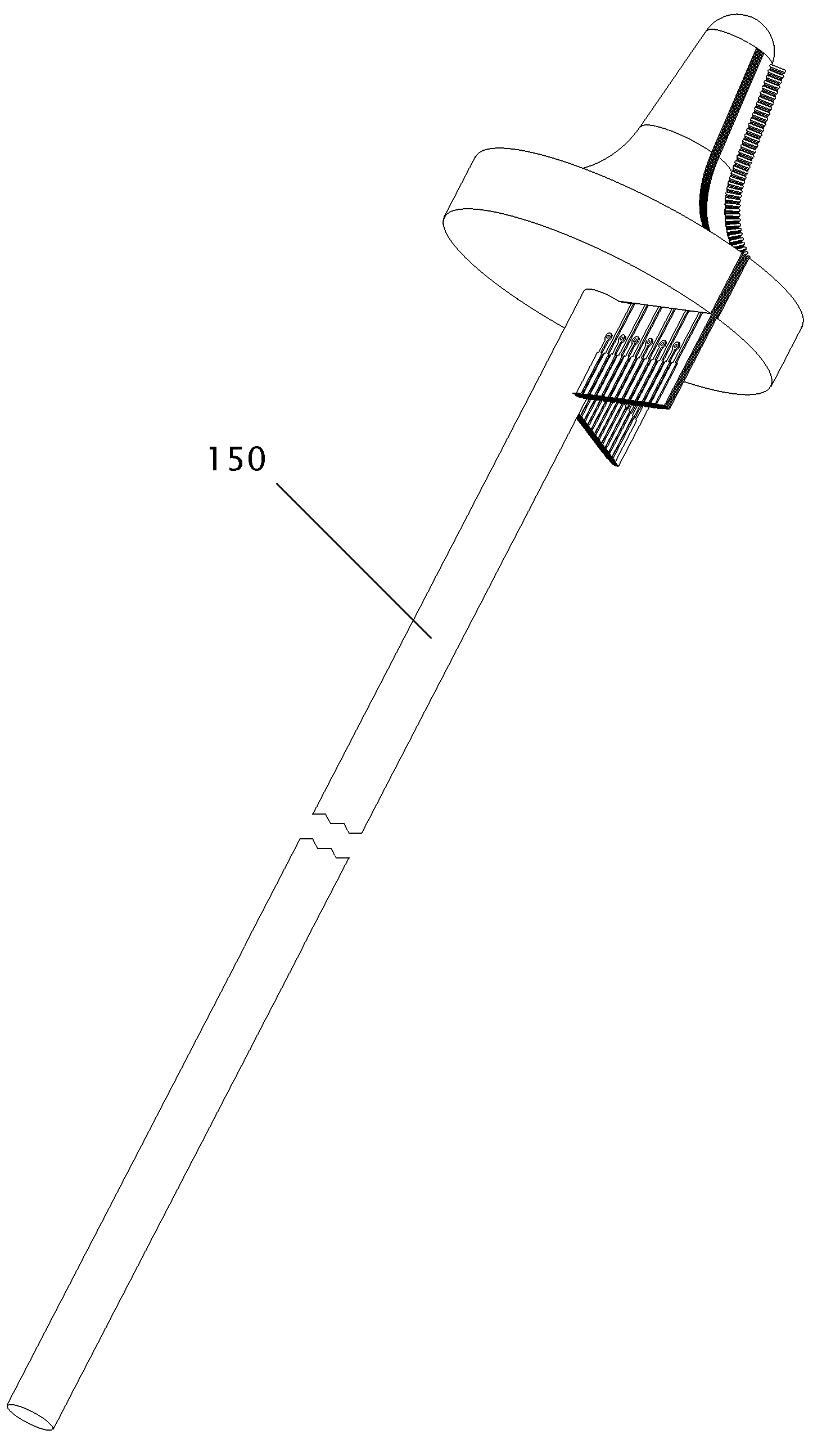
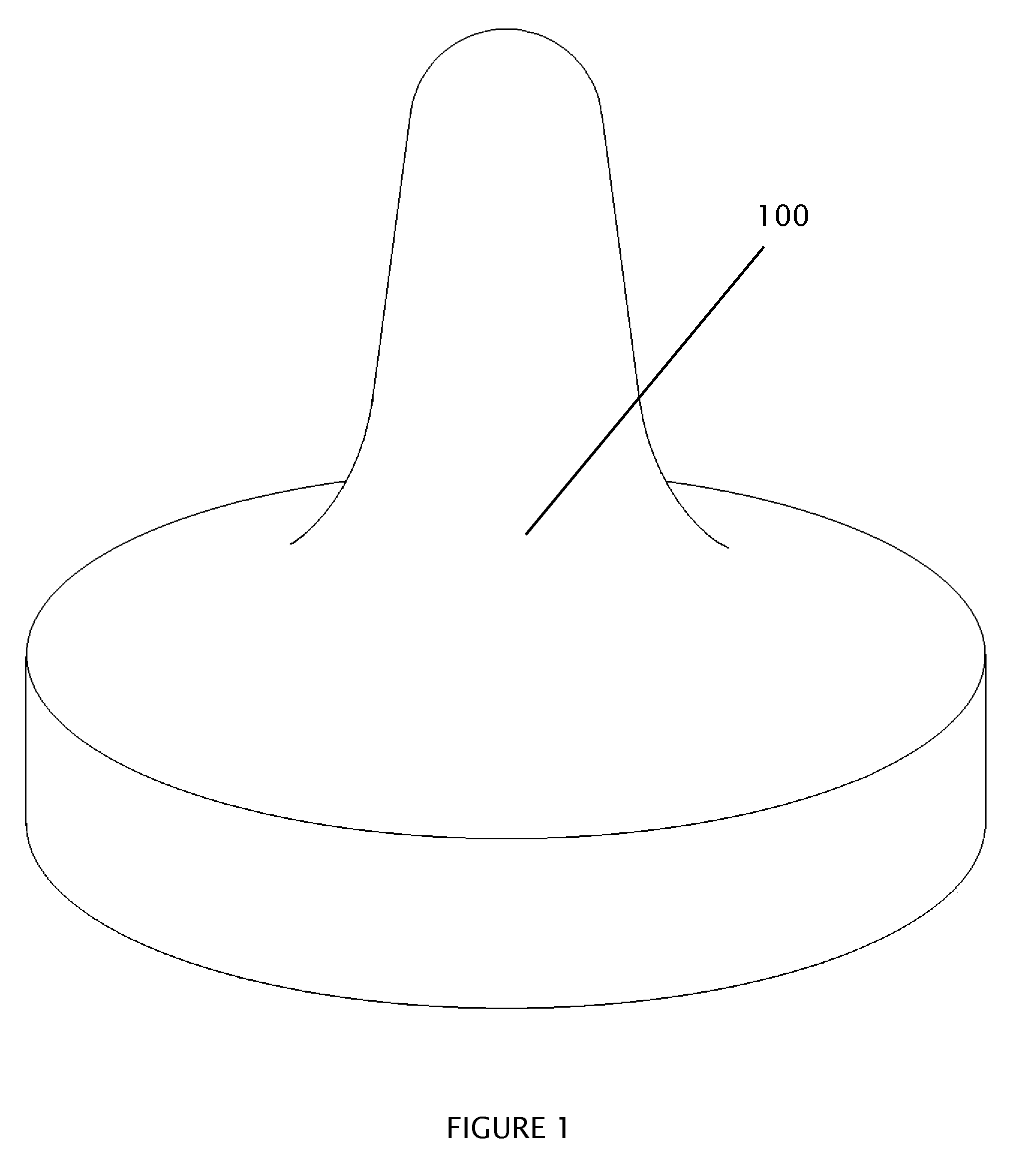
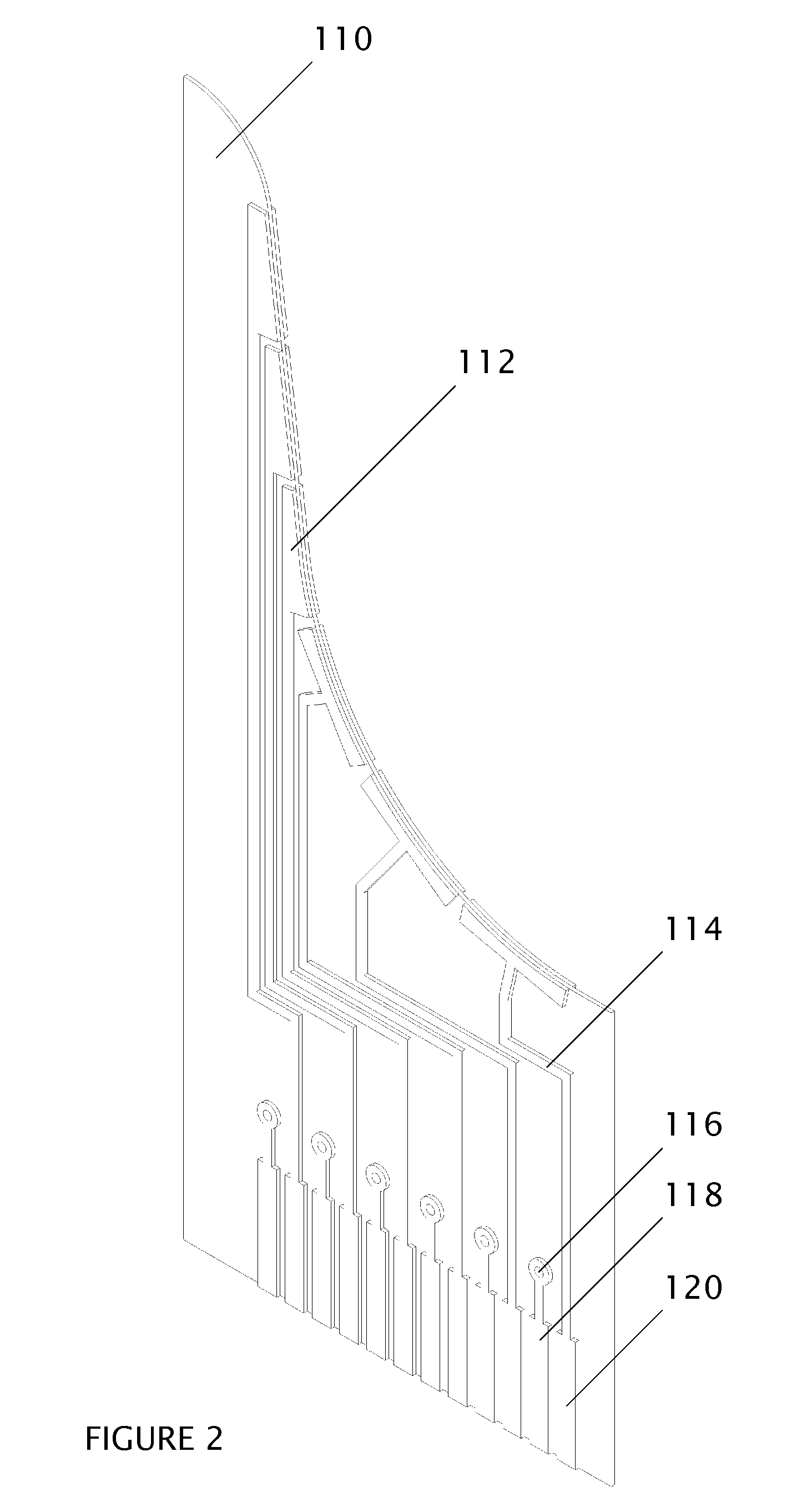
[0060]The system consists of (a) a probe tip which contains an array of electrodes and contacts the tissue directly; (b) a handpiece which is mechanically and electrically connected to the probe tip and which consists of a connecting drive shaft assembly, motors or other kinetic devices to position the probe tip precisely, and an electrical connection to the electrodes in the tip; (c) an electrical signal generation device to stimulate the tissue by means of electrical waveforms; (d) a data acquisition device to measure the electrical signal response from the tissue; (e) a processor which controls the signal generation device, data acquisition device, signal response storage and analysis, and the motors or other kinetic devices used to move the probe tip. The signal generation device and data acquisition device may be contained as electronic components and circuits within the handpiece or externally to the handpiece, as in a circuit board located within a compute...


PUM


Abstract

A miniature electrode array is utilized to stimulate tissue and measure tissue response. When the tissue response is used to diagnose tissue, the spacing of the electrodes in the array is approximately equal to the depth of tissue which is to be examined. The manufacture of such arrays can be accomplished by embedding printed circuit boards, for example, into a properly shaped probe tip. A kinetic device to position the probe on the surface of the tissue provides location information which is correlated with the simultaneous electrical response data in order to generate a tissue map. The tissue mapping capability can be combined with tissue removal devices in order to direct the excision of tissue. When the tissue response is used simply to determine that the array is in contact with the tissue, the electrodes can be widely spaced. Combined with a cell collection device, the electrodes provide feedback to the practitioner that the collection device is making proper contact over the region where cells are to be collected.

Description

CROSS REFERENCE TO RELATED APPLICATIONS[0001]Provisional Patent Application[0002]U.S. Application No. 60 / 684,881[0003]Filing Date: May 26, 2005[0004]Name of Applicant: Craig James Miller[0005]Title of Invention: Cervical Bioimpedance ProbeFEDERALLY SPONSORED RESEARCH [0006]Not ApplicableSEQUENCE LISTING OR PROGRAM [0007]Not ApplicableBACKGROUND OF THE INVENTION [0008]1. Field of the Invention[0009]This invention is intended for use in the diagnosis of tissue types of human and animal subjects. It performs electrical measurements as it is moved over the surface of the tissue and from these measurements creates maps of tissue on and below the surface. The invention can also use the electrical measurements to aid in collecting cells, sampling tissue, or treating tissue.[0010]2. Prior Art[0011](a) Many patents have been lodged that employ electrical measurements on tissue to arrive at a diagnosis. Such diagnoses typically relate to the detection of cancerous or precancerous tissue. For ...

Claims


Application Information

Patent Timeline
Patent Type & Authority Applications(United States)
IPC IPC(8): A61B10/00
CPCA61B5/053A61B5/0422A61B5/287
Inventor MILLER, CRAIG JAMES
Owner MILLER CRAIG JAMES
Why Eureka
Try Eureka