
Illumination System For A Microscope

a microscope and illumination system technology, applied in the field of microscope equipment, can solve the problems of difficult control, difficult light exposure of cells to be cultured under dark conditions, and a long time for the lighting level of xenon light to stabiliz

Inactive Publication Date: 2008-01-17
CHIP MAN TECH
View PDF6 Cites 5 Cited by
  • Summary
  • Abstract
  • Description
  • Claims
  • Application Information

AI Technical Summary

Benefits of technology

[0004] Now, a solution has been invented which makes it possible to illuminate an object briefly and precisely so that the exposure of living cells to be examined to radiation can be substantially reduced in comparison with Xenon lighting.
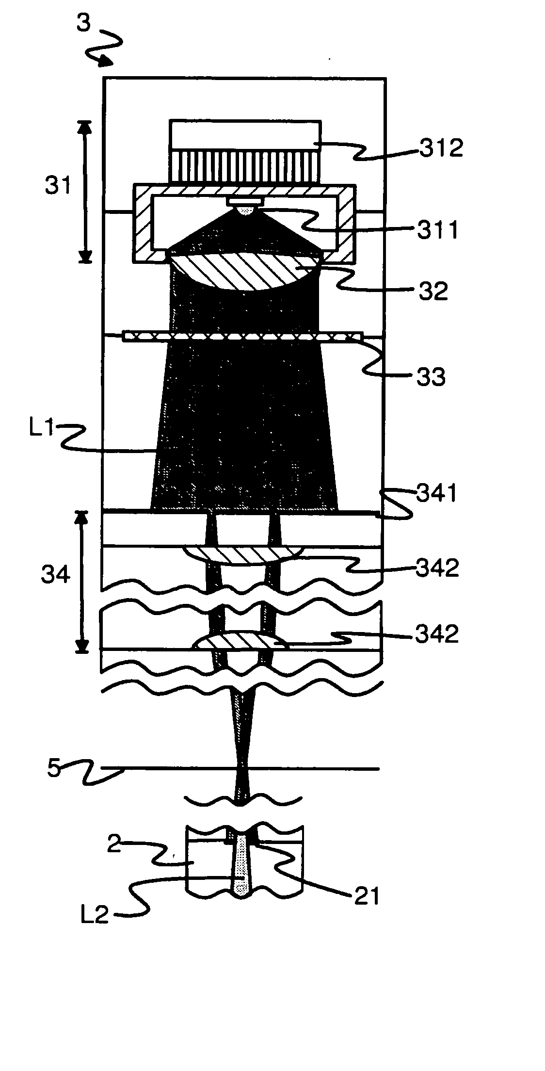
[0006] The basic idea of the invention is that the light required for imaging a living object is produced for the object only when the object is being imaged, and furthermore, the spectrum of the light used is kept narrow. During the relative movement of the imaging apparatus and the object to be imaged, no light is directed to the object. According to an advantageous embodiment of the invention, the illumination is provided by using LED (Light Emitting Diode) technology in which the functional mode is achieved very quickly and the range of emission is known with a substantial accuracy.
[0007] When the time of exposure and the spectrum of light used for the illumination are limited as well as possible, the cells are exposed to a minimum of radiation. The LED achieves its stable functional mode significantly faster than Xenon, for example. Consequently, illumination by LED can be turned on accurately for the time necessary for the imaging only, wherein the cells are not exposed to extra light. In other words, the energy of the radiation to which the cells are exposed can be controlled better, and this, in turn, has a positive effect on the overall result of the examination.
[0008] Furthermore, the light emitted by the LED is almost monochromatic, wherein the light does not cause significant chromatic distortions and the quality of the image is thus significantly improved when compared to the use of wide-spectrum light. By means of the illumination system according to the invention, it is possible to form images of high fidelity without the use of bandpass filters.
[0009] One embodiment of the invention has the advantage that the wavelength can be selected according to the need. In one embodiment, use is made of a LED which can operate at several different wavelengths. The use of several wavelengths makes it possible to take sharp multilayer images of the object easily, because the different wavelengths are focused at different distances. At present, a high-quality bandpass filter is about one decade more expensive than an efficient LED, and only one wavelength band can be separated by one component. By the above-mentioned LED, the wavelength of the light can be changed without the mechanical changing of a filter, which, for its part, makes it possible to take images fast at different wavelengths.

Problems solved by technology

For the research, however, it is problematic that the cells to be cultured under dark conditions must be exposed to light during the imaging.
The energy of the wide-spectrum Xenon light, to which the cells are exposed, is very difficult to control, and furthermore, it takes some time for the lighting level of Xenon light to stabilize.
For this reason, it is possible that when Xenon light is used, cells are exposed to too much light in terms of either the total quantity or the maximum intensity of light energy.

Method used


Image

  • Illumination System For A Microscope
  • Illumination System For A Microscope
  • Illumination System For A Microscope

Examples


Embodiment Construction

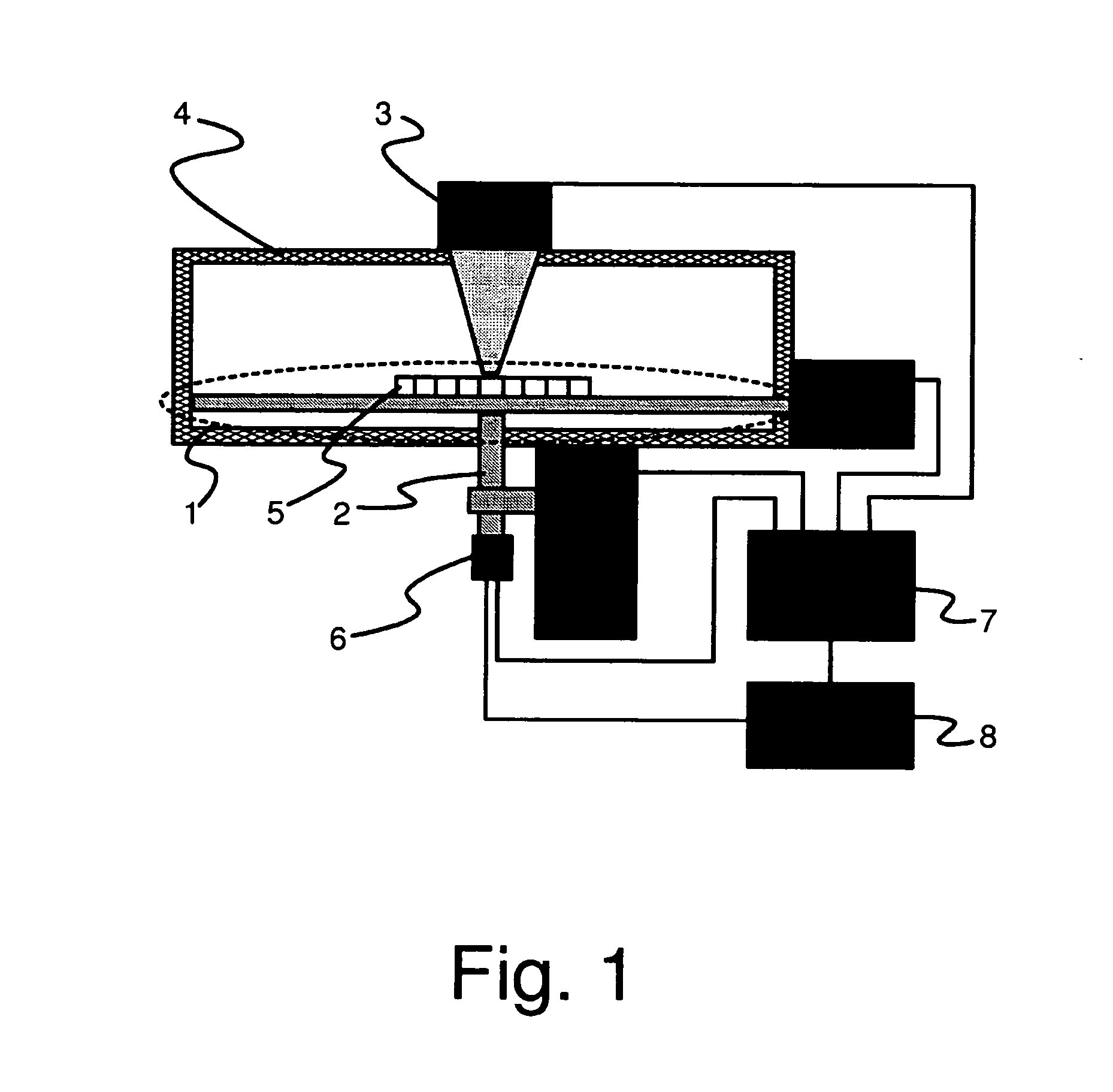
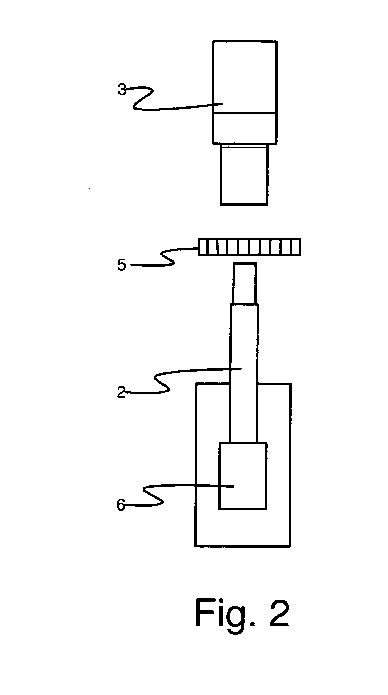
[0018]FIG. 1 shows an apparatus which is suitable, for example, for the culturing and examining of living cells. The apparatus comprises e.g. a well plate station 1, a phase contrast tube microscope 2, and an illuminating device 3. Furthermore, the figure shows a shield structure 4 providing the well plate 5 with a space whose illumination and temperature are controllable. Typically, living cells are preferably kept in the dark at the temperature of 36 to 37 degrees. Furthermore, it is often advantageous to control the composition of the ambient gas around the cells, for example by controlling the content of carbon dioxide and / or oxygen.

[0019] The well plate station 1 according to the example makes it possible to insert a well plate 5 in the apparatus in such a way that the position of the well plate can be changed in the horizontal plane (that is, in the X-Y directions) in relation to the microscope 2. The movement of the well plate 5 with respect to the microscope 2 makes it poss...


PUM


Abstract

An automatic microscope apparatus intended to be used in the phase contrast imaging of living cells. The apparatus includes a sample plate station in which a sample plate containing a sample to be examined can be fitted, as well as a microscope. The microscope includes an optical element, an imager and an illumination arrangement that can be arranged to illuminate the sample. The illumination arrangement includes an LED illuminator to provide illumination.

Description

FIELD OF THE INVENTION [0001] The invention relates to a microscope apparatus according to the preamble of the appended claim 1. Furthermore, the invention relates to the use of an illuminating component according to the appended claim 6. BACKGROUND OF THE INVENTION [0002] Cell culturing is generally used e.g. in various cell biological and biomedical analyses. Typically, the cell material to be analyzed is cultured in a Petri dish or on a well plate placed in suitable conditions with respect to the temperature, ambient gas and illumination. At various stages of the analyses, the samples are subjected to, for example, microscopy, and in arrangements of prior art, the well plate is arranged to be examined with a microscope which may be equipped with a camera. In many studies, the same samples are examined at regular intervals so that the development of the cell can be monitored. For the research, however, it is problematic that the cells to be cultured under dark conditions must be e...

Claims


Application Information

Patent Timeline
IPC IPC(8): G02B21/06G02BG02B21/14
CPCG02B21/14
Inventor KORPINEN, JUHATARVAINEN, JUSSI
Owner CHIP MAN TECH
Learn More