X-ray device having a dual energy mode and method to analyze projection images detected in the dual energy mode

Active Publication Date: 2008-02-21
SIEMENS HEALTHCARE GMBH
View PDF6 Cites 21 Cited by
  • Summary
  • Abstract
  • Description
  • Claims
  • Application Information

AI Technical Summary

Benefits of technology

[0014] The object of the present invention is to provide an evaluation method and the devices corresponding herewith by means of which a simple identification of the vascular system is possible inter alia using the projection images.

Problems solved by technology

The quantification of the blood flow through the myocardium using an angiographic procedure is problematic because the coronary vessels when angiographically observed have a diameter of barely 1 mm or more.
These classifications are, however, complex and can be used only by expending considerable time.
This presents a distinct danger of artifacts.

Method used


Image

  • X-ray device having a dual energy mode and method to analyze projection images detected in the dual energy mode
  • X-ray device having a dual energy mode and method to analyze projection images detected in the dual energy mode
  • X-ray device having a dual energy mode and method to analyze projection images detected in the dual energy mode

Examples


Embodiment Construction

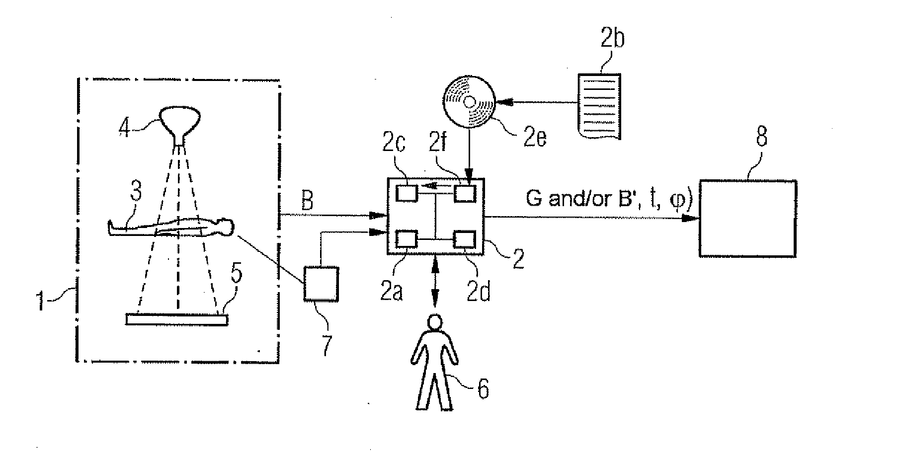
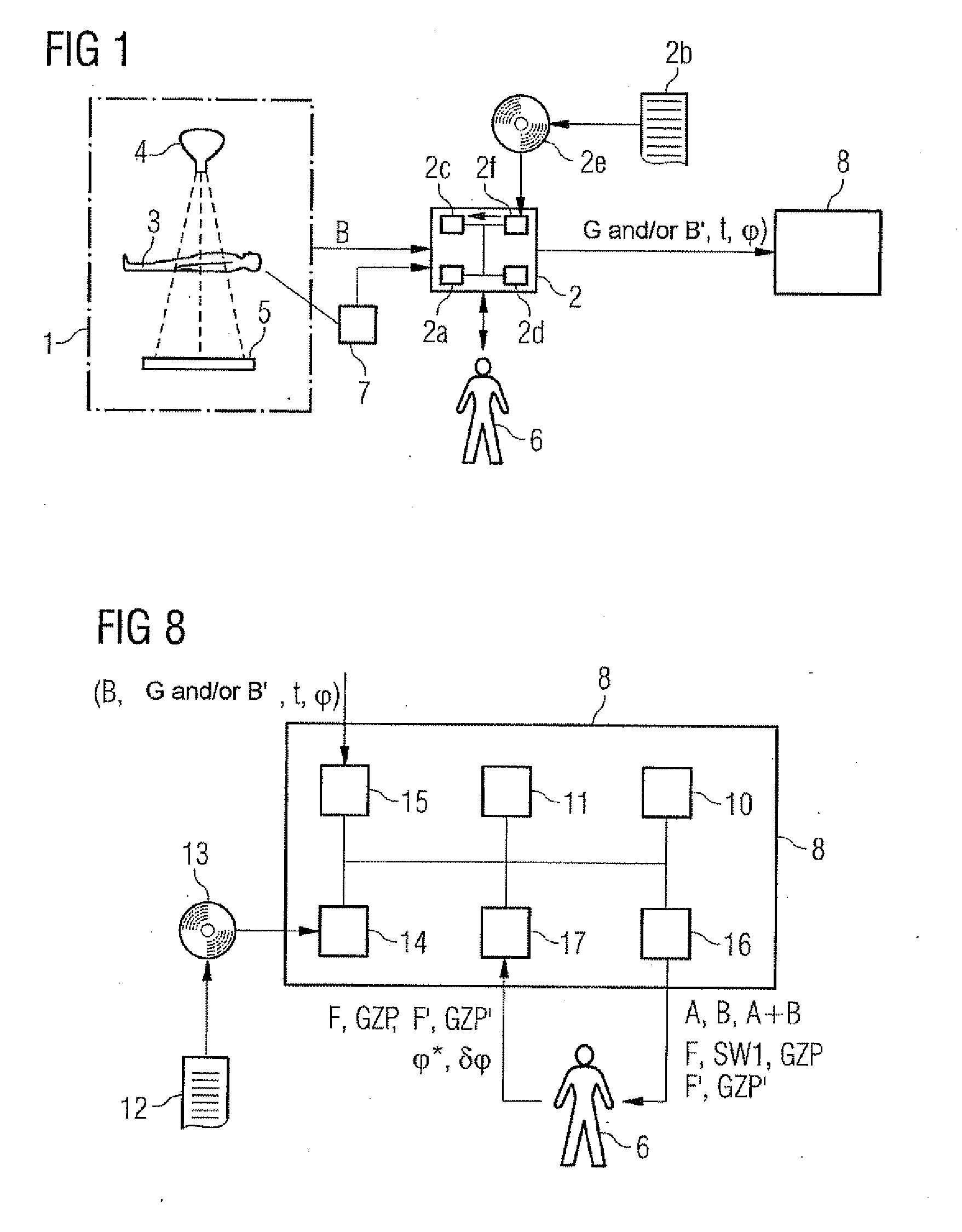
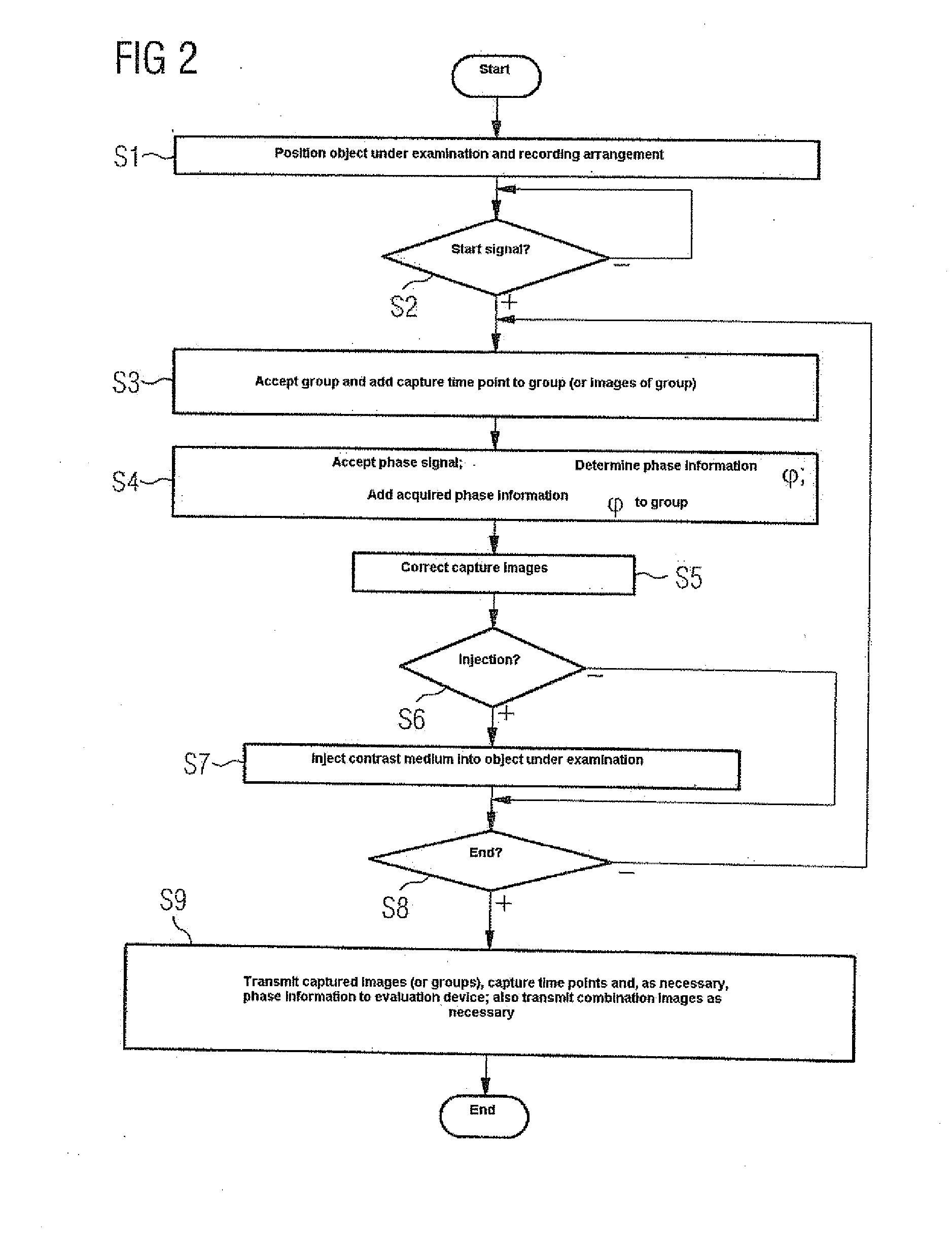
[0067]FIG. 1 is a schematic of an x-ray system. According to FIG. 1, a recording device 1 is controlled by a control device 2. Images B of an object under examination 3 are captured by means of the recording device 1. In this present case in which the object under examination 3 is a human, images B of the heart or of the brain of the human 3 are, for example, captured.

[0068] To capture the images B, the recording arrangement 1 has an x-ray source 4 and the x-ray detector 5.

[0069] The control device 2 is preferably designed as a software-programmed control device 2. It has a processor unit 2a on which in operation a control program 2b is executed. The control program 2b determines the operating mode of the control device 2 and therefore also of the overall x-ray system. The control program 2b is stored in a mass storage unit 2c of the control device 2. It can be called up through an input / output interface 2d by means of a suitable call-up command by an operator 6 of the x-ray syste...


PUM


Abstract

A sequence of groups of projection images shows an object under examination comprising a vascular system and its environment. A computer determines a 2-dimensional evaluation image having a plurality of pixels based on combination images determined from the projection images of a group. The combination images have a plurality of pixels with pixel values. The sequence of the combination images shows the time characteristic of the distribution of a contrast medium in the object. The pixels of the evaluation image correspond to those of the projection images. The computer assigns each pixel, at least in a part area of the evaluation image, a type that is characteristic of whether the respective pixel corresponds to a vessel of the vascular system, a perfusion area or a background. It performs the assignment of the type on the basis of the time characteristic of the pixel values of the combination images.

Description

CROSS REFERENCE TO RELATED APPLICATIONS [0001] The present application claims priority of German application No. 10 2006 025 423.6 filed on May 31, 2006, which is incorporated by reference herein in its entirety. FIELD OF THE INVENTION [0002] The present invention relates to an evaluation method for a sequence of groups of 2-dimensional projection images that show an object under examination that contains a vascular system and its environment, with each projection image having a plurality of pixels with pixel values, with the pixel values of corresponding pixels of the projection images being determined by areas of the object under examination that are at least essentially in the same location. BACKGROUND OF THE INVENTION [0003] Evaluation methods for projection images captured by means of x-ray systems are generally known. [0004] An evaluation method for projection images is known from the technical article “Quantitative Analyse von Koronarangio-graphisichen Bildfolgen zur Bestimmu...

Claims


Application Information

Patent Timeline
IPC IPC(8): G06K9/00
CPCA61B6/4241A61B6/481A61B6/482A61B6/504G01N23/04G06T2207/30101A61B6/507
Inventor BERNHARDT, PHILIPPCAMUS, ESTELLEHOHEISEL, MARTINREDEL, THOMAS
Owner SIEMENS HEALTHCARE GMBH
Why Eureka
Try Eureka