

Method for display of medical 3D image data on a monitor
a 3d image and monitor technology, applied in the field of medical 3d image data display on a monitor, can solve the problems of unaccepted 3d representation methods, the free 3d views available today are unknown or highly unfamiliar to most surgeons and radiologists, and the evaluation of the image data sets is therefore relatively time-consuming
- Summary
- Abstract
- Description
- Claims
- Application Information
AI Technical Summary
Benefits of technology
Problems solved by technology
Method used
Image

Examples
Embodiment Construction
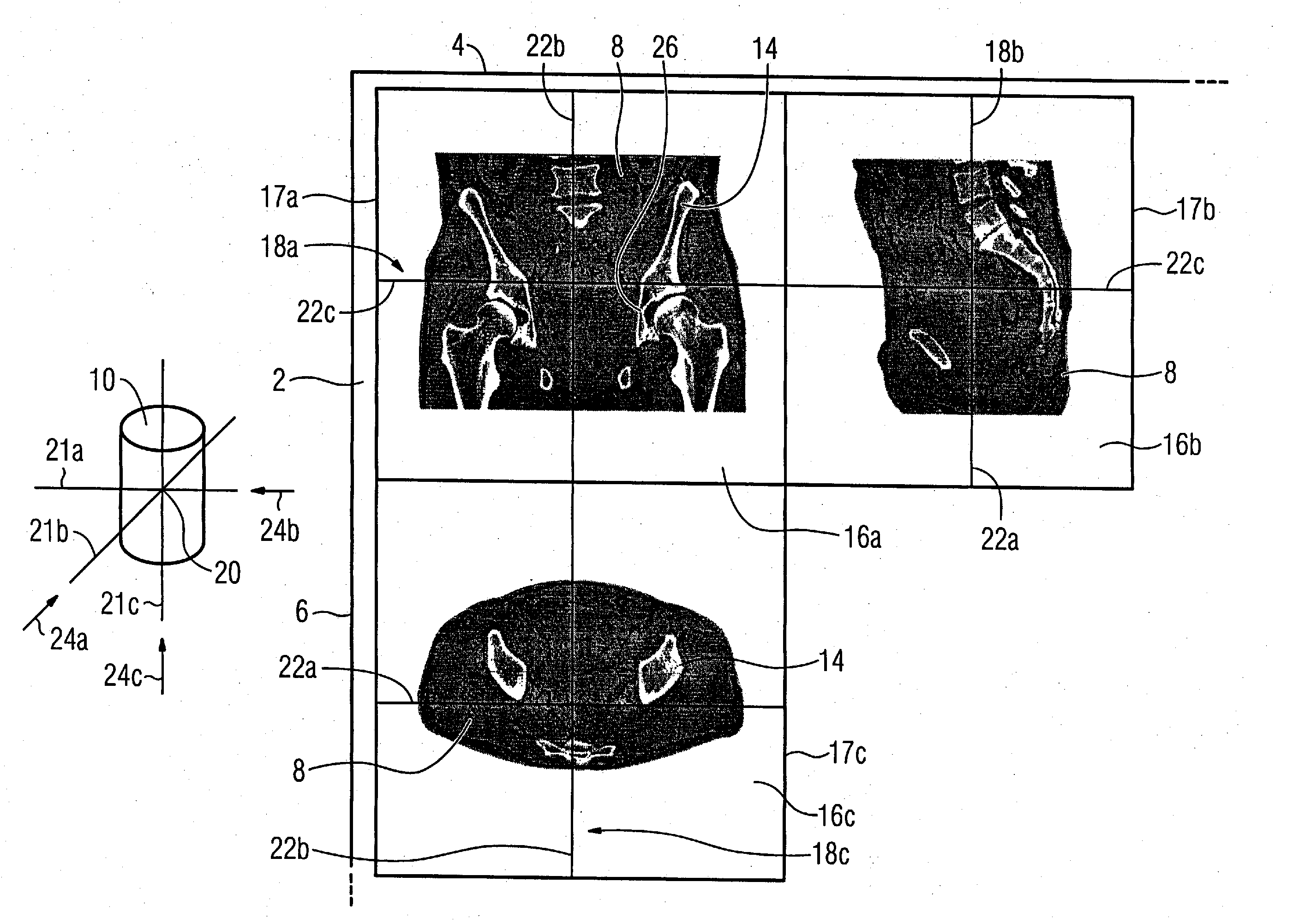
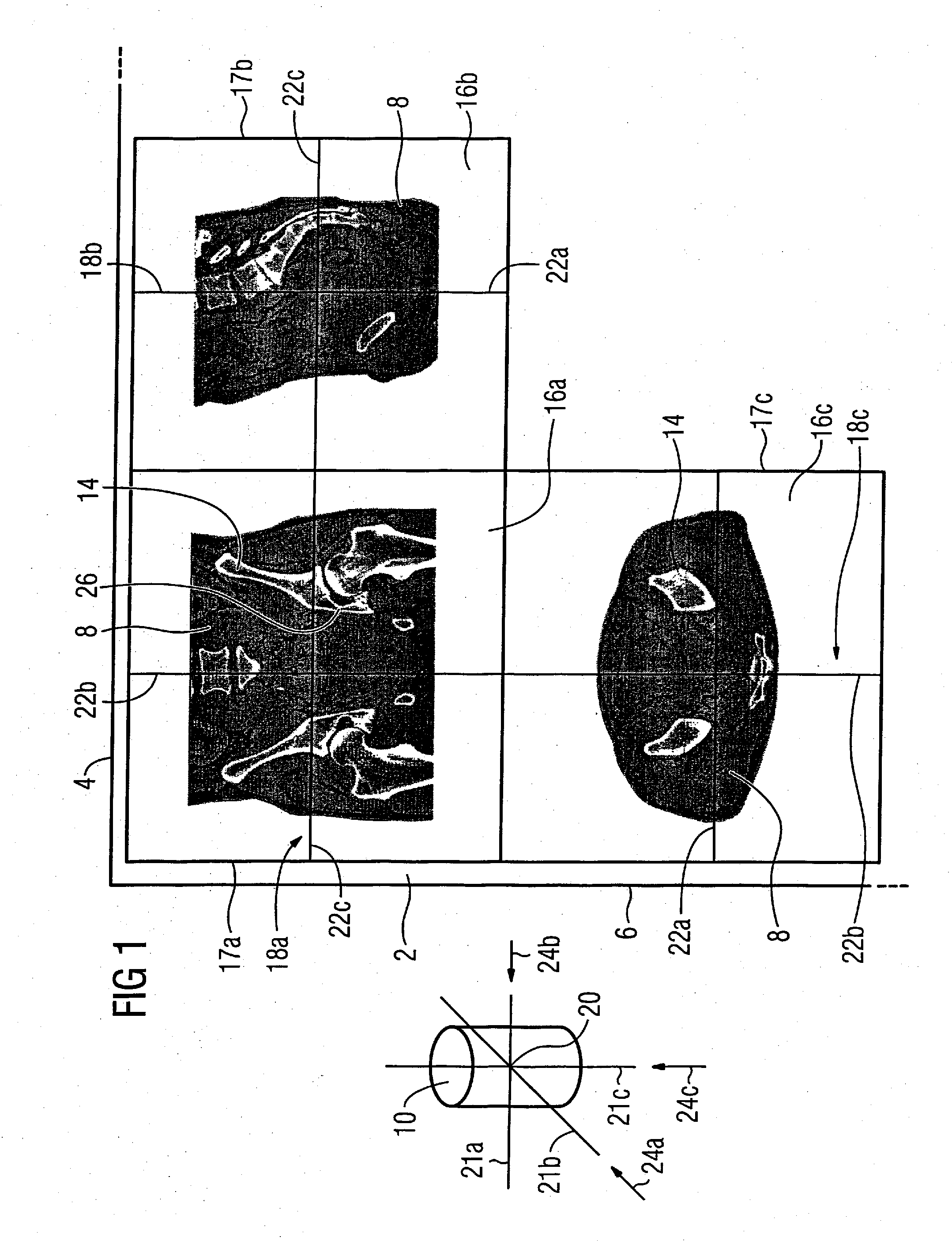
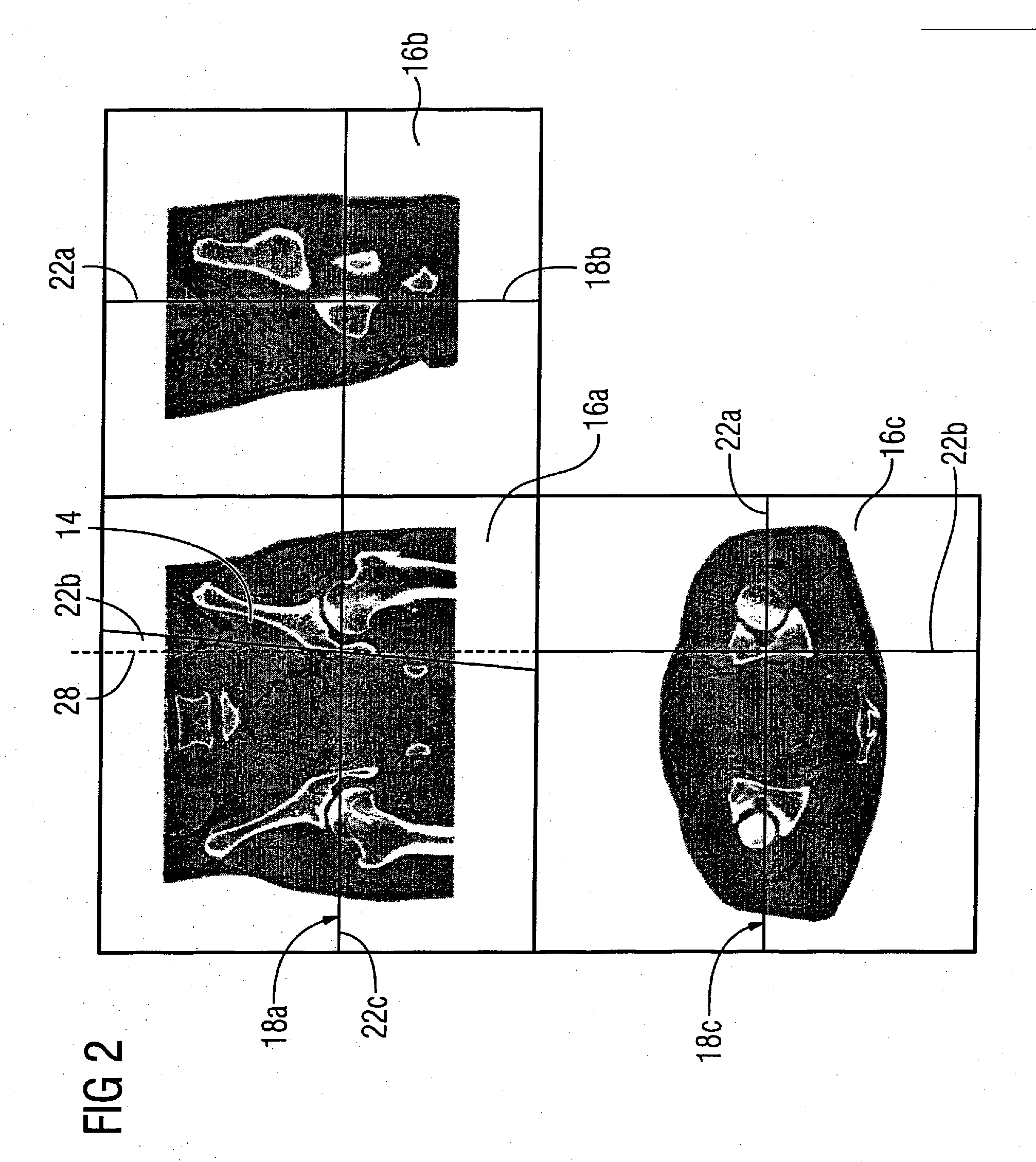
[0046]FIG. 1 shows a section of a monitor 2 of a medical computerized imaging system (not shown). The monitor 2 is proportioned such that its upper monitor edge 4 proceeds horizontally and its lateral monitor edge 6 proceeds approximately vertically. The monitor 2 serves for preoperative planning for a physician (not shown) who has acquired a three-dimensional image data set of a patient 8 in the form of 3D image data 10 by a computerized tomography. The physician wants to adapt a metal plate 12 (not shown in FIG. 1) to the left pelvic bone 14 of the patient 8 with the aid of the 3D image data 10.
[0047] From past experience the physician is accustomed to execute this procedure using two frontal and lateral 2D x-ray exposures (not shown) of the patient. However, in the present example this is executed using the 3D image data 10. Therefore MPR representations of the 3D image data 10 are presented in three windows 16a-16c on the monitor 2. The window 16a which shows a frontal view of ...
PUM
 Abstract
Description
Claims
Application Information