Device for merging a 2D radioscopy image with an image from a 3D image data record

a radioscopy image and image data technology, applied in image enhancement, image analysis, instruments, etc., can solve the problems of spatial orientation of the doctor, limited two-dimensional imaging of fluoroscopy images, and relatively little information, so as to achieve less use of x-rays

Active Publication Date: 2008-06-12
SIEMENS HEALTHCARE GMBH
View PDF8 Cites 23 Cited by
  • Summary
  • Abstract
  • Description
  • Claims
  • Application Information

AI Technical Summary

Benefits of technology

[0012]The result is an image which on the one hand shows the 3D anatomy of the structure, for example of an organ, and on the other hand a radioscopy image, in particular x-ray image, projected thereon—onto this structure. The radioscopy image is primarily used to display interventional instruments. As long as the instrument is inside the organ a projection of the instrument on the internal wall of the organ is displayed in the image displayed. It is true that the three-dimensional position cannot be determined directly from this, since a deviation arises as a result of the back-projection if the direction of view does not correspond exactly to the angulation in the recordi

Problems solved by technology

A frequent problem in these procedures is the spatial orientation of the doctor performing the procedure.
Fluoroscopy images not only have the disadvantage of being limited to two-dimensional imaging, but—unless a contrast agent is used—only provide relatively little information as regards tissue structures.
Soft tissue structures in particular cannot be mapped in fluoroscopy images, or only insufficiently.
T

Method used


Image

  • Device for merging a 2D radioscopy image with an image from a 3D image data record
  • Device for merging a 2D radioscopy image with an image from a 3D image data record
  • Device for merging a 2D radioscopy image with an image from a 3D image data record

Examples


Embodiment Construction

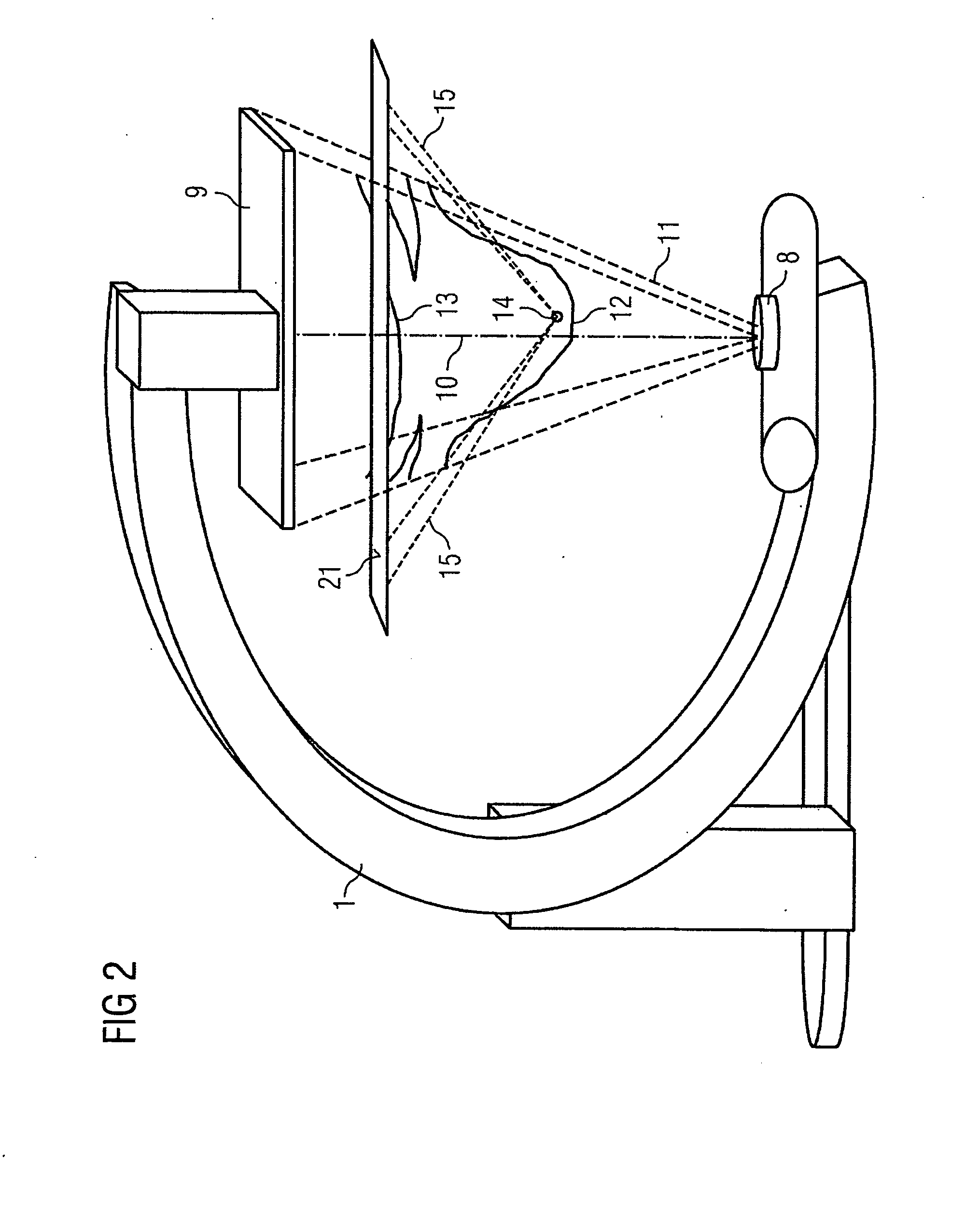
[0020]Preferably the proposed device is part of a C-arm device, with which 2D fluoroscopy images can be recorded during an interventional action on a patient. The device, which can for example be implemented in the image processor of the C-arm device, is here connected to an image display unit, for example a monitor, on which the merged images are displayed.

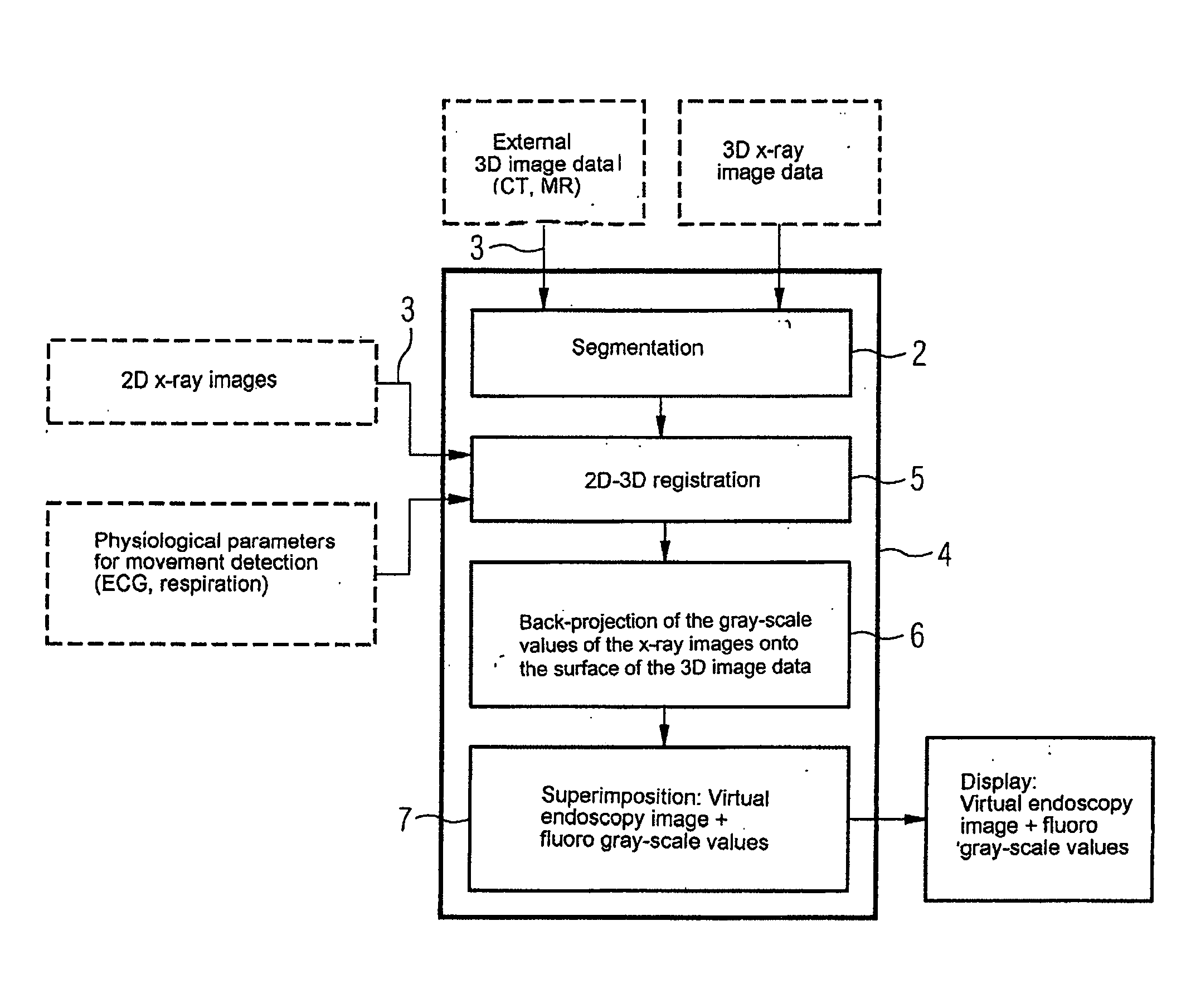
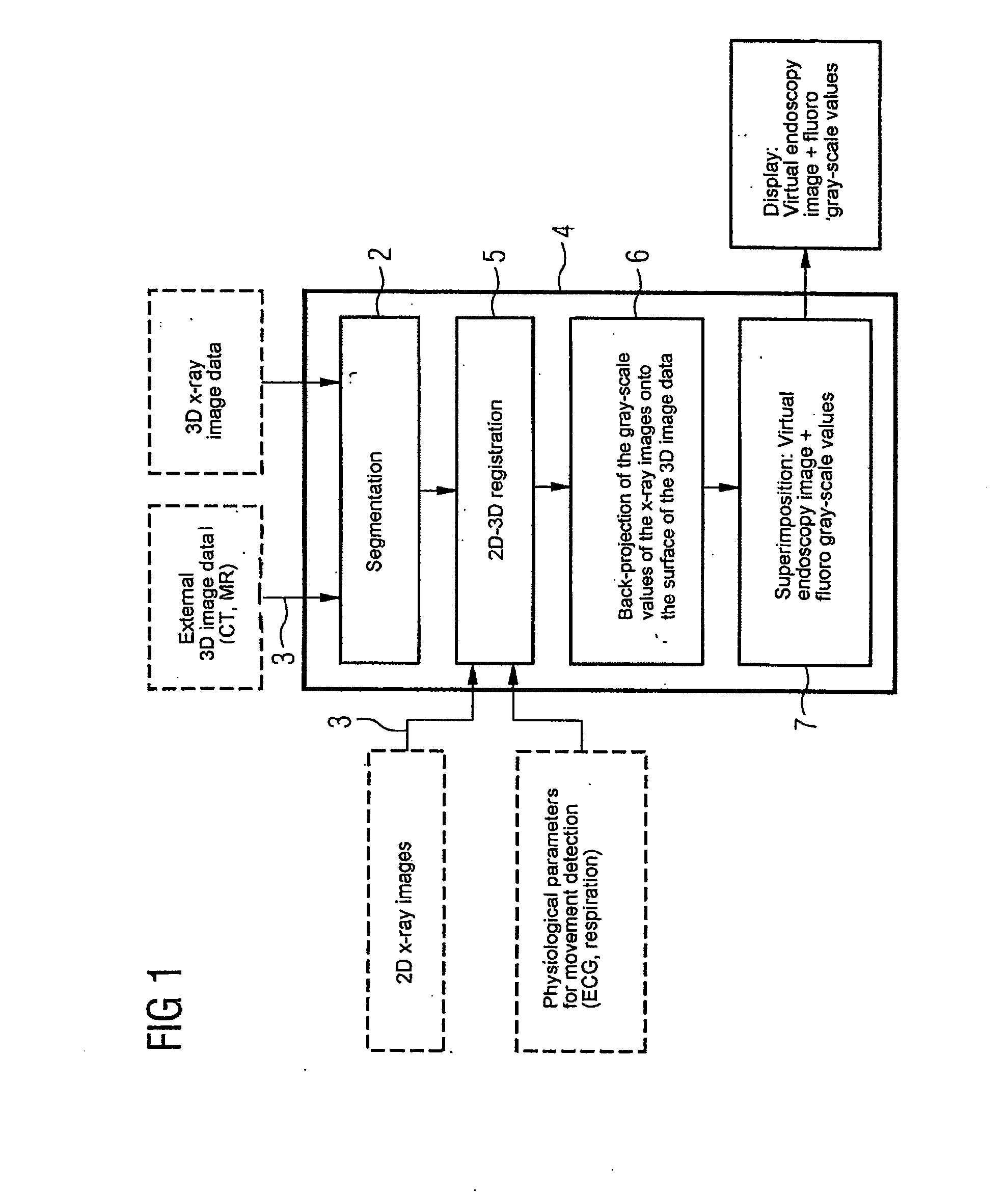
[0021]FIG. 1 shows in diagrammatic form an example of the structure of the proposed device. The device 4 has the following essential components in the example illustrated: a segmentation unit 2, a registration unit 5, a back-projection unit 6 and an image merger unit 7.

[0022]The segmentation unit 2 is designed for preprocessing the 3D image data which is fed to a memory unit (not illustrated) of the device via an interface 3. The 3D image data originates from a 3D imaging method such as, for example, a method of magnetic resonance tomography or computer tomography or was recorded by means of 3D angiography. In the segmentation un...


PUM


Abstract

The invention relates to a device for merging a 2D radioscopy image with an image obtained from a 3D image data record, having a memory unit that stores the 2D radioscopy image and the 3D image data record, a segmentation unit that segments an inner surface of a hollow organ from the 3D image data record, a registration unit that registers the 2D radioscopy image with the 3D image data record, a back-projection unit that back-projects the pixels of the 2D radioscopy image onto the segmented surface, taking account of the projection geometry of the 2D radioscopy image and the registration, and an image merger unit that generates a virtual endoscopy view of the surface from the segmented surface using the back-projected pixels. The device primarily allows 2D radioscopy images to be superimposed during interventional procedures with a fly display of the interior of an interesting organ.

Description

CROSS REFERENCE TO RELATED APPLICATIONS[0001]This application claims priority of German application No. 10 2006 046 735.3 filed Sep. 29, 2006, which is incorporated by reference herein in its entirety.FIELD OF THE INVENTION[0002]The present invention relates to a device for merging a 2D radioscopy image with an image obtained from a 3D image data record, with a memory unit in which data from the 2D radioscopy image and from the 3D image data record obtained via an input interface of the device can be stored, a registration unit that registers the 2D radioscopy image with the 3D image data record, a segmentation unit that segments a surface to be displayed of an interesting structure from the 3D image data record and an image merger unit that merges the 2D radioscopy image with the segmented surface and generates an image display that gives the impression of a 3D image.BACKGROUND OF THE INVENTION[0003]Such devices are primarily used in interventional or surgical procedures in which o...

Claims


Application Information

Patent Timeline
IPC IPC(8): G06K9/00G06T15/08
CPCG06T7/0038G06T2207/10072G06T15/08G06T11/008G06T7/38
Inventor BOESE, JANJOHN, MATTHIASRAHN, NORBERT
Owner SIEMENS HEALTHCARE GMBH
Why Eureka
Try Eureka