Method for assessing image focus quality

a technology of image focus and focus quality, applied in the field of electronic imaging systems, can solve the problems of difficult automatic assessment of focus quality by computers, difficult for computers to determine the distance between the imaged focal plane and the ideal or optimal focal height,

Active Publication Date: 2008-06-12
CYTYC CORP
View PDF15 Cites 84 Cited by
  • Summary
  • Abstract
  • Description
  • Claims
  • Application Information

AI Technical Summary

Benefits of technology

[0015]In another aspect of the invention, a method of focusing on a specimen using an automatic imaging apparatus includes the steps of obtaining three digital images of the specimen at different focal heights using the imaging apparatus. The digital image is formed from a plurality of pixels. A Brenner auto-focus score is calculated for each of the three digital images. An exponentiated quadratic function is fit to the (x, y) points formed by the three focal heights and the corresponding Brenner scores. A new image is acquired at a new focal height based on the function fitted to the data. A new Brenner score is calculated for the new image. Additional images are acquired the different new (e.g., modified) focal heights until the ideal focal height is reached. Of course, the process may stop when the imaging apparatus senses that it is within close proximity to the ideal focal height (e.g., + / − a displacement value).

Problems solved by technology

This requirement poses several technical challenges.
Computerized systems have difficulty with this task.
However, given only a single image, it is difficult for computers to automatically assess focus quality.
Furthermore, it is difficult for a computer to determine how far the imaged focal plane is from the ideal or optimal focal height.
Unfortunately, real images contain noise, and this limits the usefulness of the Fourier transform.
Fourier transform-based methods perform poorly on images having low signal-to-noise (SNR) ratios.
The Fourier transform methods used are also computationally intensive, requiring robust computational hardware and software.
However, in many medical imaging applications where large batches of samples must be processed, frequently performing this sort of auto-focus would require far too much time.

Method used


Image

  • Method for assessing image focus quality
  • Method for assessing image focus quality
  • Method for assessing image focus quality

Examples


Embodiment Construction

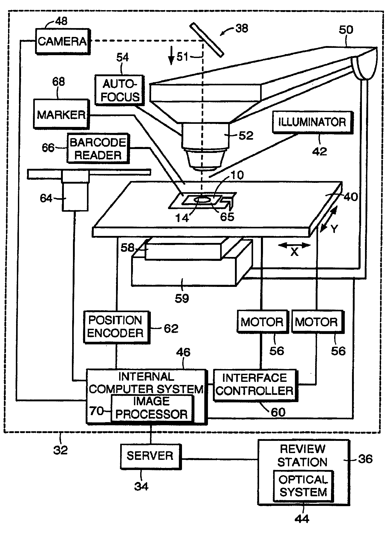
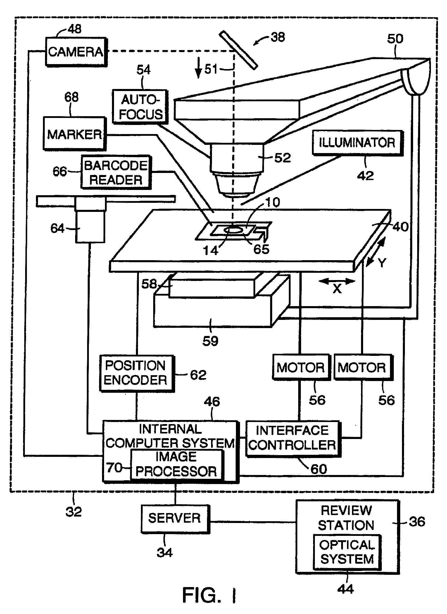
[0022]FIG. 1 illustrates an exemplary specimen imaging apparatus 32 of the present invention. The image processing apparatus 32 includes a first optical system 38, and a slide stage 40 movable relative thereto. A review station 36 is provided and includes a second optical system 44, and is connected to the image processing system 32 via the server 34. An internal computer system 46 controls the first optical system 38 and is in communication with the server 34.

[0023]The first optical system 38 includes an electronic camera 48, such as a CCD camera 48, and a microscope 50. The microscope 50 is preferably an automated microscope. The automated microscope 50 may include features to provide fast, precise imaging of an area of a slide 10 positioned in the optical path 51 of the microscope 50, such as an autofocusing mechanism 54. The first optical system 38 may include one or more lens systems 52. An illuminator 42 may provide illumination for the specimen 14 deposited on the slide 10 an...


PUM


Abstract

A method for determining the quality of focus of a digital image of a biological specimen includes obtaining a digital image of a specimen using a specimen imaging apparatus. A measure of image texture is calculated at two different scales, and the measurements are compared to determine how much high-resolution data the image contains compared to low-resolution data. The texture measurement may, for example, be a Brenner auto-focus score calculated from the means of adjacent pairs of pixels for the high-resolution measurement and from the means of adjacent triples of pixels for the low-resolution measurement. A score indicative of the quality of focus is then established based on a function of the low-resolution and high-resolution measurements. This score may be used by an automated imaging device to verify that image quality is acceptable. The device may adjust the focus and acquire new images to replace any that are deemed unacceptable.

Description

FIELD OF THE INVENTION[0001]The field of the invention generally relates to the field of electronic imaging systems and more particularly to computer-controlled microscopic imaging systems and focusing systems used therein.BACKGROUND OF THE INVENTION[0002]In the field of pathology, it is often necessary to examine tissue samples for underlying pathological or disease states. In some cases, a sample of tissue or other biological material is embedded within an embedding medium and cut into very thin slices. These slices are then placed on sample holders such as slides for subsequent imaging and analysis. In other applications, cells or other biological material are transferred to slides in a liquid-based preparation. For example, cells may be scraped from a target location (e.g., tissue) and optionally washed in a liquid solution. Alternatively, cells may be obtained directly from a bodily fluid (e.g., urine). The cells may be transferred directly to a slide, for example, blood sample...

Claims


Application Information

Patent Timeline
Patent Type & Authority Applications(United States)
IPC IPC(8): G06K9/40
CPCG02B21/244G06T2207/30168G06T7/0002
Inventor ZAHNISER, MICHAEL
Owner CYTYC CORP
Why Eureka
Try Eureka