Method and apparatus for sensitivity-encoded magnetic resonance imaging using an acquisition coil array

Active Publication Date: 2008-06-12
SIEMENS HEALTHCARE GMBH
View PDF5 Cites 8 Cited by
  • Summary
  • Abstract
  • Description
  • Claims
  • Application Information

AI Technical Summary

Benefits of technology

[0020]An object of the present invention is to provide a method in which additional measurements (calibration measurements) for precise detection of coil sensitivities can be foregone in order to shorten the total measurement time of a parallel acquisition method, in par

Problems solved by technology

In spite of the technical advances in the design of MRT apparatuses, the acquisition time of an MRT image remains the limiting factor for many applications of MRT in medical diagnostics.
The longer (in comparison to Cartesian scanning) measurement time that is required for nominally equal spatial resolution is disadvantageous in the case of a non-Cartesian radial, spiral or arbitrary further two- or three-dimensional scanning of the frequency domain.
Such a block, however, represents a limitation with regard to the reconstruction precisi

Method used


Image

  • Method and apparatus for sensitivity-encoded magnetic resonance imaging using an acquisition coil array
  • Method and apparatus for sensitivity-encoded magnetic resonance imaging using an acquisition coil array
  • Method and apparatus for sensitivity-encoded magnetic resonance imaging using an acquisition coil array

Examples


Embodiment Construction

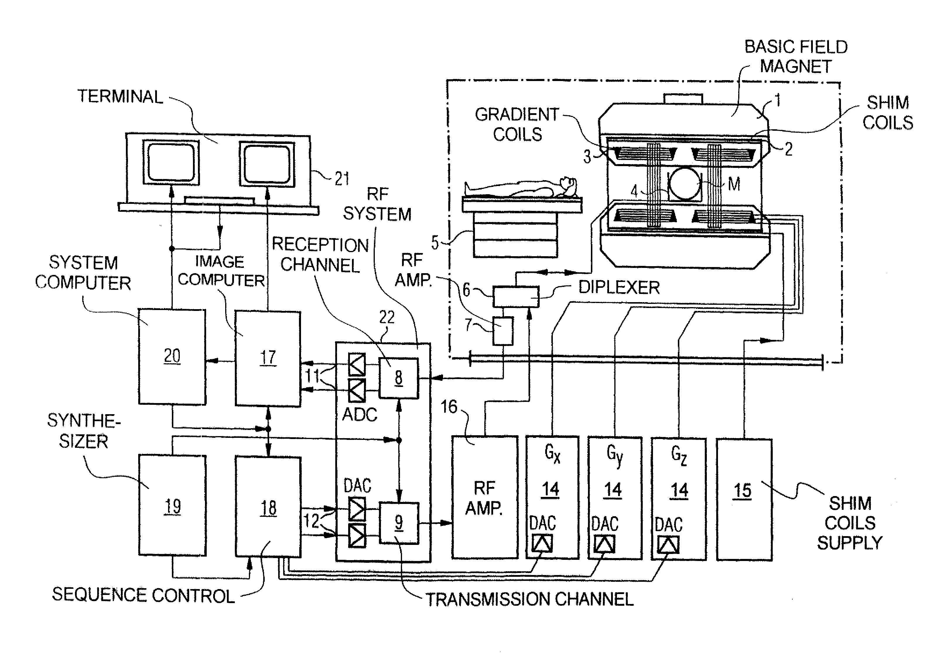
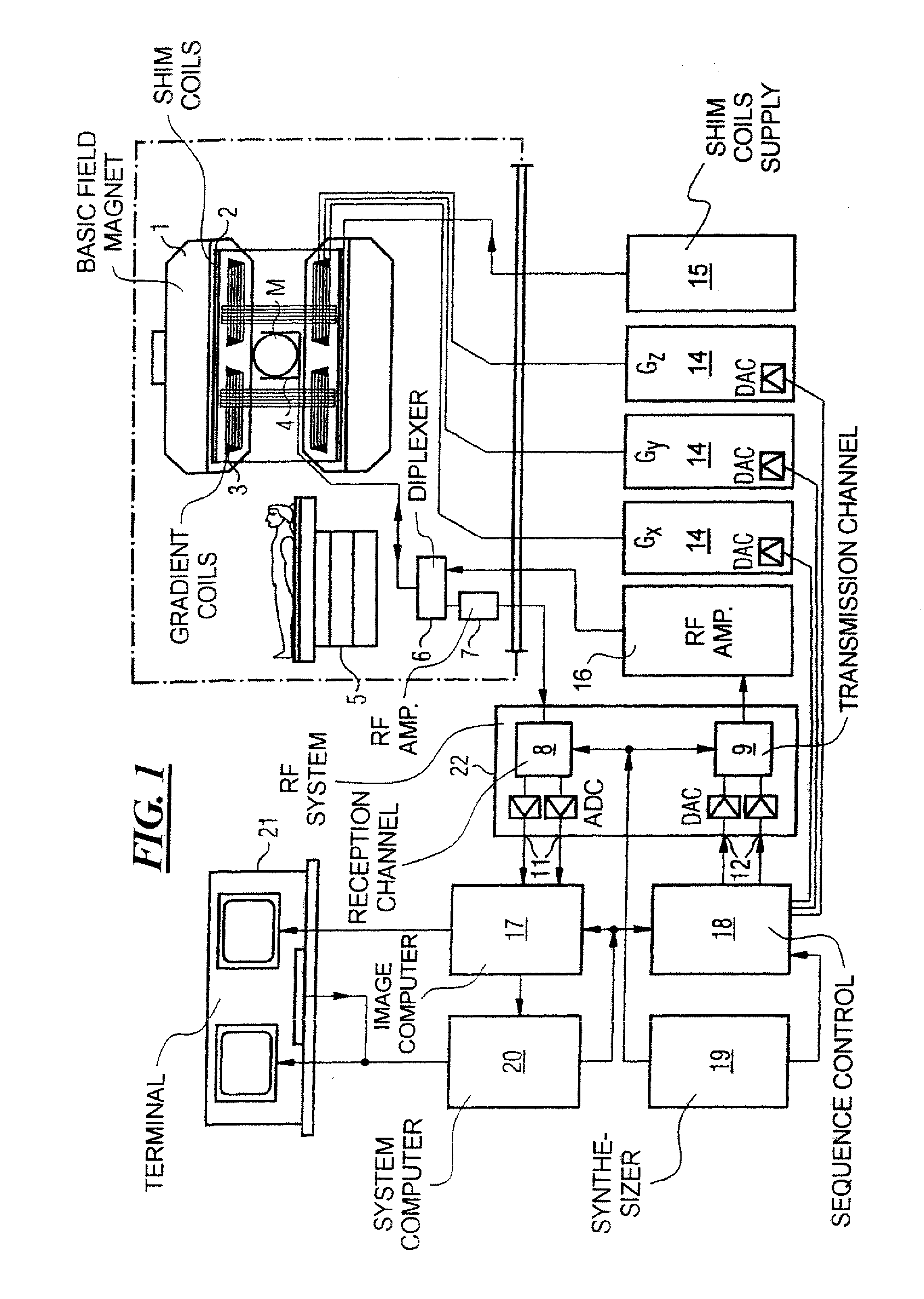
[0041]FIG. 1 schematically illustrates a magnetic resonance imaging or tomography apparatus for generation of a magnetic resonance image of a subject according to the present invention. The basic design of the magnetic resonance tomography apparatus corresponds to that of a conventional tomography apparatus, but with the important differences discussed below. A basic field magnet 1 generates a temporally-constant strong magnetic field for polarization or alignment of the nuclear spins in the examination region of a subject such as, for example, of a part of a human body to be examined. The high homogeneity of the basic magnetic field necessary for the magnetic resonance measurement is defined in a typically spherical measurement volume MV, into which the parts of the human body to be examined are introduced. To support the homogeneity requirements, and in particular to eliminate temporally invariable influences, shim plates made of ferromagnetic material are mounted at a suitable lo...


PUM


Abstract

In a method as well as a magnetic resonance tomography apparatus for implementation of such a method for improved sensitivity-encoded magnetic resonance imaging using a two-dimensional or three-dimensional acquisition coil array, two-dimensional or three-dimensional undersampling of k-space is undertaken by measurement of a number N of basic partial trajectories τn in k-space that in their entirety form a geometric arrangement of source points, a number M of different operators Cm(Δkm) are determined, with each operator representing an algebraic transformation with which unmeasured target points at an interval Δkm from one of the measured source points are synthesized from a number of measured source points, the operators Cm(Δkm) are applied to at least one subset of the measured source points for at least partial completion of the magnetic resonance data set, and a largely artifact-free image is reconstructed in three-dimensional space on the basis of the measured source points and the synthesized data points.

Description

BACKGROUND OF THE INVENTION[0001]1. Field of the Invention[0002]The present invention generally concerns magnetic resonance tomography (MRT) as used in medicine for examination of patients. The present invention particularly concerns a method as well as a magnetic resonance tomography apparatus for implementation of such a method in which two-dimensional or three-dimensional sensitivity-encoded magnetic resonance imaging using an acquisition coil array for partially parallel acquisition (PPA) is employed for completion of under-sampled magnetic resonance data sets.[0003]2. Description of the Prior Art[0004]MRT is based on the physical phenomenon of nuclear magnetic resonance and has been successfully used as an imaging method for over 20 years in medicine and biophysics. In this examination modality the subject is exposed to a strong, constant magnetic field. The nuclear spins of the atoms in the subject, which were previously randomly oriented, thereby align. Radio-frequency energy...

Claims


Application Information

Patent Timeline
IPC IPC(8): A61B5/055
CPCG01R33/5611G01R33/4824
Inventor KANNENGIESSER, STEPHANLOEFFLER, RALFMUELLER, EDGARSCHMITT, PETER
Owner SIEMENS HEALTHCARE GMBH
Why Eureka
Try Eureka